. 2000 Feb;20(4):1344-60.
doi: 10.1128/MCB.20.4.1344-1360.2000.

Epstein-Barr virus suppresses a G(2)/M checkpoint activated by genotoxins

Affiliations

Epstein-Barr virus suppresses a G(2)/M checkpoint activated by genotoxins

M Wade et al. Mol Cell Biol. 2000 Feb.

Abstract

Several Epstein-Barr virus (EBV)-negative Burkitt lymphoma-derived cell lines (for example, BL41 and Ramos) are extremely sensitive to genotoxic drugs despite being functionally null for the tumor suppressor p53. They rapidly undergo apoptosis, largely from G(2)/M of the cell cycle. 5-bromo-2'-deoxyuridine labeling experiments showed that although the treated cells can pass through S phase, they are unable to complete cell division, suggesting that a G(2)/M checkpoint is activated. Surprisingly, latent infection of these genotoxin-sensitive cells with EBV protects them from both apoptosis and cell cycle arrest, allowing them to complete the division cycle. However, a comparison with EBV-immortalized B-lymphoblastoid cell lines (which have functional p53) showed that EBV does not block apoptosis per se but rather abrogates the activation of, or signalling from, the checkpoint in G(2)/M. Furthermore, analyses of BL41 and Ramos cells latently infected with P3HR1 mutant virus, which expresses only a subset of the latent viral genes, showed that LMP-1, the main antiapoptotic latent protein encoded by EBV, is not involved in the protection afforded here by viral infection. This conclusion was confirmed by analysis of clones of BL41 stably expressing LMP-1 from a transfected plasmid, which respond like the parental cell line. Although steady-state levels of Bcl-2 and related proteins varied between BL41 lines and clones, they did not change significantly during apoptosis, nor was the level of any of these anti- or proapoptotic proteins predictive of the outcome of treatment. We have demonstrated that a subset of EBV latent gene products can inactivate a cell cycle checkpoint for monitoring the fidelity and timing of cell division and therefore genomic integrity. This is likely to be important in EBV-associated growth transformation of B cells and perhaps tumorigenesis. Furthermore, this study suggests that EBV will be a unique tool for investigating the intimate relationship between cell cycle regulation and apoptosis.


Figures
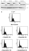
FIG. 1
FIG. 1
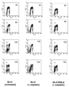
Various genotoxic drugs induce PARP cleavage but not necessarily a sub-G1 population in FACS analysis. BL41 cells were treated for 16 h with genotoxins (cisplatin, 10 μg/ml; doxorubicin, 5 μg/ml; etoposide, 10 μg/ml; camptothecin, 3 μg/ml). (A) Cells were harvested at 4-h intervals and assessed for PARP cleavage by Western blotting: (0 h [tracks 1, 6, 11, and 16], 4 h [tracks 2, 7, 12, and 17], 8 h [tracks 3, 8, 13, and 18], 12 h [tracks 4, 9, 14, and 19], and 16 h [tracks 5, 10, 15, and 20]). (B) Samples were also subjected to FACS analysis for cell cycle distribution. Cell cycle profiles corresponding to time points at which PARP cleavage was judged to be 100% are shown.
FIG. 2
FIG. 2
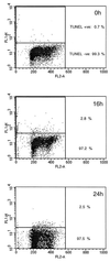
After treatment with cisplatin (10 μg/ml), BL41 cells pass through S phase but do not divide and reenter G1. BL41 cells were pulsed with BrdU and harvested at time intervals shown in the top right of each plot prior to staining with FITC-labeled anti-BrdU antibody (y axis) and PI (x axis). (A) Untreated cells. An asterisk indicates cells which have traversed S and completed mitosis since the start of the experiment. (B) Cells incubated in the presence of cisplatin. Note the absence of cells which have completed mitosis. (C) Schematic diagram of asynchronously growing cells labeled with BrdU. (i) G0/G1 cells which did not incorporate BrdU during the labeling period; (ii) labeled cells in early- and mid-S phase; (iii) cells in late S phase and G2/M; (iv) unlabeled cells in G2/M. The area above the dotted line was electronically gated to examine progression of S-phase cells through the cell cycle during the course of the subsequent experiments.
FIG. 2
FIG. 2
After treatment with cisplatin (10 μg/ml), BL41 cells pass through S phase but do not divide and reenter G1. BL41 cells were pulsed with BrdU and harvested at time intervals shown in the top right of each plot prior to staining with FITC-labeled anti-BrdU antibody (y axis) and PI (x axis). (A) Untreated cells. An asterisk indicates cells which have traversed S and completed mitosis since the start of the experiment. (B) Cells incubated in the presence of cisplatin. Note the absence of cells which have completed mitosis. (C) Schematic diagram of asynchronously growing cells labeled with BrdU. (i) G0/G1 cells which did not incorporate BrdU during the labeling period; (ii) labeled cells in early- and mid-S phase; (iii) cells in late S phase and G2/M; (iv) unlabeled cells in G2/M. The area above the dotted line was electronically gated to examine progression of S-phase cells through the cell cycle during the course of the subsequent experiments.
FIG. 3
FIG. 3
BL41 cells treated with cisplatin fail to complete mitosis and instead undergo apoptosis. Cells were electronically gated as described for Fig. 2C. A significant proportion of untreated BrdU-labeled BL41 cells (A) have returned to G1 phase by 12 h, while BL41 cells treated with cisplatin (B) are unable to complete mitosis (compare graphs 5 in panels A and B). The apparent increase in cells in S phase (indicated with an arrow) in graph 6 is derived from cells which have undergone apoptosis from G2/M.
FIG. 4
FIG. 4
EBV protects against cisplatin-induced apoptosis. BL41 and BL41/B95.8 cells were incubated with cisplatin (10 μg/ml), harvested at 4-h intervals, and assessed for PARP cleavage by Western blotting. Note the complete absence of PARP cleavage even at 16 h in the EBV-positive cells. The BL41/B95.8 cells used were judged to be 100% EBV positive by immunofluorescence staining with an anti-EBNA-LP MAb (not shown).
FIG. 5
FIG. 5
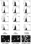
BL41/B95.8 cells complete cell division after treatment with cisplatin (10 μg/ml). While by 12 h a significant proportion of untreated BL41 cells have completed mitosis (asterisk, 12 h), cisplatin-treated cells are unable to do so and instead undergo apoptosis (middle column). However, latent infection with EBV allows BL41 cells to complete mitosis in the presence of cisplatin (right column).
FIG. 6
FIG. 6
(A) Electronically gated BrdU-labeled BL41/B95.8 cells. The data presented in Fig. 5 were electronically gated as described for Fig. 2. Note the reemergence of a BrdU-labeled G1 population of cisplatin-treated BL41/B95.8 cells (right column, 12h and 16h) which is absent in treated parental BL41 cells (middle column). (B) Morphology as shown by acridine orange staining. BL41 cells treated with cisplatin for 16 h (center) all have a characteristic apoptotic morphology. Untreated BL41 and cisplatin-treated BL41/B95.8 cells show the regular nuclear morphology of healthy proliferating B cells.
FIG. 7
FIG. 7
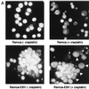
De novo infection of Ramos cells with B95.8 EBV confers resistance to cisplatin-induced apoptosis. (A) Cells were harvested and stained with acridine orange before (left) or 16 h after (right) incubation with cisplatin. Note the aggregation of cells in the infected Ramos culture which is characteristic of EBV infection of B cells (lower left). While uninfected Ramos cells treated for 16 h with cisplatin (upper right) are almost all apoptotic, cells within the aggregates in the infected cells appear morphologically normal (lower right, arrowheads), with occasional single apoptotic cells (arrows). (B) Confocal microscopy after dual staining of parental Ramos (upper panels) and infected Ramos (lower panels) cells with TUNEL for apoptosis (green) and EBNA-LP as a marker of EBV gene expression (red). The majority of parental, uninfected Ramos cells are TUNEL positive after treatment with cisplatin (upper right). In the Ramos-EBV cells treated with cisplatin, there was no colocalization of TUNEL and EBNA-LP staining (merged image, lower right panels). The split image clearly shows that cellular TUNEL and EBNA-LP staining are mutually exclusive and that the effect was not simply due to a masking of one fluorescence with the other.
FIG. 7
FIG. 7
De novo infection of Ramos cells with B95.8 EBV confers resistance to cisplatin-induced apoptosis. (A) Cells were harvested and stained with acridine orange before (left) or 16 h after (right) incubation with cisplatin. Note the aggregation of cells in the infected Ramos culture which is characteristic of EBV infection of B cells (lower left). While uninfected Ramos cells treated for 16 h with cisplatin (upper right) are almost all apoptotic, cells within the aggregates in the infected cells appear morphologically normal (lower right, arrowheads), with occasional single apoptotic cells (arrows). (B) Confocal microscopy after dual staining of parental Ramos (upper panels) and infected Ramos (lower panels) cells with TUNEL for apoptosis (green) and EBNA-LP as a marker of EBV gene expression (red). The majority of parental, uninfected Ramos cells are TUNEL positive after treatment with cisplatin (upper right). In the Ramos-EBV cells treated with cisplatin, there was no colocalization of TUNEL and EBNA-LP staining (merged image, lower right panels). The split image clearly shows that cellular TUNEL and EBNA-LP staining are mutually exclusive and that the effect was not simply due to a masking of one fluorescence with the other.
FIG. 8
FIG. 8
EBV cannot prevent genotoxin-induced apoptosis per se. In an independent measure of apoptosis, cells were treated with cisplatin (10 μg/ml) before harvesting at the indicated time points and assessed for apoptosis (y axis) using FITC-labeled nucleotide incorporation (the TUNEL assay) and DNA content using PI (x axis). Consistent with PARP cleavage data (Fig. 4), BL41/B95.8 cells are protected against cisplatin-induced apoptosis (compare middle and left columns). However, expression of the same viral latent genes in the lymphoblastoid cell line AS-LCL (right column) is unable to protect these cells from genotoxin-induced apoptosis.
FIG. 9
FIG. 9
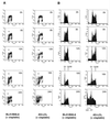
LCLs treated with cisplatin (10 μg/ml) also traverse G2/M but subsequently die from G1/S. BrdU labeling experiments were as described for Fig. 2. (A) Both BL41/B95.8 cells and AS-LCL cells are able to complete mitosis in the presence of cisplatin (indicated by asterisks). (B) The appearance of cells with G1 DNA content after electronically gating confirmed this. However, AS-LCLs (presumably due to activation of p53 at the G1/S transition) subsequently undergo apoptosis and exhibit a sub-G1 distribution (B, 24h, right column).
FIG. 10
FIG. 10
LMP-1 is not required or necessary for protection. (A) Western blots showing LMP-1 was not expressed in uninfected BL41 cells (tracks 1 to 5), cells infected with P3HR1 virus (tracks 11 to 15), or cells transfected with empty vector (tracks 26 to 30) but was expressed at constant levels in cells infected with B95.8 virus (tracks 6 to 10) and cells transfected with LMP-1 expression vectors, BL41/MTLM-5 (tracks 16 to 20) and BL41/MTLM-11 (tracks 21 to 25) (54). The level of LMP-1 expression in an LCL is shown for comparison (tracks 31 to 35). The apparent lack of expression in tracks 17 and 18 resulted from faulty transfer of protein to the nitrocellulose filter. Each set of five lanes represents a time course over 16 h after the addition of cisplatin. Samples were taken every 4 h. (B) Western blots showing PARP cleavage from the same time course. BL41/MTLM-5 and -11 cells expressed levels of LMP-1 similar to the level produced by BL41/B95.8 and were as sensitive to cisplatin-induced apoptosis as the empty vector control, BL41/gpt. Conversely, BL41/P3HR1 cells expressed no LMP-1; they were protected against cisplatin-induced apoptosis.
FIG. 11
FIG. 11
The P3HR1 strain of EBV prevents apoptosis in BL41 cells, as judged by TUNEL assay. Independent confirmation that P3HR1 virus protects against genotoxin-induced apoptosis was obtained using the TUNEL assay as described for Fig. 8. Note complete absence of TUNEL positivity even 24 h after treatment.
FIG. 12
FIG. 12
The P3HR1 strain also prevents apoptosis and allows cell division in Ramos cells treated with cisplatin (10 μg/ml). (A) A Western blot showed that although Ramos cells were sensitive to cisplatin-induced apoptosis as measured by PARP cleavage, two independent sublines of Ramos infected with the P3HR1 strain of EBV (AW- and EHRB-Ramos) were protected over the same 16-h time course. (B) After exposure to cisplatin, Ramos cells fail to complete mitosis [compare 16h Ramos (+ cisplatin) and (− cisplatin)], whereas the P3HR1 EBV-infected EHRB-Ramos cell line is able to complete mitosis [EHRB (+ cisplatin); the asterisk indicates the labeled G1 cells]. (C) Cell cycle profiles of BrdU-labeled populations were electronically gated as described for Fig. 2.
FIG. 12
FIG. 12
The P3HR1 strain also prevents apoptosis and allows cell division in Ramos cells treated with cisplatin (10 μg/ml). (A) A Western blot showed that although Ramos cells were sensitive to cisplatin-induced apoptosis as measured by PARP cleavage, two independent sublines of Ramos infected with the P3HR1 strain of EBV (AW- and EHRB-Ramos) were protected over the same 16-h time course. (B) After exposure to cisplatin, Ramos cells fail to complete mitosis [compare 16h Ramos (+ cisplatin) and (− cisplatin)], whereas the P3HR1 EBV-infected EHRB-Ramos cell line is able to complete mitosis [EHRB (+ cisplatin); the asterisk indicates the labeled G1 cells]. (C) Cell cycle profiles of BrdU-labeled populations were electronically gated as described for Fig. 2.
FIG. 13
FIG. 13
Western blots of sublines to validate the viral strains in the latently infected cells. The B95.8 strain of EBV expresses the nuclear antigens EBNA1 and EBNA2, and these were detected in both BL41/B95.8 and an LCL established with this virus (tracks 2 and 4, respectively). P3HR1 does not express EBNA2 due to a genomic deletion; the presence of virus was instead confirmed by immunoblotting infected BL41 and Ramos BL cell lines with human serum which detected EBNA1 (tracks 3, 6, and 7). It should be noted that an EBNA1 protein with an electrophoretic mobility slightly faster than that from B95.8 (X) is characteristic of a cross-reactive cellular protein recognized by the human serum.
FIG. 14
FIG. 14
Western blots showing the expression of Bcl-2 family proteins. BL41 and BL41 cell lines infected with the B95.8 or P3HR1 strain of EBV were exposed to cisplatin (10 μg/ml) for 16 h, and levels of Bcl-2 and related proteins was determined from samples taken at 4-h intervals. Levels of antiapoptotic Bcl-2 and Bcl-XL and the proapoptotic protein Bax are similar and remain unchanged in all three cell lines during the experiment. The levels of the antiapoptotic protein Mcl-1 decreased slightly in all three cell lines.

Similar articles

Cited by

References

    1. Agami R, Blandino G, Oren M, Shaul Y. Interaction of c-Abl and p73alpha and their collaboration to induce apoptosis. Nature. 1999;399:809–813. - PubMed
    1. Allday M J, Inman G J, Crawford D H, Farrell P J. DNA damage in human B cells can induce apoptosis, proceeding from G1/S when p53 is transactivation competent and G2/M when it is transactivation defective. EMBO J. 1995;14:4994–5005. - PMC - PubMed
    1. Allday M J, Sinclair A, Parker G, Crawford D H, Farrell P J. Epstein-Barr virus efficiently immortalizes human B cells without neutralizing the function of p53. EMBO J. 1995;14:1382–1391. - PMC - PubMed
    1. Blasina A, Paegle E S, McGowan C H. The role of inhibitory phosphorylation of CDC2 following DNA replication block and radiation-induced damage in human cells. Mol Biol Cell. 1997;8:1013–1023. - PMC - PubMed
    1. Brown J M, Wouters B G. Apoptosis, p53, and tumor cell sensitivity to anticancer agents. Cancer Res. 1999;59:1391–1399. - PubMed

Publication types

MeSH terms