

Involvement of both the V2 and V3 regions of the CCR5-tropic human immunodeficiency virus type 1 envelope in reduced sensitivity to macrophage inflammatory protein 1alpha
- PMID: 10644351
- PMCID: PMC111656
- DOI: 10.1128/jvi.74.4.1787-1793.2000
Involvement of both the V2 and V3 regions of the CCR5-tropic human immunodeficiency virus type 1 envelope in reduced sensitivity to macrophage inflammatory protein 1alpha
Abstract
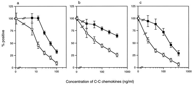
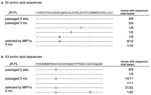
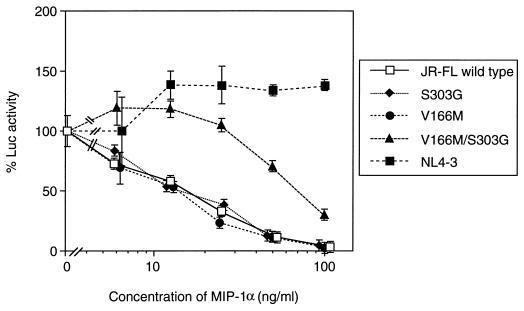
To determine whether C-C chemokines play an important role in the phenotype switch of human immunodeficiency virus (HIV) from CCR5 to CXCR4 usage during the course of an infection in vivo, macrophage inflammatory protein (MIP)-1alpha-resistant variants were isolated from CCR5-tropic (R5) HIV-1 in vitro. The selected variants displayed reduced sensitivities to MIP-1alpha (fourfold) through CCR5-expressing CD4-HeLa/long terminal repeat-beta-galactosidase (MAGI/CCR5) cells. The variants were also resistant to other natural ligands for CCR5, namely, MIP-1beta (>4-fold) and RANTES (regulated upon activation, normal T-cell expressed and secreted) (6-fold). The env sequence analyses revealed that the variants had amino acid substitutions in V2 (valine 166 to methionine) and V3 (serine 303 to glycine), although the same V3 substitution appeared in virus passaged without MIP-1alpha. A single-round replication assay using a luciferase reporter HIV-1 strain pseudotyped with mutant envelopes confirmed that mutations in both V2 and V3 were necessary to confer the reduced sensitivity to MIP-1alpha, MIP-1beta, and RANTES. However, the double mutant did not switch its chemokine receptor usage from CCR5 to CXCR4, indicating the altered recognition of CCR5 by this mutant. These results indicated that V2 combined with the V3 region of the CCR5-tropic HIV-1 envelope modulates the sensitivity of HIV-1 to C-C chemokines without altering the ability to use chemokine receptors.
Figures
Similar articles
-
Role of the HIV type 1 glycoprotein 120 V3 loop in determining coreceptor usage.AIDS Res Hum Retroviruses. 1999 May 20;15(8):731-43. doi: 10.1089/088922299310827. AIDS Res Hum Retroviruses. 1999. PMID: 10357469
-
In vivo evolution of HIV-1 co-receptor usage and sensitivity to chemokine-mediated suppression.Nat Med. 1997 Nov;3(11):1259-65. doi: 10.1038/nm1197-1259. Nat Med. 1997. PMID: 9359702
-
Identification of determinants on a dualtropic human immunodeficiency virus type 1 envelope glycoprotein that confer usage of CXCR4.J Virol. 1998 Mar;72(3):2509-15. doi: 10.1128/JVI.72.3.2509-2515.1998. J Virol. 1998. PMID: 9499115 Free PMC article.
-
Macrophage inflammatory protein-1.Cytokine Growth Factor Rev. 2002 Dec;13(6):455-81. doi: 10.1016/s1359-6101(02)00045-x. Cytokine Growth Factor Rev. 2002. PMID: 12401480 Review.
-
Synthetic peptides for study of human immunodeficiency virus infection.Appl Biochem Biotechnol. 2002 Jul-Dec;102-103(1-6):41-7. doi: 10.1385/abab:102-103:1-6:041. Appl Biochem Biotechnol. 2002. PMID: 12396109 Review.
Cited by
-
A novel human antibody against human immunodeficiency virus type 1 gp120 is V1, V2, and V3 loop dependent and helps delimit the epitope of the broadly neutralizing antibody immunoglobulin G1 b12.J Virol. 2003 Jun;77(12):6965-78. doi: 10.1128/jvi.77.12.6965-6978.2003. J Virol. 2003. PMID: 12768015 Free PMC article.
-
Highly potent inhibition of human immunodeficiency virus type 1 replication by TAK-220, an orally bioavailable small-molecule CCR5 antagonist.Antimicrob Agents Chemother. 2005 Aug;49(8):3474-82. doi: 10.1128/AAC.49.8.3474-3482.2005. Antimicrob Agents Chemother. 2005. PMID: 16048963 Free PMC article.
-
[CCR5 antagonists and HIV-1 infection: Bases and consequences of this therapeutic approach].Antibiotiques (Paris). 2010 Mar;12(1):27-41. doi: 10.1016/j.antib.2010.01.006. Epub 2010 Feb 18. Antibiotiques (Paris). 2010. PMID: 32288525 Free PMC article. French.
-
Novel reporter T-cell line highly susceptible to both CCR5- and CXCR4-using human immunodeficiency virus type 1 and its application to drug susceptibility tests.J Clin Microbiol. 2003 Jun;41(6):2515-21. doi: 10.1128/JCM.41.6.2515-2521.2003. J Clin Microbiol. 2003. PMID: 12791875 Free PMC article.
-
Potent anti-R5 human immunodeficiency virus type 1 effects of a CCR5 antagonist, AK602/ONO4128/GW873140, in a novel human peripheral blood mononuclear cell nonobese diabetic-SCID, interleukin-2 receptor gamma-chain-knocked-out AIDS mouse model.J Virol. 2005 Feb;79(4):2087-96. doi: 10.1128/JVI.79.4.2087-2096.2005. J Virol. 2005. PMID: 15681411 Free PMC article.
References
-
- Alkhatib G, Combadiere C, Broder C C, Feng Y, Kennedy P E, Murphy P M, Berger E A. CC CKR5: a RANTES, MIP-1α, MIP-1β receptor as a fusion cofactor for macrophage-tropic HIV-1. Science. 1996;272:1955–1958. - PubMed
-
- Atchison R E, Gosling J, Monteclaro F S, Franci C, Digilio L, Charo I F, Goldsmith M A. Multiple extracellular elements of CCR5 and HIV-1 entry: dissociation from response to chemokines. Science. 1996;274:1924–1926. - PubMed
-
- Aukrust P, Muller F, Froland S S. Circulating levels of RANTES in human immunodeficiency virus type 1 infection: effect of potent antiretroviral therapy. J Infect Dis. 1998;177:1091–1096. - PubMed
Publication types
MeSH terms
Substances
LinkOut - more resources
Full Text Sources
Other Literature Sources
Research Materials
