. 2000 Jan;12(1):151-64.
doi: 10.1105/tpc.12.1.151.

A lipid transfer-like protein is necessary for lily pollen tube adhesion to an in vitro stylar matrix

Affiliations

A lipid transfer-like protein is necessary for lily pollen tube adhesion to an in vitro stylar matrix

S Y Park et al. Plant Cell. 2000 Jan.

Abstract

Flowering plants possess specialized extracellular matrices in the female organs of the flower that support pollen tube growth and sperm cell transfer along the transmitting tract of the gynoecium. Transport of the pollen tube cell and the sperm cells involves a cell adhesion and migration event in species such as lily that possess a transmitting tract epidermis in the stigma, style, and ovary. A bioassay for adhesion was used to isolate from the lily stigma/stylar exudate the components that are responsible for in vivo pollen tube adhesion. At least two stylar components are necessary for adhesion: a large molecule and a small (9 kD) protein. In combination, the two molecules induced adhesion of pollen tubes to an artificial stylar matrix in vitro. The 9-kD protein was purified, and its corresponding cDNA was cloned. This molecule shares some similarity with plant lipid transfer proteins. Immunolocalization data support its role in facilitating adhesion of pollen tubes to the stylar transmitting tract epidermis.


Figures

Figure 1.
Figure 1.
Lily Transmitting Tract and Pollen Tube Adhesion in Vivo. (A) and (C) show chemical fixation, (B) shows a cryofixed section, and (D) shows a section prepared by high-pressure frozen/freeze substitution. (A) Cross-section of transmitting tract epidermal cells. Note the transfer cell–like outer walls. (B) Cross-section of pollinated style stained with toluidine blue O. Note adhesion of pollen tubes to transmitting tract epidermal cells. (C) Pollen tube adhesion to transmitting tract epidermal cell and another pollen tube. Ruthenium red was added to the fixative. Note fibrillar extracellular matrix retained at the juncture of the tube cell wall and transmitting tract epidermal cell wall (arrowhead). (D) Stylar extracellular matrix embedded between adhering pollen tubes. Note the tube cell wall adhesion (arrowhead). This amount of stylar extracellular matrix was retained only in high-pressure frozen/freeze substituted preparations. ECM, extracellular matrix; PT, pollen tube; TTE, transmitting tract epidermis. formula image; formula image; formula image.
Figure 2.
Figure 2.
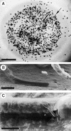
Views of the Adhesion Assay Showing Lily Pollen Tubes Adhered to an in Vitro Stylar Matrix Bound to Nitrocellulose Membrane. (A) Toluidine blue O staining. The 2-hr in vitro–germinated pollen tubes (arrowheads) adhered to the in vitro matrix made from stylar exudate; the incubation time was 5 hr. Pollen grains (arrow) were stained with toluidine blue O to quantify the adhesion assay. (B) and (C) Scanning electron microscope images of pollen tubes adhered to the in vitro matrix made from petal homogenate and filtered stigma exudate. (B) shows pollen tube tip adhesion. (C) shows the area of tube wall tightly adhered to the matrix. formula image; formula image; formula image.
Figure 3.
Figure 3.
Transmitting Electron Microscope Images of Adhesion Assay. (A) Pollen tube adhesion to the stylar extracellular matrix and to other pollen tubes (arrowhead). (B) and (C) Pollen tube adhesion to the nitrocellulose membrane surface impregnated with stylar exudate. NC, nitrocellulose membrane; PT, pollen tube; SE, stylar exudate. formula image; formula image.
Figure 4.
Figure 4.
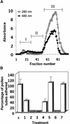
Protein (280 nm) and Carbohydrate (485 nm) Profiles of the Stylar Exudate Fractions and Adhesion Assay Using Groups of Fractions. (A) Fractionation. Stylar exudate was fractionated over a Sephadex G200 column, and the total fractions were combined into three groups: group I (fractions 8 to 30), group II (fractions 31 to 52), and group III (fractions 53 to 100). (B) Adhesion assays. The groups were used for adhesion assays separately or in combinations of equivalent amounts. For each assay, 75 μg (dry weight) of total material was used. The adhesion percentage was obtained by comparison with the control stylar exudate, which contained 200 to 300 adhered pollen tubes. c, control; 1, group I; 2, group II; 3, group III; 4, groups I and II; 5, groups I and III; 6, groups II and III; 7, groups I to III.
Figure 5.
Figure 5.
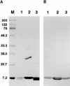
Cation Exchanger Purification of 9-kD Protein. (A) SDS gel stained with Coomassie blue. The gel shows partial purification of a 9-kD protein from stigma exudate. Lane 1 contains 5 μg of stigma exudate protein; lane 2, CM–Sephadex cation exchange–unbound protein (5 μg of protein); and lane 3, 2 M NaCl eluate of cation exchanger (1 μg of protein). The arrowhead indicates the position of the 9-kD protein. Molecular mass standards (in kilodaltons) are shown at left. (B) Two-dimensional gel of stigma exudate cation exchange–bound protein stained with Coomassie blue. Three micrograms of protein was loaded on the first-dimension gel, nonequilibrium pH gel electrophoresis (NEPHGE), and the second-dimensional separation (SDS-PAGE) was done on a 12 to 15% gradient gel. The arrowhead indicates the 9-kD protein. Molecular mass standards (in kilodaltons) are shown at left. (C) Immunoblot of gel shown in (B), using polyclonal antibodies to lily lipid transfer–like protein (arrowhead).
Figure 6.
Figure 6.
Immunoblot Analysis of Proteins from Stigma and Style Exudates. (A) Proteins were separated on a 13% acrylamide gel and stained with Coomassie blue. Lane 1 contains stylar exudate group III protein of a Sephadex G200 column fraction (1 μg); lane 2, 100-kD centricon filtrate of stigma exudate (5 μg); and lane 3, stylar exudate (10 μg). Molecular mass standards (in kilodaltons) are shown at left and in lane M. (B) Immunoblot of gel as given in (A) but with half the amount of protein in each lane. The 9-kD LTP was detected with polyclonal antibodies to lily lipid transfer–like protein.
Figure 7.
Figure 7.
Amino Acid Sequence Alignments of Lily 9-kD Protein with Plant LTPs. The deduced amino acid sequence (GenBank accession number AF171094) was aligned with rice (GenBank accession number AF017361), maize (GenBank accession number M57249), and Arabidopsis (Swiss-Prot accession number Q42589) LTPs. The amino acids determined by microsequencing of the lily 9-kD protein are underlined. The arrowhead indicates the N terminus of the mature lily 9-kD protein. Conserved cysteine residues are in boldface. Identical amino acids are indicated by colons, and spaces introduced to maximize alignment are indicated by dashes.
Figure 8.
Figure 8.
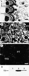
Immunogold Labeling of Pollinated Lily Style Tissue and Protein Gel Blot of Pollen Proteins. (A) Section of lily pollinated style stained with toluidine blue O. (B) Dark-field immunogold localization of protein in pollinated style using antibodies against lily lipid transfer–like protein. (C) Control preimmune serum. (D) Protein gel blot of protein extracted from in vivo and in vitro pollen tubes using antibodies to lily lipid transfer–like protein. All lanes contain 15 μg of protein. Lanes 1 and 2 show protein extracted from in vitro–grown pollen tubes. Pollen tubes were cultured for 2 hr in germination medium and continued to grow for another 5 hr without (lane 1) or with (lane 2) the addition of cation exchange–bound purified lily lipid transfer–like protein added to the germination medium. Lane 3 shows protein extracted from in vivo–grown pollen tubes removed from the style 24 hr after pollination. The molecular mass standard (in kilodaltons) is at left. PT, pollen tube; TTE, transmitting tract epidermis. formula image; formula image.
Figure 9.
Figure 9.
Model for the Role of Cell Adhesion Molecules in Pollen Tube Growth in the Lily Style. The pollen tube tip is illustrated as it progresses along the stylar transmitting tract epidermis. The cell wall of the pollen tube is thin at the tip, where vesicles fuse, contributing esterified pectins to the tube cell wall. The male germ unit (MGU) is composed of the tube cell nucleus and the generative cell (or two sperm). The putative adhesion molecules secreted from the style (open squares) may act between the pollen tube cell wall and the stylar transmitting tract epidermis. Solid squares denote membrane-bound arabinogalactan protein, solid circles indicate unesterified pectins, and open circles represent esterified pectins. (Reproduced with permission from Lord et al. [1996].)

Similar articles

Cited by

References

    1. Baker, B., Zambryski, P.L., Staskawicz, B., and Dinesh-Kumar, S.P. (1997). Signaling in plant–microbe interactions. Science 276 726–733. - PubMed
    1. Becraft, P.W., Stinard, P.S., and McCarty, D.R. (1996). CRINKLY4: A TNFR-like receptor kinase involved in maize epidermal differentiation. Science 273 1406–1409. - PubMed
    1. Bernhard, W.R., Thoma, S., Botella, J., and Somerville, C.R. (1991). Isolation of a cDNA clone for a spinach lipid transfer protein and evidence that the protein is synthesized by the secretory pathway. Plant Physiol. 95 164–170. - PMC - PubMed
    1. Blumenkrantz, N., and Asboe-Hansen, G. (1973). New method for quantitative determination of uronic acids. Anal. Biochem. 54 484–489. - PubMed
    1. Broekaert, W.F., Cammue, B., De Bolle, M., Thevissen, K., and De Samblanx, G. (1997). Antimicrobial peptides from plants. Crit. Rev. Plant Sci. 16 297–323.

Publication types

LinkOut - more resources