. 2000 Jan;20(2):617-27.
doi: 10.1128/MCB.20.2.617-627.2000.

Activation of p38 mitogen-activated protein kinase and c-Jun NH(2)-terminal kinase by double-stranded RNA and encephalomyocarditis virus: involvement of RNase L, protein kinase R, and alternative pathways

Affiliations

Activation of p38 mitogen-activated protein kinase and c-Jun NH(2)-terminal kinase by double-stranded RNA and encephalomyocarditis virus: involvement of RNase L, protein kinase R, and alternative pathways

M S Iordanov et al. Mol Cell Biol. 2000 Jan.

Abstract

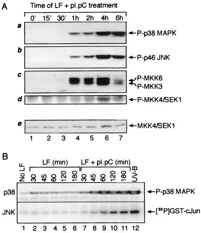
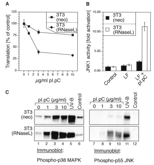
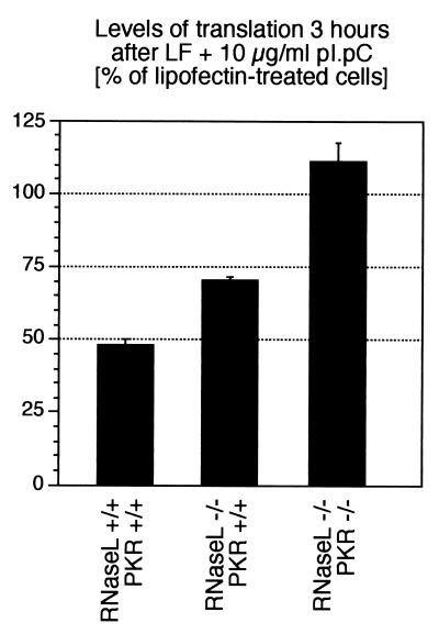
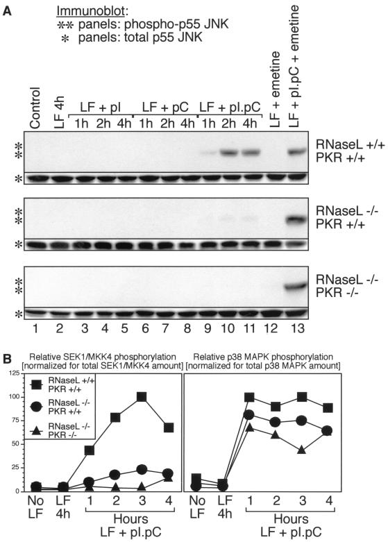
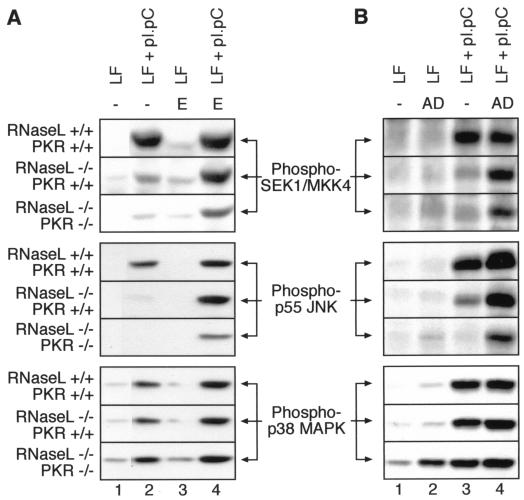
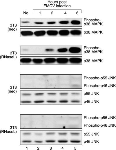
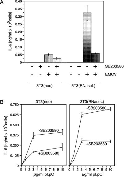
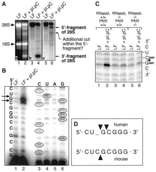
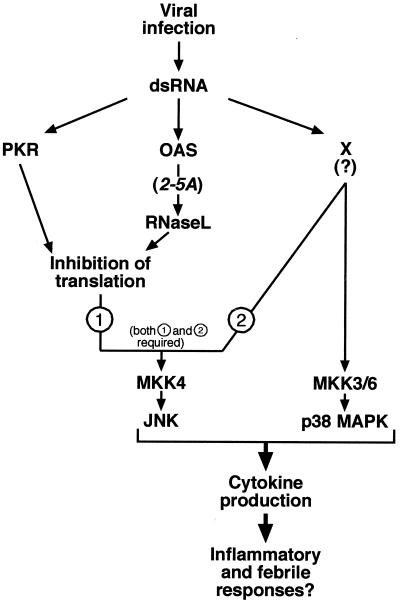
Double-stranded RNA (dsRNA) accumulates in virus-infected mammalian cells and signals the activation of host defense pathways of the interferon system. We describe here a novel form of dsRNA-triggered signaling that leads to the stimulation of the p38 mitogen-activated protein kinase (p38 MAPK) and the c-Jun NH(2)-terminal kinase (JNK) and of their respective activators MKK3/6 and SEK1/MKK4. The dsRNA-dependent signaling to p38 MAPK was largely intact in cells lacking both RNase L and the dsRNA-activated protein kinase (PKR), i. e., the two best-characterized mediators of dsRNA-triggered antiviral responses. In contrast, activation of both MKK4 and JNK by dsRNA was greatly reduced in cells lacking RNase L (or lacking both RNase L and PKR) but was restored in these cells when introduction of dsRNA was followed by inhibition of ongoing protein synthesis or transcription. These results are consistent with the notion that the role of RNase L and PKR in the activation of MKK4 and JNK is the elimination, via inhibition of protein synthesis, of a labile negative regulator(s) of the signaling to JNK acting upstream of SEK1/MKK4. In the course of these studies, we identified a long-sought site of RNase L-mediated cleavage in the 28S rRNA, which could cause inhibition of translation, thus allowing the activation of JNK by dsRNA. We propose that p38 MAPK is a general participant in dsRNA-triggered cellular responses, whereas the activation of JNK might be restricted to cells with reduced rates of protein synthesis. Our studies demonstrate the existence of alternative (RNase L- and PKR-independent) dsRNA-triggered signaling pathways that lead to the stimulation of stress-activated MAPKs. Activation of p38 MAPK (but not of JNK) was demonstrated in mouse fibroblasts in response to infection with encephalomyocarditis virus (ECMV), a picornavirus that replicates through a dsRNA intermediate. Fibroblasts infected with EMCV (or treated with dsRNA) produced interleukin-6, an inflammatory and pyrogenic cytokine, in a p38 MAPK-dependent fashion. These findings suggest that stress-activated MAPKs participate in mediating inflammatory and febrile responses to viral infections.


Figures

FIG. 1
FIG. 1
Phosphorylation of p38 MAPK, JNK, MKK3, MKK6, and SEK1/MKK4 and increased kinase activity of JNK in response to dsRNA. (A) Immunoblot analysis. HeLa cells were grown to ∼80% confluence in normal growth medium. The cells were then treated with pI · pC (3 μg/ml) in the presence of Lipofectin (LF; 10 μg/ml). At indicated times, the cells were harvested and cell lysates representing equal number of cells were subjected to immunoblot analyses with antibodies specific for the phosphorylated forms of p38 MAPK, JNK, MKK3, MKK6, and SEK1/MKK4 (see Materials and Methods). Panel e shows an immunoblot analysis of the levels of total (phosphorylated and nonphosphorylated) SEK1/MKK4 run in a parallel gel. (B) Analogous analysis of pI · pC action in Rat-1 fibroblasts, demonstrating also that pI · pC, and not Lipofectin, is the kinase-activating agent. A direct determination of JNK activity (rather than phosphorylation of JNK) after pI · pC treatment is presented. JNK1 was immunoprecipitated from Rat-1 cells, and the activity of the kinase was determined in immunocomplex kinase reactions using glutathione S-transferase–c-Jun as the substrate for phosphorylation (see Materials and Methods).
FIG. 2
FIG. 2
dsRNA-induced inhibition of protein synthesis, activation of JNK, and phosphorylation of p38 MAPK and JNK in 3T3(neo) and 3T3(RNaseL) cells. (A) The cells were grown in 12-well plates to ∼70% confluence in normal growth medium and then leucine deprived for 1.5 h in leucine- and serum-free DMEM. Lipofectin mixes were made in leucine- and serum-free DMEM to contain 0, 1, 3, or 10 μg of pI · pC per ml and were given to the cells. Between 2.5 and 3 h after the pI · pC addition, the cells were pulse-labeled with 1 μCi of [3H]leucine per ml and processed further as described in Materials and Methods. Error bars indicate standard deviation from experimental points in triplicate determinations. (B) Immunocomplex kinase assay. The cells were grown in 10-cm-diameter plates to ∼70% confluence in normal growth medium. The growth medium was then exchanged, where indicated, with serum-free DMEM (control) or Lipofectin (LF)-containing serum-free DMEM, without or with 10 μg of pI · pC per ml. JNK activity was determined 3 h later in immunocomplex kinase assays as described for Fig. 1B. Error bars indicate standard deviation from experimental points in triplicate determinations. (C) Immunoblot analyses of p38 MAPK and JNK phosphorylation, using phosphoepitope-specific antibodies. The cells were grown and treated as for panel B with the indicated concentrations of pI · pC. For the UV-B irradiation, the cells were given a 1,200-J/m2 dose of UV-B as described previously (26) and harvested 30 min later.
FIG. 3
FIG. 3
dsRNA-induced inhibition of protein synthesis in RNase L+/+ PKR+/+, RNase L−/− PKR+/+, and RNase L−/− PKR−/− cells. The cells were grown in 12-well plates to ∼100% confluence in normal growth medium and then serum deprived for 24 hours in serum-free DMEM. Lipofectin (LF) mixes were made in leucine- and serum-free DMEM to contain 10 μg of pI · pC per ml and were given to the cells. Between 2.5 and 3 h after the pI · pC addition, the cells were pulse-labeled with 1 μCi of [3H]leucine per ml and processed further as described in Materials and Methods. Error bars indicate standard deviation from experimental points in triplicate determinations.
FIG. 4
FIG. 4
dsRNA-induced phosphorylation of p38 MAPK, JNK, and SEK/MKK4 in RNase L+/+ PKR+/+, RNase L−/− PKR+/+, and RNase L−/− PKR−/− cells. (A) Immunoblot analyses of JNK phosphorylation (∗∗ panels) and total levels of JNK (∗ panels). The cells were grown in 10-cm-diameter plates to ∼100% confluence in normal growth medium and then serum deprived for 24 h in serum-free DMEM. Lipofectin (LF) mixes were made in serum-free DMEM to contain no nucleic acid or 10 μg of either pI, pC, or pI · pC per ml and were given to the cells for the indicated times. Where indicated, the cells were treated with emetine (100 μg/ml) 1 h after the addition of Lipofectin mixes. (B) Graphic presentation of immunoblot analyses. Cells were treated as indicated in the graphs and as for panel A, and analyses were performed with antibodies against the phosphorylated forms of SEK1/MKK4 and p38 MAPK. The membranes were stripped and rehybridized with antibodies recognizing total (phosphorylated and nonphosphorylated) SEK1/MKK4 and p38 MAPK. For each immunoblot, appropriately nonsaturated film exposures were selected and scanned, and the scanned images were imported into IP Lab Gel software for quantification. The maximum level of phosphorylation of each kinase (after normalization for total amount of the kinase) was expressed as 100%.
FIG. 5
FIG. 5
Effects of emetine and actinomycin D posttreatments on the dsRNA-induced phosphorylation of SEK/MKK4, JNK, and p38 MAPK in RNase L+/+ PKR+/+, RNase L−/− PKR+/+, and RNase L−/− PKR−/− cells determined by immunoblot analyses. The cells were grown and treated with pI · pC for 4 h as for Fig. 4A. Where indicated, emetine (E; 100 μg/ml) or actinomycin D (AD; 25 μg/ml) was added either 1 h after (emetine) or 15 min after (actinomycin D) the treatment with pI · pC. Note the lack of effect of both emetine and actinomycin D on dsRNA-induced phosphorylation of p38 MAPK. The mechanism of actinomycin D-induced p38 MAPK phosphorylation in RNase L−/− PKR−/− cells (B, bottom panel, lane 2) is unknown. LF, lipofectin.
FIG. 6
FIG. 6
Phosphorylation of p38 MAPK, but not JNK, in EMCV-infected cells. 3T3(neo) and 3T3(RNaseL) cells were grown in 10-cm-diameter dishes and incubated with IFN-α BBDB (2,000 U/ml) for 20 h. The medium was replaced twice with serum-free, antibiotic-free DMEM; cells were infected with EMCV at an MOI of 10 and incubated at 37°C. At indicated times, the cell were harvested and processed for immunoblot analyses. Appropriate positive controls (not shown) were performed for the immunoblot analysis of JNK phosphorylation to demonstrate that, when present, phosphorylated JNK was detectable.
FIG. 7
FIG. 7
p38 MAPK-dependent expression of IL-6 in fibroblasts in response to EMCV infection or dsRNA. (A) IL-6 detection by ELISA. 3T3(neo) and 3T3(RNaseL) cells were grown and infected as described in Materials and Methods, and the appearance of IL-6 in the cell culture medium was assessed by ELISA as described in Materials and Methods. Error bars indicate standard deviation from experimental points in triplicate determinations. (B) Identical assessment of IL-6 expression 24 h after pI · pC treatment (0, 1, 3, or 10 μg/ml). Error bars indicate standard deviation from experimental points in triplicate determinations.
FIG. 8
FIG. 8
Identification of a major site of dsRNA-induced cleavage of 28S rRNA in human and mouse cells. (A) Northern blot analysis. HeLa cells were treated for 3 h with pI · pC (3 μg/ml). Total RNA was isolated, and 2-μg aliquots were used for Northern blot detection of cleavage fragments of 28S rRNA, using DNA oligonucleotides hybridizing specifically to 28S rRNA and fragments thereof. One probe hybridized to the fragment located 3′ of the site of cleavage (panel b), whereas the other hybridized to the fragment located 5′ of the site of cleavage (panel c). LF, Lipofectin. (B) The RNA preparation used for panel A was subjected to a reverse transcriptase-driven primer extension using the primer 5′-GAGTAGTGGTATTTCAC-3′. Direct RNA sequencing using the same primer is shown to the right. The sites of cleavage are represented by arrows. (C) Analysis identical to that shown in panel B was performed with RNase L+/+ PKR+/+, RNase L−/− PKR+/+, and RNase L−/− PKR−/−cells, except that 10 μg of pI · pC per ml was used. (D) Alignment of the human and mouse sequences within the region of dsRNA-induced cleavage of the 28S rRNA. The sites of cleavage are represented by arrowheads.
FIG. 9
FIG. 9
Model for the mechanisms of activation of stress-activated MAP kinases by dsRNA. See the text for explanation.

Similar articles

Cited by

References

    1. Baldassare J J, Bi Y, Bellone C J. The role of p38 mitogen-activated protein kinase in IL-1 beta transcription. J Immunol. 1999;162:5367–5373. - PubMed
    1. Beyaert R, Cuenda A, Vanden Berghe W, Plaisance S, Lee J C, Haegeman G, Cohen P, Fiers W. The p38/RK mitogen-activated protein kinase pathway regulates interleukin-6 synthesis response to tumor necrosis factor. EMBO J. 1996;15:1914–1923. - PMC - PubMed
    1. Brimacombe R. The structure of ribosomal RNA: a three-dimensional jigsaw puzzle. Eur J Biochem. 1995;230:365–383. - PubMed
    1. Cavigelli M, Li W W, Lin A, Su B, Yoshioka K, Karin M. The tumor promoter arsenite stimulates AP-1 activity by inhibiting a JNK phosphatase. EMBO J. 1996;15:6269–6279. - PMC - PubMed
    1. Chebath J, Benech P, Hovanessian A, Galabru J, Revel M. Four different forms of interferon-induced 2′,5′-oligo(A) synthetase identified by immunoblotting in human cells. J Biol Chem. 1987;262:3852–3857. - PubMed

Publication types

MeSH terms