. 1999 Dec;10(12):4283-98.
doi: 10.1091/mbc.10.12.4283.

Nuclear accumulation of S-adenosylhomocysteine hydrolase in transcriptionally active cells during development of Xenopus laevis

Affiliations
Free PMC article

Nuclear accumulation of S-adenosylhomocysteine hydrolase in transcriptionally active cells during development of Xenopus laevis

N Radomski et al. Mol Biol Cell. 1999 Dec.
Free PMC article

Abstract

The oocyte nuclear antigen of the monoclonal antibody 32-5B6 of Xenopus laevis is subject to regulated nuclear translocation during embryogenesis. It is distributed in the cytoplasm during oocyte maturation, where it remains during cleavage and blastula stages, before it gradually reaccumulates in the nuclei during gastrulation. We have now identified this antigen to be the enzyme S-adenosylhomocysteine hydrolase (SAHH). SAHH is the only enzyme that cleaves S-adenosylhomocysteine, a reaction product and an inhibitor of all S-adenosylmethionine-dependent methylation reactions. We have compared the spatial and temporal patterns of nuclear localization of SAHH and of nuclear methyltransferase activities during embryogenesis and in tissue culture cells. Nuclear localization of Xenopus SAHH did not temporally correlate with DNA methylation. However, we found that SAHH nuclear localization coincides with high rates of mRNA synthesis, a subpopulation colocalizes with RNA polymerase II, and inhibitors of SAHH reduce both methylation and synthesis of poly(A)(+) RNA. We therefore propose that accumulation of SAHH in the nucleus may be required for efficient cap methylation in transcriptionally active cells. Mutation analysis revealed that the C terminus and the N terminus are both required for efficient nuclear translocation in tissue culture cells, indicating that more than one interacting domain contributes to nuclear accumulation of Xenopus SAHH.


Figures
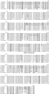
Figure 1
Figure 1
Protein sequence alignment of vertebrate SAHH. The cDNA-derived amino acid sequences of X. laevis (xSAHH 1, GenBank accession number [gb] L35559 and this communication; xSAHH 2, gb AJ007835 and this communication), mouse (mSAHH, gb L32836), rat (rSAHH, gb M15185) and human (hSAHH, gb M61832) have been aligned together with the consensus sequence, using the GCG programs Pileup and Pretty. The two isoforms of X. laevis are distinguished from one another by eight conservative changes, each marked by shading. Sequences obtained by protein microsequencing of tryptic peptides of the X. laevis oocyte protein are underlined in xSAHH. Lysine and arginine residues are shown in bold. The NAD+ binding site is printed in italics and underlined in the consensus. An amphiphatic helical domain near the C terminus is underlined twice, and a lysine residue (K427) shown to be essential for tetramer formation and for catalytic activity of the hSAHH (Ault-Riche et al., 1994) is printed in italics and shaded.
Figure 2
Figure 2
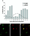
The amount of nuclear xSAHH in XTC cells is dependent on the cell cycle. (A) XTC cells arrested in G0 by serum starvation were stimulated to exit G0 by addition of 10% serum. Cells were pulse labeled with BrdU 1 h before fixation, and parallel samples were immunostained for BrdU incorporated in DNA and for xSAHH. The percentage of significant nuclear staining is plotted against the time after serum stimulation (not drawn to scale). (B–D) Nonsynchronous XTC cells were pulse labeled for 1 h with BrdU, fixed, and double stained for de novo–synthesized DNA (B) and for xSAHH (C). Superposition of confocal images shows limited coincidence of both labels (D). The example shown was selected for maximal overlap of both labels. In the nucleus marked with an arrowhead, the peripheral BrdU label likely represents replicating heterochromatin. Bar, 10 μm.
Figure 3
Figure 3
xSAHH translocates into interphase nuclei during gastrulation. Intracellular localization of endogenous xSAHH was analyzed on Technovit sections after whole-mount immunofluoresescent staining using mAb 32-5B6 (A, C, E, G, I, and K), and counterstaining of the same sections for DNA with DAPI is shown in B, D, F, H, J, and L. Details of sections cut in the animal to vegetal direction were selected that show the marginal zone of a late blastula at stage 9 (A and B), the vegetal area (C and D), and the dorsal lip of an early gastrula at stage 10 (E and F), a marginal area of a gastrula at stage 10.5, with mesoderm to the left and endoderm to the right (G and H), and all three germ layers anteriorly of a very early neurula at stage 13 (I and J). Single interphase nuclei showing nuclear xSAHH are marked with arrowheads in A–H. Mitotic cells are marked with arrows, and arrows point at prometaphase in C and D. On a complete transverse section of a gastrula at stage 11.5 (K) the endoderm of the blastoporus (bp) is flanked by the two blastoporal lips, containing mesoderm and ectoderm. bc, remnant of the blastocoel. Cytoplasmic xSAHH is excluded from yolk platelets and is concentrated at the cell boundaries. Bars: A–J, 50 μm; L, 200 μm. Sections were 2 μm thick in G and H and 5 μm thick in all other panels.
Figure 4
Figure 4
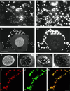
Nuclear run-on transcriptional assay in A6 cells and embryos. Transcriptional activity was monitored by a nuclear run-on assay with incorporation of Br-UTP in permeabilized A6 cells (A) and in embryos (D), as detailed in MATERIALS AND METHODS. A6 cells were fixed after 10 min of labeling and double stained for incorporated Br-UTP (A), and xSAHH (B). The confocal images shown in A and B are superimposed in C. Bar, 10 μm. (D and E) A midgastrula embryo at stage 10.5 was coinjected with digitonin and ribonucleotides including Br-UTP and fixed after 60 min of labeling at stage 11. The gastrula was dissected in halves along the animal to vegetal axis, and halves were double stained as whole mounts for the Br-UTP label and for xSAHH. Confocal images of the surface show coincident nuclear staining with anti BrU (D) and with mAb 32-5B6 for xSAHH (E). Bar, 200 μm.
Figure 5
Figure 5
Immunohistological localization of xSAHH in the nuclei of germ cells. xSAHH was detected on 1.5-μm thin sections after whole-mount immunofluorescent staining of adult testis (A) and ovary from a 4-cm-long young female (C) with mAb 32-5B6. (B and D) Counterstaining of the same sections with DAPI. In the testis (A and B), nuclear staining with 32-5B6 predominates in transcriptionally active Sertoli cells (S) and spermatogonia (G). In the ovary (C and D), xSAHH is nuclear in all cells. In the single diplotene (stage I) oocyte nucleus shown, the xSAHH antigen appears most highly concentrated around the lampbrush chromosomes, which are weakly stained with DAPI in D. (E–H) Frozen sections of an ovary were stained with mAb 32-5B6 (E) and with anti-RNA Polymerase II mAb H14 (G). (F and H) DAPI counterstain of the sections shown in E and G. Follicle cell nuclei stained with DAPI are overexposed in D, F, and H to visualize lampbrush chromosomes. Bar, 50 μm. (I–K) Lampbrush chromosome spread from a stage V oocyte double stained for xSAHH (I) and for RNA polymerase II (J). The confocal images shown in I and J are superimposed in K. Bar in K, 10 μm.
Figure 6
Figure 6
Inhibitors of SAHH interfere with methylation and elongation of poly(A)+ RNA. XTC cells were simultaneously pulse labeled with l-[methyl-3H]methionine and [U-14C]uridine in the presence or absence of 2-deoxyadenosine (2dAde), tubercidine, or DRB, and the incorporation of radioactive precursors in poly(A)+ RNA was analyzed as detailed in MATERIALS AND METHODS. The results of two independent experiments are combined and shown with error bars. Incorporation of [U-14C]uridine in poly(A)+ RNA of control cells was between 290 and 373 cpm, and incorporation of [3H]methyl was between 30 and 74 cpm.
Figure 7
Figure 7
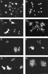
Nuclear translocation of full-length and mutated xSAHH in transfected cells. (A) Endogenous nucleoplasmic xSAHH stained with mAb 32-5B6 in untransfected A6 cells. A mitotic cell is marked by a long arrow. Full-length (B and C) and C-terminally (D-F) and N-terminally deleted (G and H) MT-xSAHH was detected in Xenopus A6 cells using mAb 9E10 after transient transfection as detailed in MATERIALS AND METHODS. Cells were fixed and processed for immunofluorescence 120 h (B) or 40–48 h (C–H) after transfection. Cells were transfected with pCS2+MT containing cDNA encoding full-length xSAHH (B and C), xSAHHΔ425–433 (D), xSAHHΔ402–433 (E), xSAHHΔ361–433 (F), xSAHHΔ1–13 (G), or xSAHHΔ1–21(H). Transfected cells with predominantly nuclear localization of MT-xSAHH are marked with arrowheads, and cells with predominantly cytoplasmic localization are marked with short arrows. Bar, 50 μm.
Figure 8
Figure 8
Influence of N-terminal and C-terminal deletions on intracellular localization of MT-xSAHH in transfected A6 cells. (A) Full-length MT-xSAHH or the truncated forms explained in B were expressed in A6 cells after transient transfection. The percentages of transfected cells showing localization of the MT-xSAHH exclusively (N) or predominantly in the nucleus (N>C), in the cytoplasm (C>N), or at similar levels in both compartments (N=C) are compiled from two or three independent experiments and plotted. Several hundred cells were analyzed for each individual fusion protein, except for xSAHHΔ361–433, where n was 165, because this product was either toxic or unstable, resulting in a low percentage of evaluable cells. (B) The N terminus and the C terminus of the fusion proteins encoded by the cDNAs used for transfection are shown, with numbers referring to the wt-xSAHH2 aa sequence. Only the last of six N-terminal myc tags are shown in italics. The original xSAHH sequence is shown in bold letters; linker sequence is shown in plain uppercase letters; and aa exchanged by mutagenesis are underlined.

Similar articles

Cited by

References

    1. Araujo FD, Knox JD, Szyf M, Price GB, Zannis-Hadjopoulos M. Concurrent replication and methylation at mammalian origins of replication. Mol Cell Biol. 1998;18:3475–3482. - PMC - PubMed
    1. Ault-Riche DB, Yuan CS, Borchardt RT. A single mutation at lysine 426 of human placental S-adenosylhomocysteine hydrolase inactivates the enzyme. J Biol Chem. 1994;269:31472–31478. - PubMed
    1. Bachvarova R, Davidson EH. Nuclear activation at the onset of amphibian gastrulation. J Exp Zool. 1966;163:285–296.
    1. Bergmann S, Shatrov V, Ratter F, Schiemann S, Schulze-Osthoff K, Lehmann V. Adenosine and homocysteine together enhance TNF-mediated cytotoxicity but do not alter activation of nuclear factor-kappa B in L929 cells. J Immunol. 1994;153:1736–1743. - PubMed
    1. Bestor TH. Activation of mammalian DNA methyltransferase by cleavage of a Zn binding regulatory domain. EMBO J. 1992;11:2611–2617. - PMC - PubMed

MeSH terms

LinkOut - more resources