Emergence of a highly pathogenic simian/human immunodeficiency virus in a rhesus macaque treated with anti-CD8 mAb during a primary infection with a nonpathogenic virus
- PMID: 10570196
- PMCID: PMC24188
- DOI: 10.1073/pnas.96.24.14049
Emergence of a highly pathogenic simian/human immunodeficiency virus in a rhesus macaque treated with anti-CD8 mAb during a primary infection with a nonpathogenic virus
Abstract
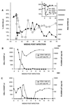
Although simian/human immunodeficiency virus (SHIV) strain DH12 replicates to high titers and causes immunodeficiency in pig-tailed macaques, virus loads measured in SHIV(DH12)-infected rhesus monkeys are consistently 100-fold lower and none of 22 inoculated animals have developed disease. We previously reported that the administration of anti-human CD8 mAb to rhesus macaques at the time of primary SHIV(DH12) infection resulted in marked elevations of virus loads. One of the treated animals experienced rapid and profound depletions of circulating CD4(+) T lymphocytes. Although the CD4(+) T cell number partially recovered, this monkey subsequently suffered significant weight loss and was euthanized. A tissue culture virus stock derived from this animal, designated SHIV(DH12R), induced marked and rapid CD4(+) cell loss after i.v. inoculation of rhesus monkeys. Retrospective analyses of clinical specimens, collected during the emergence of SHIV(DH12R) indicated: (i) the input cloned SHIV remained the predominant virus during the first 5-7 months of infection; (ii) variants bearing only a few of the SHIV(DH12R) consensus changes first appeared 7 months after the administration of anti-CD8 mAb; (iii) high titers of neutralizing antibody directed against the input SHIV were detected by week 10 and persisted throughout the infection; and (iv) no neutralizing antibody against SHIV(DH12R) ever developed.
Figures


Similar articles
-
Short- and long-term clinical outcomes in rhesus monkeys inoculated with a highly pathogenic chimeric simian/human immunodeficiency virus.J Virol. 2000 Aug;74(15):6935-45. doi: 10.1128/jvi.74.15.6935-6945.2000. J Virol. 2000. PMID: 10888632 Free PMC article.
-
Chronology of genetic changes in the vpu, env, and Nef genes of chimeric simian-human immunodeficiency virus (strain HXB2) during acquisition of virulence for pig-tailed macaques.Virology. 1998 Sep 1;248(2):275-83. doi: 10.1006/viro.1998.9300. Virology. 1998. PMID: 9721236
-
A highly pathogenic simian/human immunodeficiency virus with genetic changes in cynomolgus monkey.J Gen Virol. 1999 May;80 ( Pt 5):1231-1240. doi: 10.1099/0022-1317-80-5-1231. J Gen Virol. 1999. PMID: 10355770
-
Protection of neonatal macaques against experimental SHIV infection by human neutralizing monoclonal antibodies.Transfus Clin Biol. 2001 Aug;8(4):350-8. doi: 10.1016/s1246-7820(01)00187-2. Transfus Clin Biol. 2001. PMID: 11642027 Review.
-
Understanding the basis of CD4(+) T-cell depletion in macaques infected by a simian-human immunodeficiency virus.Vaccine. 2002 May 6;20(15):1934-7. doi: 10.1016/s0264-410x(02)00072-5. Vaccine. 2002. PMID: 11983249 Review.
Cited by
-
Identification of interdependent variables that influence coreceptor switch in R5 SHIV(SF162P3N)-infected macaques.Retrovirology. 2012 Dec 13;9:106. doi: 10.1186/1742-4690-9-106. Retrovirology. 2012. PMID: 23237529 Free PMC article.
-
Mucosal transmissibility, disease induction and coreceptor switching of R5 SHIVSF162P3N molecular clones in rhesus macaques.Retrovirology. 2013 Jan 31;10:9. doi: 10.1186/1742-4690-10-9. Retrovirology. 2013. PMID: 23369442 Free PMC article.
-
Protection of macaques against AIDS with a live attenuated SHIV vaccine is of finite duration.Virology. 2008 Feb 20;371(2):238-45. doi: 10.1016/j.virol.2007.10.008. Epub 2007 Nov 7. Virology. 2008. PMID: 17988702 Free PMC article.
-
In vivo Serial Passaging of Human-Simian Immunodeficiency Virus Clones Identifies Characteristics for Persistent Viral Replication.Front Microbiol. 2021 Nov 18;12:779460. doi: 10.3389/fmicb.2021.779460. eCollection 2021. Front Microbiol. 2021. PMID: 34867922 Free PMC article.
-
Peripheral edema with hypoalbuminemia in a nonhuman primate infected with simian-human immunodeficiency virus: a case report.J Am Assoc Lab Anim Sci. 2008 Jan;47(1):42-8. J Am Assoc Lab Anim Sci. 2008. PMID: 18210998 Free PMC article.
References
-
- Shibata R, Maldarelli F, Siemon C, Matano T, Parta M, Miller G, Fredrickson T, Martin M A. J Infect Dis. 1997;176:362–373. - PubMed
MeSH terms
Substances
LinkOut - more resources
Full Text Sources
Other Literature Sources
Research Materials

