Evidence that Myb-related CDC5 proteins are required for pre-mRNA splicing
- PMID: 10570151
- PMCID: PMC24143
- DOI: 10.1073/pnas.96.24.13789
Evidence that Myb-related CDC5 proteins are required for pre-mRNA splicing
Abstract
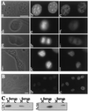
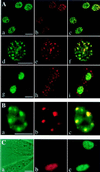
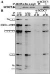
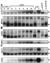
The conserved CDC5 family of Myb-related proteins performs an essential function in cell cycle control at G(2)/M. Although c-Myb and many Myb-related proteins act as transcription factors, herein, we implicate CDC5 proteins in pre-mRNA splicing. Mammalian CDC5 colocalizes with pre-mRNA splicing factors in the nuclei of mammalian cells, associates with core components of the splicing machinery in nuclear extracts, and interacts with the spliceosome throughout the splicing reaction in vitro. Furthermore, genetic depletion of the homolog of CDC5 in Saccharomyces cerevisiae, CEF1, blocks the first step of pre-mRNA processing in vivo. These data provide evidence that eukaryotic cells require CDC5 proteins for pre-mRNA splicing.
Figures


Similar articles
-
Removal of a single alpha-tubulin gene intron suppresses cell cycle arrest phenotypes of splicing factor mutations in Saccharomyces cerevisiae.Mol Cell Biol. 2002 Feb;22(3):801-15. doi: 10.1128/MCB.22.3.801-815.2002. Mol Cell Biol. 2002. PMID: 11784857 Free PMC article.
-
Myb-related Schizosaccharomyces pombe cdc5p is structurally and functionally conserved in eukaryotes.Mol Cell Biol. 1998 Jul;18(7):4097-108. doi: 10.1128/MCB.18.7.4097. Mol Cell Biol. 1998. PMID: 9632794 Free PMC article.
-
Proteomics analysis reveals stable multiprotein complexes in both fission and budding yeasts containing Myb-related Cdc5p/Cef1p, novel pre-mRNA splicing factors, and snRNAs.Mol Cell Biol. 2002 Apr;22(7):2011-24. doi: 10.1128/MCB.22.7.2011-2024.2002. Mol Cell Biol. 2002. PMID: 11884590 Free PMC article.
-
Pre-mRNA splicing in Schizosaccharomyces pombe: regulatory role of a kinase conserved from fission yeast to mammals.Curr Genet. 2003 Feb;42(5):241-51. doi: 10.1007/s00294-002-0355-2. Epub 2002 Dec 13. Curr Genet. 2003. PMID: 12589463 Review.
-
Connections between pre-mRNA processing and regulation of the eukaryotic cell cycle.Front Horm Res. 1999;25:59-82. doi: 10.1159/000060995. Front Horm Res. 1999. PMID: 10941402 Review. No abstract available.
Cited by
-
Transcriptional regulation: a genomic overview.Arabidopsis Book. 2002;1:e0085. doi: 10.1199/tab.0085. Epub 2002 Apr 4. Arabidopsis Book. 2002. PMID: 22303220 Free PMC article.
-
Identification of conserved gene structures and carboxy-terminal motifs in the Myb gene family of Arabidopsis and Oryza sativa L. ssp. indica.Genome Biol. 2004;5(7):R46. doi: 10.1186/gb-2004-5-7-r46. Epub 2004 Jun 29. Genome Biol. 2004. PMID: 15239831 Free PMC article.
-
Highly conserved features of DNA binding between two divergent members of the Myb family of transcription factors.Nucleic Acids Res. 2001 Jan 15;29(2):527-35. doi: 10.1093/nar/29.2.527. Nucleic Acids Res. 2001. PMID: 11139623 Free PMC article.
-
Removal of a single alpha-tubulin gene intron suppresses cell cycle arrest phenotypes of splicing factor mutations in Saccharomyces cerevisiae.Mol Cell Biol. 2002 Feb;22(3):801-15. doi: 10.1128/MCB.22.3.801-815.2002. Mol Cell Biol. 2002. PMID: 11784857 Free PMC article.
-
α-tubulin regulation by 5' introns in Saccharomyces cerevisiae.Genetics. 2023 Dec 6;225(4):iyad163. doi: 10.1093/genetics/iyad163. Genetics. 2023. PMID: 37675603 Free PMC article.
References
Publication types
MeSH terms
Substances
Grants and funding
LinkOut - more resources
Full Text Sources
Molecular Biology Databases

