. 1999 Nov;10(11):3549-65.
doi: 10.1091/mbc.10.11.3549.

Shr3p mediates specific COPII coatomer-cargo interactions required for the packaging of amino acid permeases into ER-derived transport vesicles

Affiliations
Free PMC article

Shr3p mediates specific COPII coatomer-cargo interactions required for the packaging of amino acid permeases into ER-derived transport vesicles

C F Gilstring et al. Mol Biol Cell. 1999 Nov.
Free PMC article

Abstract

The SHR3 gene of Saccharomyces cerevisiae encodes an integral membrane component of the endoplasmic reticulum (ER) with four membrane-spanning segments and a hydrophilic, cytoplasmically oriented carboxyl-terminal domain. Mutations in SHR3 specifically impede the transport of all 18 members of the amino acid permease (aap) gene family away from the ER. Shr3p does not itself exit the ER. Aaps fully integrate into the ER membrane and fold properly independently of Shr3p. Shr3p physically associates with the general aap Gap1p but not Sec61p, Gal2p, or Pma1p in a complex that can be purified from N-dodecylmaltoside-solubilized membranes. Pulse-chase experiments indicate that the Shr3p-Gap1p association is transient, a reflection of the exit of Gap1p from the ER. The ER-derived vesicle COPII coatomer components Sec13p, Sec23p, Sec24p, and Sec31p but not Sar1p bind Shr3p via interactions with its carboxyl-terminal domain. The mutant shr3-23p, a nonfunctional membrane-associated protein, is unable to associate with aaps but retains the capacity to bind COPII components. The overexpression of either Shr3p or shr3-23p partially suppresses the temperature-sensitive sec12-1 allele. These results are consistent with a model in which Shr3p acts as a packaging chaperone that initiates ER-derived transport vesicle formation in the proximity of aaps by facilitating the membrane association and assembly of COPII coatomer components.


Figures

Figure 1
Figure 1
Membrane topology of Gap1p in SHR3 and shr3Δ6 strains. (A) Hydrophilicity plot of Gap1p calculated according to the method of Kyte and Doolittle (1982) (window size of 11). The black boxes depict the 12 hydrophobic putative membrane-spanning domains (I–XII) present in Gap1p. In the schematic representation of the CT Gap1–Suc2p fusion protein, the white box represents Gap1p, and the hatched box represents the Suc2p reporter construct. Asterisks indicate potential glycosylation sites present within Suc2p. The black oval symbol in the C-terminal portion of Suc2p indicates the location of the HA3 epitope-tag. The arrows depict the locations of the junctions of the six (PT7–PT12) Gap1–Suc2p fusion proteins. (B) GAP1-SUC2 gene fusions (pFG32–pFG38) expressed in FGY84 (SHR3) and FGY85 (shr3Δ6). Transformants were grown in SUD (plus lysine and adenine), and extracts of total cell protein were prepared. Protein preparations were solubilized in SDS-PAGE sample buffer, treated with endoH where indicated, and resolved by SDS-PAGE in 7.5% polyacrylamide gels, immunoblotted, and analyzed as described in MATERIALS AND METHODS. The topological orientation of each Suc2p reporter and the positions of the molecular mass markers in kilodaltons are indicated.
Figure 2
Figure 2
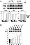
Transcription levels of ER stress response proteins in SHR3 and shr3Δ6 strains. (A) Total RNA preparations (5 μg) isolated from cultures of FGY58 (SHR3) and FGY60 (shr3Δ6) transformed with pPL257 (GAP1) were separated by gel electrophoresis, transferred to nylon filters, and hybridized with radiolabeled DNA probes specific for KAR2, PDI1, EUG1, and ACT1. Cultures were grown at 30°C for 2.5 h in SUD in the absence or presence of 4 μg/ml tunicamycin (Tuni) or 3 mM DTT. Phosphorimager quantitations from two independent experiments are presented as ratios of basal transcription levels with respect to the SHR3 strain (FGY58) normalized to 1; error bars represent 1 standard deviation. (B) The stress response pathway is induced in strains expressing mutant alleles of gap1. Cultures of strains FGY58 (SHR3) transformed with plasmid pPL257 (GAP1, black bar, lane 1) and FGY60 (shr3Δ6) transformed with plasmids pPL257 (GAP1, hatched bar, lane 2), pFG80 (gap1–119), pFG81 (gap1–159), pFG82 (gap1–171), pFG83 (gap1–411), and pFG84 (gap1–417) (white bars, lanes 3–7, respectively) were grown at 30°C for 4 h in SPD (plus adenine and lysine). RNA was isolated and analyzed using radiolabeled DNA probes specific for KAR2 and ACT1. Phosphorimager quantitations from three independent experiments are presented as ratios of the transcription levels with respect to the SHR3 strain (FGY58, lane 1) normalized to 1; error bars represent 1 standard deviation.
Figure 3
Figure 3
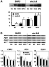
Gap1p and KAR2, PDI1 and EUG1 transcript levels in strains grown on alternative nitrogen sources. Overnight cultures of FGY58 (SHR3) and FGY60 (shr3Δ6) transformed with pPL257 (GAP1-FLU1), pregrown at 30°C under GAP1-repressing conditions in SC (minus uracil), were harvested, washed once with H2O, and used to inoculate SC (minus uracil), SD (plus adenine and lysine), SUD (plus adenine and lysine), and SPD (plus adenine and lysine) media at a starting OD600 of 0.5. The cultures were incubated for 4 h at 30°C, and extracts of total cell protein and RNA were prepared. (A) Proteins from an equivalent of 0.2 OD600 units were analyzed by immunoblotting as described in MATERIALS AND METHODS. Gap1p expression levels were quantitated by phosphorimaging. Values from two independent experiments have been corrected for background and normalized to the Gap1p expression level in the FGY58 strain grown in SC (minus uracil); error bars indicate 1 standard deviation. (B) Five-microgram RNA aliquots were separated by gel electrophoresis and analyzed by Northern analysis as described in Figure 2. Phosphorimager quantitations from two independent experiments are presented; transcript levels are normalized to the expression found in the SHR3 strain (FGY58) grown in SC (minus uracil), the medium with the highest basal transcription levels; error bars indicate 1 standard deviation.
Figure 4
Figure 4
shr3Δ1 interacts genetically with sec13-1 and sec31-1. (A) Serial dilutions of strains with the indicated SHR3 and SEC13 genotypes were grown on SD (plus uracil) incubated at 20°C for 8 d (permissive temperature, upper panel) and on SC incubated at 30°C for 4 d (semipermissive temperature, lower panel). Dilution series 1–8 correspond to strains MAS35-6A (SHR3 SEC13), MAS35-9C (shr3Δ1 SEC13), MAS35-6B (SHR3 sec13-1), MAS35-3A (shr3Δ1 sec13-1), MAS35-10B (shr3Δ1 sec13-1), MAS35-14B (shr3Δ1 sec13-1), MAS35-1C (shr3Δ1 SEC13), and MAS35-19D (SHR3 sec13-1), respectively. (B) Serial dilutions of strains with the indicated SHR3 and SEC31 genotypes were grown on SPD (plus uracil, adenine, and lysine) incubated at 30°C for 5 d (permissive temperature, upper panel) and on SPD (plus uracil, adenine, and lysine) containing 0.6 mM histidine incubated at 35°C for 5 d (semipermissive temperature, lower panel). Dilution series 1–8 correspond to strains MAS203-1B (SHR3 SEC31), MAS203-1A (SHR3 sec31-1), MAS203-6C (shr3Δ1 SEC31), MAS203-12D (shr3Δ1 sec31-1), MAS203-20C (shr3Δ1 sec31-1), MAS203-1C (shr3Δ1 sec31-1), MAS202-3C (SHR3 sec31-1), and MAS203-5B (shr3Δ1 SEC31), respectively.
Figure 5
Figure 5
Multicopy expression of SHR3 partially suppresses the temperature-sensitive phenotypes of sec12-1, sec13-1, sec13-4, and sec31-1 mutations. (A) Serial dilutions of strains transformed with pRS202 (−) and pPL250 (+) were incubated at the indicated temperatures for 3 d and photographed. Strains from left to right are PLY144 (SEC+), MAS26-1A (sec12-1), MAS35-6B (sec13-1), MAS242-7B (sec13-4), MAS203-1A (sec31-1), and MAS297-2B (sec62-1). Strains were grown on SC (minus uracil), with the exception of MAS242-7B (sec13-4), which was grown on SPD medium containing 0.6 mM histidine. (B) Multicopy expression of SHR3 suppresses the inositol requirement of sec13-1 mutations. Serial dilutions of strains transformed with pRS202 (−) and pPL250 (+) were spotted onto media without (left and center panels) and with (right panel) inositol. Culture plates were incubated at the indicated temperature for 3 d and photographed. Dilution series 1 and 2, strain MAS35-6A (SHR3 SEC13); series 3 and 4, strain MAS35-6B (SHR3 sec13-1); series 5, MAS35-3A (shr3Δ1 sec13-1).
Figure 6
Figure 6
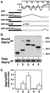
Shr3p physically associates with Gap1p. (A) Schematic diagram of the pGAL1-promoted GST-containing proteins and hydrophilicity plot of Shr3p calculated according to the method of Kyte and Doolittle (1982) (window size of 11). (B) Immunoblot analysis of Gap1p-associated (Bound Gap1p) with GST fusion proteins expressed in FGY145, GST alone (lane 1), GST-CT-shr3(160–210) (lane 2), GST-SHR3 (lane 3), GST-shr3-23 (lane 4), and GST-shr3ΔCT(163–201) (lane 5). GST fusion proteins were purified by affinity chromatography to glutathione-conjugated Sepharose beads. The beads were washed four times using FGB buffer containing 0.1% DM, and SDS-PAGE sample buffer was added to a total of 20 μl of beads. Proteins were denatured at 37°C for 10 min and resolved by SDS-PAGE in a 10% polyacrylamide gel. Gap1p and GST proteins were visualized using antiserum from rabbits immunized with the amino-terminal portion of Gap1p fused to GST (Springael and André, 1998). Protein bands corresponding to each of the GST constructs are marked with an asterisk. The total amount of Gap1p present in protein extracts before GST protein purification is shown in the lower gel panel; the amount of total protein loaded into each lane corresponds to 1/25 of that incubated with glutathione beads. (C) Quantification of chemiluminescent signals (LAS1000; Fuji) from two independent experiments. Values represent the ratio of the amount of Gap1p copurifying (bound) per unit of GST protein; the error bars indicate 1 standard deviation.
Figure 7
Figure 7
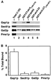
Shr3p specifically associates with Gap1p. (A) Immunoblot analysis of Gap1p, Sec61p, Gal2p, and Pma1p associated with purified GST fusion proteins. GST proteins were expressed in FGY145, purified, and analyzed as described in Figure 6. Protein bands corresponding to Sec61p, Gal2p, and Pma1p were visualized using rabbit antiserum as described in MATERIALS AND METHODS. For purposes of comparison, the appropriate portion of the Gap1p immunoblot depicted in Figure 6 is included. Lane 1 (Total) contains an aliquot of extract, corresponding to 1/25 of that incubated with glutathione beads, prepared from strain FGY145 expressing the GST-SHR3 construct. (B) The chemiluminescent signals derived from proteins present in the total extract (lane 1) and from proteins copurifying with GST-SHR3 construct (lane 4) were quantitated (LAS1000; Fuji). The percentages of proteins bound to GST-SHR3 were calculated (Gap1p, Sec61p, Gal2p, or Pma1p bound to GST-SHR3 [lane 4]/total Gap1p, Sec61p, Gal2p, or Pma1p present in the extract [lane 1, multiplied by 25 to correct for loading] × 100). Values from two independent experiments are plotted; error bars indicate 1 standard deviation.
Figure 8
Figure 8
Shr3p interacts transiently with Gap1p. The expression of GST-SHR3 (pFG117) and Gap1p in strain FGY145 was induced as described in MATERIALS AND METHODS. Cultures were labeled with [35S]methionine for 5 min and chased by the addition of excess unlabeled methionine and cysteine. Gap1p was immunoprecipitated from labeled extracts using anti-Gap1p antibodies directly (1o anti-Gap1p) or in a second round of immunoprecipitation from resolubilized immunoprecipitates obtained using glutathione-Sepharose beads (1o anti-GST and 2o anti-Gap1p).
Figure 9
Figure 9
COPII coatomer components copurify with GST-SHR3. The indicated GST fusion constructs were expressed in FGY145 and purified as in Figure 6, except that after affinity chromatography the glutathione-conjugated beads were washed four times with low-potassium FGB containing 0.02% DM (see MATERIALS AND METHODS). Proteins were denatured at 65°C for 5 min and resolved by SDS-PAGE in a 10% polyacrylamide gel. Protein bands corresponding to Sec31p, Sec24p, Sec23p, Sec13p, and Sar1p were visualized using rabbit antiserum as described in MATERIALS AND METHODS.
Figure 10
Figure 10
Multicopy expression of shr3-23 partially suppresses the temperature-sensitive phenotype of sec12-1. Serial dilutions of strain MAS26-1A (sec12-1) transformed with pRS202 (−), pPL250 (SHR3), and pMB42 (shr3-23) were spotted on SC (minus uracil), incubated at the indicated temperatures for 3 d, and photographed.

Similar articles

Cited by

References

    1. Allen JB, Elledge SJ. A family of vectors that facilitate transposon and insertional mutagenesis of cloned genes in yeast. Yeast. 1994;10:1267–1272. - PubMed
    1. André B. An overview of membrane transport proteins in Saccharomyces cerevisiae. Yeast. 1995;11:1575–1611. - PubMed
    1. Annaert WG, Becker B, Kistner U, Reth M, Jahn R. Export of cellubrevin from the endoplasmic reticulum is controlled by BAP31. J Cell Biol. 1997;139:1397–1410. - PMC - PubMed
    1. Antebi A, Fink GR. The yeast Ca2+-ATPase homologue, PMR1, is required for normal Golgi function and localizes in a novel Golgi-like distribution. Mol Biol Cell. 1992;3:633–654. - PMC - PubMed
    1. Aridor M, Weissman J, Bannykh S, Nuoffer C, Balch WE. Cargo selection by the COPII budding machinery during export from the ER. J Cell Biol. 1998;141:61–70. - PMC - PubMed

Publication types

MeSH terms