. 1999 Dec;73(12):10458-71.
doi: 10.1128/JVI.73.12.10458-10471.1999.

The human cytomegalovirus IE2 and UL112-113 proteins accumulate in viral DNA replication compartments that initiate from the periphery of promyelocytic leukemia protein-associated nuclear bodies (PODs or ND10)

Affiliations

The human cytomegalovirus IE2 and UL112-113 proteins accumulate in viral DNA replication compartments that initiate from the periphery of promyelocytic leukemia protein-associated nuclear bodies (PODs or ND10)

J H Ahn et al. J Virol. 1999 Dec.

Abstract

During human cytomegalovirus (HCMV) infection, the periphery of promyelocytic leukemia protein (PML)-associated nuclear bodies (also known as PML oncogenic domains [PODs] or ND10) are sites for both input viral genome deposition and immediate-early (IE) gene transcription. At very early times after infection, the IE1 protein localizes to and subsequently disrupts PODs, whereas the IE2 protein localizes within or adjacent to PODs. This process appears to be required for efficient viral gene expression and DNA replication. We have investigated the initiation of viral DNA replication compartment formation by studying the localization of viral IE proteins, DNA replication proteins, and the PML protein during productive infection. Localization of IE2 adjacent to PODs between 2 and 6 h after infection was confirmed by confocal microscopy of human fibroblasts (HF cells) infected with both wild-type HCMV(Towne) and with an IE1-deletion mutant HCMV(CR208) that fails to disrupt PODs. In HCMV(Towne)-infected HF cells at 24 to 48 h, IE2 also accumulated in newly formed viral DNA replication compartments containing the polymerase processivity factor (UL44), the single-stranded DNA binding protein (SSB; UL57), the UL112-113 accessory protein, and newly incorporated bromodeoxyuridine (BrdU). Double labeling of the HCMV(CR208)-infected HF cells demonstrated that formation of viral DNA replication compartments initiates within granular structures that bud from the periphery of some of the PODs and subsequently coalesce into larger structures that are flanked by PODs. In transient DNA transfection assays, both the N terminus (codons 136 to 290) and the C terminus (codons 379 to 579) of IE2 exon 5, but not the central region between them, were found to be necessary for both the punctate distribution of IE2 and its association with PODs. Like IE2, the UL112-113 accessory replication protein was also distributed in a POD-associated pattern in both DNA-transfected and virus-infected cells beginning at 6 h. Furthermore, when all six replication core machinery proteins (polymerase complex, SSB, and helicase-primase complex) were expressed together in the presence of UL112-113, they also accumulated at POD-associated sites, suggesting that the UL112-113 protein (but not IE2) may play a role in recruitment of viral replication fork proteins into the periphery of PODs. These results show that (i) subsequent to accumulating at the periphery of PODs, IE2 is incorporated together with the core proteins into viral DNA replication compartments that initiate from the periphery of PODs and then grow to fill the space between groups of PODs, and (ii) the UL112-113 protein appears to have a key role in assembling and recruiting the core replication machinery proteins in the initial stages of viral replication compartment formation.


Figures

FIG. 1
FIG. 1
Both IE2 and UL112-113 localize adjacent to PODs at very early times after infection. HF cells were infected with HCMV(Towne) at a low MOI (<1.0 PFU per cell) and fixed in methanol at 2 h (a) or 6 h (d) after infection, or they were infected with HCMV(CR208) at an MOI of 2.0 and fixed with paraformaldehyde at 6 h after infection (b and c). Confocal double-label IFA was carried out to detect IE2 and PML (a and b), UL112-113 and PML (c), or UL112-113 and IE2 (d). IE2 was detected with mouse MAb 12E2 and FITC-labeled donkey anti-mouse IgG, and UL112-113 was detected with rabbit PAb UL112-113(C) and rhodamine-coupled donkey anti-rabbit IgG. For PML, either mouse MAb 5E10 and FITC-labeled donkey anti-mouse IgG or rabbit PAb PML(C) and rhodamine-coupled donkey anti-rabbit IgG were used. Confocal images from each fluorochrome were recorded, and only the superimposed merged images are shown. Inserts show high-power magnification of some PODs. Note that association of IE2 and PML in PODs in wild-type virus-infected cells (a) is very transient because of IE1-induced displacement of PML from PODs.
FIG. 2
FIG. 2
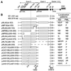
Domain requirements within the IE2 protein for POD association. (A and B) Summary of the localization patterns of both IE2 and PML in Vero cells transiently transfected with genomic plasmids expressing deleted versions of IE2. (A) The overlapping five-exon structure (solid bar) of the MIE gene transcription unit in the inverted (i.e., viral) genomic orientation is illustrated at the top. Positions of key restriction sites used to generate the deleted or truncated versions of IE2 (Bc, BclI; Ev, EcoRV; Sa, SalI; Sm, SmaI; St, StuI; Xh, XhoI) are indicated. The enhancer/promoter region of the MIE locus (ENH; hatched bar) and the translation start (ATG) and termination (TAA) sites as well as polyadenylation sites (pA) are also indicated. (B) Open bars represent coding regions with gaps denoting in-frame deletions; the diamond indicates inserted triple-terminator oligonucleotides; horizontal dashed lines indicate in-frame IE1-IE2 fusion proteins (pRL72 and pRL84). The estimated map locations for the epitopes recognized by the 12E2 and CH810 Mab are shown at the bottom (hatched bars). To detect IE2, mouse MAb 12E2 or CH810 and FITC-labeled donkey anti-mouse IgG were used. PML was detected as described for Fig. 1. IFA patterns: ND, nuclear diffuse; P, punctate; ND/P, a mixture of nuclear diffuse and punctate patterns; MP/P, nuclear micropunctate with concentrated punctate bodies; NAG, nuclear aggregation; NAG/C, NAG pattern with cytoplasmic diffuse; ND/C, nuclear and cytoplasmic diffuse pattern. a, IE2 was detected with mouse MAb CH810; b, reduced number of punctate bodies or numerous micropunctate bodies. aa, amino acids. (C) Double-label IFA images of Vero cells transfected with pMP88 encoding IE2(Δ136-290), pRL72 encoding IE2(Δ86-135), pCJC110 encoding IE2(Δ290-313), pJHA106 encoding IE2(Δ376-404), or pJHA105 encoding IE2(Δ403-419).
FIG. 2
FIG. 2
Domain requirements within the IE2 protein for POD association. (A and B) Summary of the localization patterns of both IE2 and PML in Vero cells transiently transfected with genomic plasmids expressing deleted versions of IE2. (A) The overlapping five-exon structure (solid bar) of the MIE gene transcription unit in the inverted (i.e., viral) genomic orientation is illustrated at the top. Positions of key restriction sites used to generate the deleted or truncated versions of IE2 (Bc, BclI; Ev, EcoRV; Sa, SalI; Sm, SmaI; St, StuI; Xh, XhoI) are indicated. The enhancer/promoter region of the MIE locus (ENH; hatched bar) and the translation start (ATG) and termination (TAA) sites as well as polyadenylation sites (pA) are also indicated. (B) Open bars represent coding regions with gaps denoting in-frame deletions; the diamond indicates inserted triple-terminator oligonucleotides; horizontal dashed lines indicate in-frame IE1-IE2 fusion proteins (pRL72 and pRL84). The estimated map locations for the epitopes recognized by the 12E2 and CH810 Mab are shown at the bottom (hatched bars). To detect IE2, mouse MAb 12E2 or CH810 and FITC-labeled donkey anti-mouse IgG were used. PML was detected as described for Fig. 1. IFA patterns: ND, nuclear diffuse; P, punctate; ND/P, a mixture of nuclear diffuse and punctate patterns; MP/P, nuclear micropunctate with concentrated punctate bodies; NAG, nuclear aggregation; NAG/C, NAG pattern with cytoplasmic diffuse; ND/C, nuclear and cytoplasmic diffuse pattern. a, IE2 was detected with mouse MAb CH810; b, reduced number of punctate bodies or numerous micropunctate bodies. aa, amino acids. (C) Double-label IFA images of Vero cells transfected with pMP88 encoding IE2(Δ136-290), pRL72 encoding IE2(Δ86-135), pCJC110 encoding IE2(Δ290-313), pJHA106 encoding IE2(Δ376-404), or pJHA105 encoding IE2(Δ403-419).
FIG. 3
FIG. 3
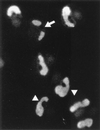
Localization pattern of IE2 at late times after infection. HF cells were infected with HCMV(Towne) at an MOI of 2.0, fixed in methanol at 96 h, and stained with mouse MAb 12E2 as described for Fig. 1. In most cells, IE2 was stained as large irregular oval structures that occupy most of the nucleus. In some cells, IE2 appeared as two distinct nuclear globular structures (arrow), but these may be connected to each other around the outside of the nucleolus (arrowheads).
FIG. 4
FIG. 4
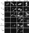
Comparison of distribution patterns of five viral proteins at different time points after infection. HF cells were infected with HCMV(Towne) at a low MOI (<1.0) in the absence or presence of PAA (200 μg/ml). Cells were BrdU pulse-labeled and fixed in methanol at 6, 24, and 72 h after infection, and single labeling IFA for IE1, IE2, UL44, and SSB (a to e) or double labeling for both UL112-113 and BrdU (e and f) was carried out as described in Materials and Methods.
FIG. 5
FIG. 5
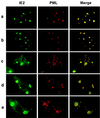
Double-label IFA images demonstrating that formation of viral DNA replication compartments initiates from the periphery of PODs. Double-label IFA images for IE2 and PML representing sequential intermediate stages in formation of early replication compartments are shown. HF cells were infected with IE1-defective mutant HCMV(CR208) at an MOI of 5.0 and fixed in methanol at 48 h (a to d) or 72 h (e) after infection. IE2 was detected with MAb 12E2 (FITC; green), and PODs were detected with PAb PML(C) (rhodamine; red). To produce merge images, each fluorochrome was recorded and the superimposed images were generated with Image-Pro software.
FIG. 6
FIG. 6
Double-label IFA images for UL112-113 with either IE2, UL44, or PML at intermediate stages of replication compartment formation. HF cells were infected with HCMV(CR208) and fixed at 48 h after infection as described for Fig. 5. For detection of UL112-113 (a to c), rabbit PAb and FITC-labeled donkey anti-rabbit IgG were used; for detection of IE2 (a), UL44 (b), and PML (c), mouse MAbs 12E2 (for IE2), ABI44 (for UL44), and 5E10 (for PML) and rhodamine-coupled donkey anti-mouse IgG were used. Merge images were obtained as described for Fig. 5.
FIG. 7
FIG. 7
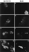
Localization of newly synthesized DNA in intermediate-stage viral DNA replication compartments. HF cells were infected with HCMV(Towne) at a low MOI (<1.0), pulse-labeled with BrdU, and fixed in methanol at 48 h after infection. Double-label IFA for both UL112-113 and BrdU was carried out as described for Fig. 4. Four independent microscopic images are shown. The large granular structures containing UL112-113 but not BrdU are indicated by arrows.
FIG. 8
FIG. 8
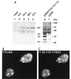
Specificity of the antipeptide PAb generated against UL112-113. (A) Immunoblot assay of extracts prepared from virus-infected HF cells and plasmid-transfected Vero cells. For lanes 1 to 5, HF cells were mock infected or infected with HCMV(Towne) at an MOI of 5.0, and total cell extracts were prepared at 6, 24, 48, and 72 h. For lanes 6 and 7, Vero cells were transfected with either a plasmid expressing 5′ F-tagged UL112-113 (pSG5-F/UL112-113, pJHA309) or the empty DNA vector (pSG5), and total extracts were prepared at 48 h after transfection. Equal amounts of each extract were subjected to SDS-PAGE (8% gel) for Western blotting with anti-UL112-113(C) PAb (lanes 1 to 5) or with anti-F MAb (lanes 6 and 7). The positions of the molecular size markers and the four F-tagged UL112-113 protein products (34, 43, 50, and 84 kDa) are indicated. (B and C) IFA images of UL112-113 in DNA-transfected cells. Vero cells were transiently transfected with plasmid pJHA309 expressing the 5′ F-tagged UL112-113 protein. Cells were fixed by the paraformaldehyde procedure at 48 h, and double-label IFA was carried out with mouse MAb directed against the F epitope and rabbit PAb UL112-113(C). (B) F-tagged UL112-113 protein products detected with mouse anti-F MAb and FITC-labeled donkey anti-mouse IgG. (C) The 84-kDa form of UL112-113 detected with rabbit anti-UL112-113(C) PAb and rhodamine-coupled anti-rabbit IgG.
FIG. 9
FIG. 9
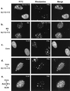
Role of UL112-113 in recruiting core replication proteins to the PODs. Double-label IFA images were obtained after the following combinations of plasmids were cotransfected into Vero cells: (a) F-tagged UL112-113 encoded by pJHA309; (b) IE2 in pMP18 and untagged UL112-113 in pRTS26; (c) the six HCMV core replication machinery proteins UL44 in pRTS22, UL54 in pRTS6, UL57 in pRTS7, UL70 in pRTS9, UL102 in pRTS29, and UL105 in pRTS10; (d) the six core machinery proteins and UL112-113 in pRTS26; and (e) the six core machinery proteins, five auxiliary replication-promoting factors UL36-38 in p302, IRS1 in pRTS18, IE2 in pMP18, UL112-113 in pRTS26, and UL84 in pRTS5, as well as the stimulatory factor UL69 in pRTS28. At 48 h after transfection, the cells were fixed with paraformaldehyde and double labeled as indicated in each panel. To detect F-tagged UL112-113 (a), IE2 (b), and UL44 (c to e), mouse MAbs and FITC-labeled donkey anti-mouse IgG were used. To detect PML (a, c, d, and e) and UL112-113 (b), rabbit PAbs and rhodamine-coupled anti-rabbit IgG were used. Merge images were obtained as described for Fig. 5.

Similar articles

Cited by

References

    1. Ahn J-H, Brignole III E J, Hayward G S. Disruption of PML subnuclear domains by the acidic IE1 protein of human cytomegalovirus is mediated through interaction with PML and may modulate a RING finger-dependent cryptic transactivator function of PML. Mol Cell Biol. 1998;18:4899–4913. - PMC - PubMed
    1. Ahn J-H, Chiou C-J, Hayward G S. Evaluation and mapping of the DNA binding and dimerization domains of the IE2 regulatory protein of human cytomegalovirus using yeast one and two hybrid interaction assays. Gene. 1997;210:25–36. - PubMed
    1. Ahn J-H, Hayward G S. The major immediate-early proteins IE1 and IE2 of human cytomegalovirus colocalize with and disrupt PML-associated nuclear bodies at very early times in infected permissive cells. J Virol. 1997;71:4599–4613. - PMC - PubMed
    1. Ahn, J.-H., and G. S. Hayward. Disruption of PML-associated nuclear bodies by IE1 is required for efficient early stages of viral gene expression and replication in human cytomegalovirus infection. Submitted for publication. - PubMed
    1. Ahn, J.-H., and G. S. Hayward. Unpublished data.

Publication types

MeSH terms

Substances