. 1999 Oct 18;147(2):221-34.
doi: 10.1083/jcb.147.2.221.

PML is critical for ND10 formation and recruits the PML-interacting protein daxx to this nuclear structure when modified by SUMO-1

Affiliations

PML is critical for ND10 formation and recruits the PML-interacting protein daxx to this nuclear structure when modified by SUMO-1

A M Ishov et al. J Cell Biol. .

Abstract

Nuclear domain 10 (ND10), also referred to as nuclear bodies, are discrete interchromosomal accumulations of several proteins including promyelocytic leukemia protein (PML) and Sp100. In this study, we investigated the mechanism of ND10 assembly by identifying proteins that are essential for this process using cells lines that lack individual ND10-associated proteins. We identified the adapter protein Daxx and BML, the RecQ helicase missing in Bloom syndrome, as new ND10-associated proteins. PML, but not BLM or Sp100, was found to be responsible for the proper localization of all other ND10-associated proteins since they are dispersed in PML-/- cells. Introducing PML into this cell line by transient expression or fusion with PML-producing cells recruited ND10-associated proteins into de novo formed ND10 attesting to PMLs essential nature in ND10 formation. In the absence of PML, Daxx is highly enriched in condensed chromatin. Its recruitment to ND10 from condensed chromatin requires a small ubiquitin-related modifier (SUMO-1) modification of PML and reflects the interaction between the COOH-terminal domain of Daxx and PML. The segregation of Daxx from condensed chromatin in the absence of PML to ND10 by increased accumulation of SUMO-1-modified PML suggests the presence of a variable equilibrium between these two nuclear sites. Our findings identify the basic requirements for ND10 formation and suggest a dynamic mechanism for protein recruitment to these nuclear domains controlled by the SUMO-1 modification state of PML.


Figures
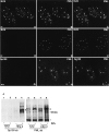
Figure 1
Figure 1
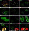
The new component of ND10, BLM helicase, as well as Sp100 are not necessary for ND10 maintenance. Confocal micrographs of various cells are presented with the labeled proteins in the upper corners. The cell type is indicated at the lower part of the image. (A–C) HF double labeled for BLM protein (A) and PML (B); the merged image shows that both proteins colocalize (C). (D–F) Bloom syndrome fibroblasts (BF) double labeled for BLM (D) and PML (E); there is no ND10 labeling with the BLM antibodies (F). (G–I) NT2 cells double labeled for Sp100 (G) and PML (H); there is no ND10 labeling for Sp100 (I). (J) Western blot analysis to determine the presence of Sp100 (lanes 1–4) and PML (lanes 5–8) in NT2 (lanes 1, 2, 5, and 6) and HEp-2 cells (lanes 3, 4, 7, and 8). IFNα + represents interferon α–treated cells. Asterisk marks unspecific protein recognized by anti–PML antibody. It indicates equal protein load. Arrowheads mark most abundant Sp100 and PML signals. Higher molecular mass polypeptides represent alternative splicing or posttranscriptional modification (by SUMO-1 or phosphorylation) of proteins. It is clearly seen that NT2 cells do not express Sp100 in normal and IFNα–upregulated conditions (lanes 1 and 2), whereas in HEp-2 cells IFNα upregulation results in a dramatic increase of Sp100 signal (lanes 3 and 4). Endogenous as well as IFNα–upregulated PML are present in both cell lines (lanes 5–8).
Figure 2
Figure 2
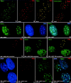
PML is responsible for the proper localization of all other ND10-associated proteins. Confocal micrographs of immunolabeled proteins are presented with the respective colors in the upper corners of the image. Cell type and overexpressed protein are indicated at the lower part of the image. (A) MPEF cells labeled for Sp100 and PML; the major ND10-associated proteins are present in mouse ND10. (B) MPEF cells double labeled for NDP55; mouse ND10 contain NDP55. C. MPEF cells double labeled for SUMO-1 and PML; mouse ND10 contain SUMO-1–modified proteins. D–H represent the same fields as in pictures above emphasizing the highest concentration of the corresponding proteins in ND10. (G) PML−/− MPEF cells labeled for Sp100 showing this protein only is dispersed throughout the nucleus in the absence of PML. (H) PML−/− MPEF cells labeled for NDP55; NDP55 is dispersed in the nucleus. (I) PML−/− MPEF cells labeled for SUMO-1; SUMO-1 is dispersed in the nucleus. (J) PML-transfected (left upper and lower cells) PML−/− MPEF labeled for Sp100 and PML showing that Sp100 is recruited to ND10-like sites. (K) PML transfected (right, upper and lower cells) PML−/− MPEF labeled for NDP55 and PML; NDP55 is recruited to ND10-like sites. (L) PML transfected PML−/− MPEF (left upper and lower cells) labeled for SUMO-1 and PML; SUMO-1 is recruited to ND10-like sites. Note exclusive nuclear localization of SUMO-1 even when PML is accumulated in the cytoplasm.
Figure 3
Figure 3
The new ND10-associated protein Daxx and its localization upon ND10 destruction and reconstitution. Confocal micrographs of immunolabeled proteins are presented with the respective colors in the upper corners of the image. Cell type and overexpressed proteins are indicated in the lower part of the image. (A–C) MPEF cells labeled for Daxx (A), PML (B), and both proteins (C) shown to colocalize at ND10. (D) PML−/− MPEF cells double labeled for centromeres and PML; some centromeres are associated with Daxx-positive sites, but mostly Daxx is not present in the same space as centromeres. (E–G) PML−/− MPEF cells double labeled for Daxx (E) and DNA (F), and together (G) show Daxx located at the condensed chromatin in the absence of PML. (H–J) PML−/− MPEF cells transfected to express PML (upper cell) and labeled for PML (H), Daxx (I), and both proteins (J); Daxx is located in PML-positive sites and contrary to the untransfected cells, very little is in the nucleoplasm and is unrecognizable at sites that may resemble condensed chromatin. (K–N) PML−/− MPEF cells fused to HF and stained for DNA (K) identifying the lower and left upper cell as of mouse origin by their DNA staining of condensed chromatin, and the right upper cell as human. Staining for PML is shown in L and for Daxx in M. In the merged image (N), the left upper mouse cell is shown to contain human PML and is, therefore, fused to HF. In the fused mouse cell, Daxx is present not only at condensed chromatin as in the lower mouse cell, but also in ND10-like domains.
Figure 4
Figure 4
Localization of GFP-fused wild-type Daxx and Daxx mutants upon transfection. Confocal micrographs presenting HEp-2 cells transiently transfected by plasmids expressing Daxx and Daxx mutants fused with GFP and stained by PML (A, D, and G) and GFP (B, E, and H). Merged images are presented in C, F, and I. Overexpressed proteins are indicated in the lower part of the image. (A–C) HEp-2 cells 16 h after transfection with wild-type Daxx fused to GFP-NLS; transiently expressed Daxx accumulates in PML-positive sites. (D–F) HEp-2 cells 16 h after transfection with Daxx 1-595 fused to GFP-NLS; this COOH-terminal deletion mutant does not accumulate in PML-positive sites. (G-I) HEp-2 cells 16 h after transfection with Daxx 624-740 fused to GFP-NLS; the transiently expressed Daxx COOH-terminal fragment accumulates in PML-positive sites but also floods the nucleoplasm.
Figure 5
Figure 5
SUMO-1 modification of PML is important for Daxx localization. Confocal micrographs of immunolabeled proteins are presented with the respective colors in the upper corners of the image. Cell type and overexpressed proteins are indicated in the lower part of the image. (A and E) PML-transfected HEp-2 cells double labeled for SUMO-1 and PML (A) or SUMO-1 only (E). SUMO-1 and PML colocalized only in the nucleus. (B and F) HEp-2 cells transfected to express the triple SUMO-1 site deletion PML mutant (PMLΔSUMO) double labeled for His-tagged PMLΔSUMO and SUMO-1 (B) or SUMO-1 only (F); PMLΔSUMO is accumulating in ND10-like structures in the upper two transfected cells; endogenous SUMO-1 indicating the location of ND10. There is no SUMO-1 in large nuclear PMLΔSUMO aggregates of the lower right cell as compared with the large aggregates of wild-type PML in A. The lower left cell is not transfected. (C and G) PML-transfected HEp-2 cells double labeled for Daxx and PML (C) or Daxx only (G); Daxx is accumulated in PML-positive sites. (D and H) HEp-2 cells transfected by His-tagged PMLΔSUMO and double labeled for Daxx and PMLΔSUMO (D) or Daxx only (H); there is no Daxx in the mutant PML accumulations as compared with the wild-type PML transfection in C. The upper left cell is not transfected. (I and J) HEp-2 cells double transfected to express PML and Daxx and double labeled for Daxx and PML (I) or Daxx only (J); Daxx and PML colocalized, but in the nucleus only. (K and L) Daxx and PMLΔSUMO double-transfected HEp-2 cells double labeled for Daxx and His-tagged PMLΔSUMO (K) or Daxx only (L); Daxx and PMLΔSUMO are deposited in different spaces.
Figure 6
Figure 6
Hierarchical scheme of ND10 formation and maintenance. PMLS indicates PML conjugated to SUMO-1; DNA M-Tase, DNA methyltransferase.

Similar articles

Cited by

References

    1. Ahn J.H., Brignole E.J., III, Hayward G.S. Disruption of PML subnuclear domains by the acidic IE1 protein of human cytomegalovirus is mediated through interaction with PML and may modulate a RING finger-dependent cryptic transactivator function of PML. Mol. Cell Biol. 1998;18:4899–4913. - PMC - PubMed
    1. Ascoli C.A., Maul G.G. Identification of a novel nuclear domain. J. Cell Biol. 1991;112:785–795. - PMC - PubMed
    1. Boddy M.N., Howe K., Etkin L.D., Solomon E., Freemont P.S. PIC 1, a novel ubiquitin-like protein which interacts with the PML component of a multiprotein complex that is disrupted in acute promyelocytic leukaemia. Oncogene. 1996;13:971–982. - PubMed
    1. Candau R., Moore P.A., Wang L., Barlev N., Ying C.Y., Rosen C.A., Berger S.L. Identification of human proteins functionally conserved with the yeast putative adaptors ADA2 and GCN5. Mol. Cell Biol. 1996;16:593–602. - PMC - PubMed
    1. Chelbi-Alix M.K., de The H. Herpes virus induced proteasome-dependent degradation of the nuclear bodies-associated PML and Sp100 proteins. Oncogene. 1999;18:935–941. - PubMed

Publication types

MeSH terms