. 1999 Oct;19(10):7216-27.
doi: 10.1128/MCB.19.10.7216.

ATP-Dependent inactivation and sequestration of ornithine decarboxylase by the 26S proteasome are prerequisites for degradation

Affiliations

ATP-Dependent inactivation and sequestration of ornithine decarboxylase by the 26S proteasome are prerequisites for degradation

Y Murakami et al. Mol Cell Biol. 1999 Oct.

Abstract

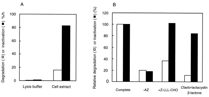
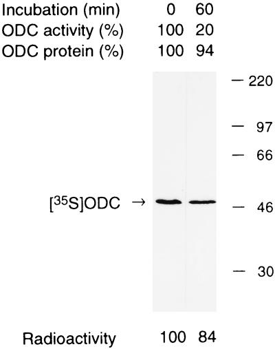
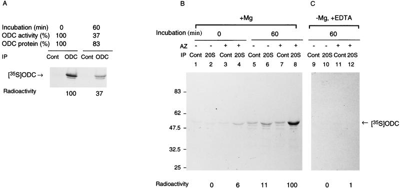
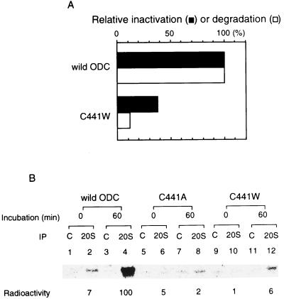
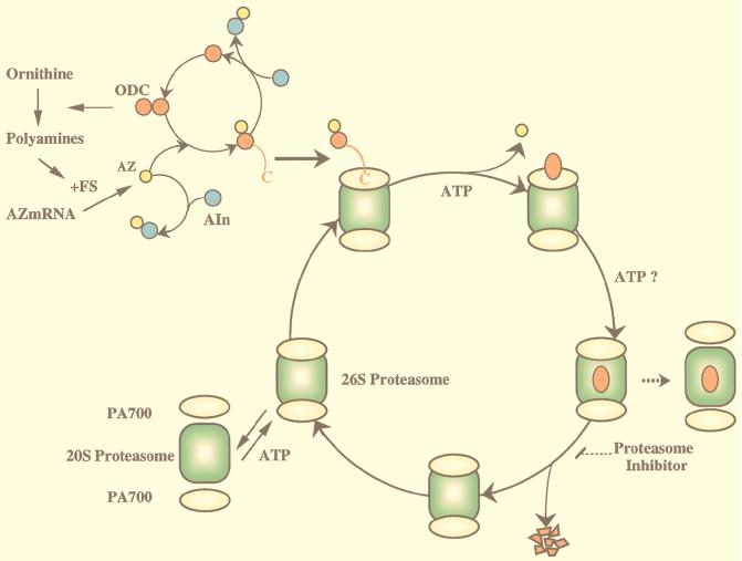
The 26S proteasome is a eukaryotic ATP-dependent protease, but the molecular basis of its energy requirement is largely unknown. Ornithine decarboxylase (ODC) is the only known enzyme to be degraded by the 26S proteasome without ubiquitinylation. We report here that the 26S proteasome is responsible for the irreversible inactivation coupled to sequestration of ODC, a process requiring ATP and antizyme (AZ) but not proteolytic activity. Neither the 20S proteasome (catalytic core) nor PA700 (the regulatory complex) by itself contributed to this ODC inactivation. Analysis with a C-terminal mutant ODC revealed that the 26S proteasome recognizes the C-terminal degradation signal of ODC exposed by attachment of AZ, and subsequent ATP-dependent sequestration of ODC in the 26S proteasome causes irreversible inactivation, possibly unfolding, of ODC and dissociation of AZ. These processes may be linked to the translocation of ODC into the 20S proteasomal inner cavity, centralized within the 26S proteasome, for degradation.


Figures

FIG. 1
FIG. 1
Irreversible inactivation of ODC by cell extracts. (A) ODC metabolically labeled with [35S]methionine in HTC cells was incubated with or without cell extracts that had been supplemented with AZ and ATP. Irreversible inactivation and degradation of ODC were determined as described in Materials and Methods. (B) ODC (a mixture of in vitro-translated labeled rat ODC and cold ODC purified from rat liver) was incubated with cell extracts in an inactivation/degradation reaction mixture. The assay was the same as for Fig. 1A, except that AZ was withdrawn or Z-LLL-CHO (100 μM) was added. When the effect of clasto-lactacystin β-lactone was examined, cell extracts were preincubated with the inhibitor (200 μM) at 37°C for 15 min in an inactivation/degradation reaction mixture without ODC, and then a one-sixth volume of ODC was added. The inactivation and degradation of ODC are represented by filled and open columns, respectively. Results are shown as percentages of the values obtained in the complete reaction mixture without inhibitors.
FIG. 2
FIG. 2
Molecular size of irreversibly inactivated ODC. ODC (mixture of purified rat ODC and metabolically labeled 35S-ODC from FM3A mouse cells) was incubated for 60 min at 37°C in an inactivation/degradation reaction mixture containing an extract of HTC cells that had been treated with Z-LLL-CHO. The decreases in ODC activity and ODC protein during incubation were 80 and 6%, respectively. Aliquots of the reaction mixture were subjected to SDS-PAGE and analyzed by autoradiography. Relative intensities of ODC bands determined with an image analyzer (Fujix BAS2000) are shown below each lane.
FIG. 3
FIG. 3
Effects of immunodepletion of the proteasome on inactivation of ODC by an HTC cell extract. Samples (300 μg) of HTC cell extracts were treated with anti-proteasome IgG (■) or control IgG (◊), and aliquots of the supernatants were assayed for Suc-LLVY-AMC degradation (A), ODC degradation (B), and ODC inactivation (C). Values are percentages of the activities measured with no added IgG, namely, 600 nmol/h for peptide hydrolysis, 25%/h for ODC inactivation, and 10%/h for ODC degradation.
FIG. 4
FIG. 4
PA700 inactivates ODC in the presence of the 20S proteasome but not in the absence of the proteasome. Purified PA700 was separated by glycerol density gradient centrifugation. The gradient was separated into 30 fractions of 1 ml each. Samples of 150 μl of the gradient fractions were precipitated with 750 μl of acetone, and the precipitates were subjected to SDS-PAGE and stained with Coomassie blue (A). Twenty microliters of the samples were assayed for proteasome activity (B) and ODC degradation (C) and inactivation (D) in the presence (■) and absence (□) of purified 20S proteasome. For the ODC inactivation assay, samples were preincubated with clasto-lactacystin β-lactone (200 μM) at 37°C for 15 min. The inset in panel B shows SDS-PAGE of the 20S proteasome used.
FIG. 5
FIG. 5
Energy- and AZ-dependent binding of ODC to the 26S proteasome. (A) ODC was incubated with a crude cell extract of HTC cells in the inactivation assay mixture for 60 min at 37°C. Aliquots of the reaction mixture before and after incubation were examined for ODC activity and ODC protein, and aliquots were immunoprecipitated with monoclonal antibody to ODC (HO101) or control IgG followed by SDS-PAGE and autoradiography. Intensities of ODC bands were determined with an image analyzer (Fujix BAS2000), and specific bindings were obtained by subtracting the value with control IgG and are expressed relative to the amount of time zero control. (B and C) ODC was incubated with partially purified 26S proteasome in the inactivation assay mixture. Where indicated, AZ was removed from the reaction mixture or EDTA was added to it instead of MgCl2. The incubated mixture was immunoprecipitated with anti-20S proteasome antibody (20S) or control IgG (Cont). The mixture without incubation was similarly treated with antibodies. The immunoprecipitates were washed extensively with buffer containing 0.1% SDS and 0.1% Triton X-100 and subjected to SDS-PAGE and then to autoradiography. Panels B and C represent separate experiments. The control (with MgCl2, without EDTA) is not shown in panel C but was almost the same as in panel B. Intensities of ODC bands were determined by an image analyzer, and the specific precipitation of ODC with anti-20S proteasome antibody was obtained by subtracting the value with control IgG and is expressed relative to the amount immunoprecipitated when ODC was incubated in the presence of AZ and MgCl2.
FIG. 6
FIG. 6
Cosedimentation of inactivated ODC with the 26S proteasome in glycerol density gradient centrifugation. 35S-ODC was incubated for 1 h with a crude extract of HTC cells (640 μg of protein) in the inactivation assay mixture (500 μl), and the mixture was centrifuged at 128,000 × g for 5 h. The precipitates were resuspended and subjected to glycerol density gradient centrifugation as for Fig. 4. Fraction 1 represents the top of the gradient (10% glycerol), and fraction 30 represents the bottom of the gradient (40% glycerol). Samples (0.8 ml) of the gradient fractions were used for determinations of ODC radioactivity, and proteasome activities in the presence and absence of SDS were determined for each 20 μl of the fractions. Arrows indicate the positions of elution of purified 20S and 26S proteasomes.
FIG. 7
FIG. 7
Time courses of ODC inactivation, degradation, and binding. ODC was incubated for the indicated times at 37°C in an inactivation/degradation assay mixture containing partially purified 26S proteasome that had been treated (B) or not treated (A) with clasto-lactacystin β-lactone (200 μM). ODC inactivation, degradation, and binding were determined as described in Materials and Methods. The inset (B) shows SDS-PAGE of ODC coimmunoprecipitated with anti-20S proteasome antibody after the inactivation reaction at the indicated times. The relative amounts of coimmunoprecipitated ODC (ODC binding) were quantitated with an image analyzer.
FIG. 8
FIG. 8
Resistance of inactivated ODC to PK digestion. 35S-ODC was preincubated for 1 h with a crude extract of HTC cells in the inactivation assay mixture. The mixtures were treated with PK (80 μg/ml) for varying times at the indicated temperatures (lanes 4 to 9 and 13 to 16). Digestion was halted by the addition of PMSF. ODCs bound to the proteasome and unbound (shown by P and S, respectively) were separated with anti-20S proteasome antibody, and both ODCs were analyzed by SDS-PAGE followed by autoradiography. As another control, unpreincubated ODC was similarly treated with PK in the presence of BSA instead of a crude cell extract and then analyzed by SDS-PAGE (shown in lanes 1 to 3, 11, and 12). Purified substrate 35S-ODC is shown in lane 10.
FIG. 9
FIG. 9
The C-terminal region of ODC is necessary for binding to and inactivation by the 26S proteasome. (A) Recombinant ODC with replacement of cysteine at position 441 by tryptophan (C441W) was obtained by using a pET vector expression system, purified by immunoaffinity chromatography, and tested for inactivation by the 26S proteasome. mRNA encoding ODC (C441W) was translated in a rabbit reticulocyte lysate in the presence of [35S]methionine. The protein product was purified by immunoaffinity chromatography and tested for degradation by the 26S proteasome. Wild-type ODC was similarly treated for comparison. (B) Purified 35S-labeled stable ODCs were prepared as described above, and binding to the 26S proteasome was examined as in Fig. 5B. Results with wild-type ODC treated similarly are shown for comparison.
FIG. 10
FIG. 10
Model of ODC degradation by the 26S proteasome. ODC is active as a homodimer complex. AZ, which is induced by translational frameshifting, binds to inactive monomeric ODC to form an ODC-AZ complex. Two terminal regulatory subcomplexes, termed PA700, are attached in an ATP-dependent manner to both ends of the 20S proteasome (central catalytic machinery) in opposite orientations to form enzymatically active proteasomes. The 26S proteasome may attack the exposed ODC C-terminal region and pull the ODC molecule, but not AZ, into the interior, which is associated with ATP-dependent substrate unfolding. The continuous translocation of unfolded ODC (inactivated ODC) may be required for further continuous unfolding of ODC (ODC inactivation), and the two processes may proceed in concert with each other. The unfolded ODC translocated into the cavity of the 20S proteasome, which harbors proteolytically active sites, is degraded, but it is unknown whether ATP consumption is needed for the degradative process itself. After degradation, the 26S proteasome traps another substrate(s) or may be in part dissociated into its constituents, the 20S proteasome and PA700. See the text for details.

Similar articles

Cited by

References

    1. Armon T, Ganoth D, Hershko A. Assembly of the 26S complex that degrades proteins ligated to ubiquitin is accompanied by the formation of ATPase activity. J Biol Chem. 1990;265:20723–20726. - PubMed
    1. Auvinen M, Paasinen A, Andersson L C, Hölttä E. Ornithine decarboxylase activity is critical for cell transformation. Nature. 1992;360:355–359. - PubMed
    1. Baumeister W, Walz J, Zuhl F, Seemuller E. The proteasome: paradigm of a self-compartmentalizing protease. Cell. 1998;92:367–380. - PubMed
    1. Bercovich Z, Kahana C. Involvement of the 20S proteasome in the degradation of ornithine decarboxylase. Eur J Biochem. 1993;213:205–210. - PubMed
    1. Chu-Ping M, Vu J H, Proske R J, Slaughter C A, DeMartino G N. Identification, purification, and characterization of a high molecular weight ATP-dependent activator (PA700) of the 20S proteasome. J Biol Chem. 1994;269:3539–3547. - PubMed

Publication types

MeSH terms