. 1999 Oct;19(10):7088-95.
doi: 10.1128/MCB.19.10.7088.

Regulation of RelA subcellular localization by a putative nuclear export signal and p50

Affiliations

Regulation of RelA subcellular localization by a putative nuclear export signal and p50

E W Harhaj et al. Mol Cell Biol. 1999 Oct.

Abstract

Nuclear factor kappaB (NF-kappaB) represents a family of dimeric DNA binding proteins, the pleotropic form of which is a heterodimer composed of RelA and p50 subunits. The biological activity of NF-kappaB is controlled through its subcellular localization. Inactive NF-kappaB is sequestered in the cytoplasm by physical interaction with an inhibitor, IkappaBalpha. Signal-mediated IkappaBalpha degradation triggers the release and subsequent nuclear translocation of NF-kappaB. It remains unknown whether the NF-kappaB shuttling between the cytoplasm and nucleus is subjected to additional steps of regulation. In this study, we demonstrated that the RelA subunit of NF-kappaB exhibits strong cytoplasmic localization activity even in the absence of IkappaBalpha inhibition. The cytoplasmic distribution of RelA is largely mediated by a leucine-rich sequence homologous to the recently characterized nuclear export signal (NES). This putative NES is both required and sufficient to mediate cytoplasmic localization of RelA as well as that of heterologous proteins. Furthermore, the cytoplasmic distribution of RelA is sensitive to a nuclear export inhibitor, leptomycin B, suggesting that RelA undergoes continuous nuclear export. Interestingly, expression of p50 prevents the cytoplasmic expression of RelA, leading to the nuclear accumulation of both RelA and p50. Together, these results suggest that the nuclear and cytoplasmic shuttling of RelA is regulated by both an intrinsic NES-like sequence and the p50 subunit of NF-kappaB.


Figures

FIG. 1
FIG. 1
Cytoplasmic expression of RelA is mediated by its C-terminal sequences located beyond the transactivation domain. (A) Primary domain structure of RelA WT and its truncation mutants. The locations of the DNA binding domain (DB) and the transactivation domain (TA) are presented according to previous studies (6, 21, 45). The DNA binding and transactivation activities of the mutants were as determined in a previous study (21) and for Fig. 1C and D. (B) Immunofluorescence assays to determine the subcellular localization of RelA WT and truncation mutants. COS cells were transfected with the indicated cDNA expression vectors. After 48 h, the transfected cells were subjected to indirect immunofluorescence assays with antisera recognizing the C terminus (WT and 31-551) or N terminus (1-450, 1-420, and 1-312) of RelA (21) and Texas red-conjugated anti-rabbit Ig secondary antibody (upper panels). To localize the nuclei of the transfected cells, the cells were counterstained with Hoechst 33258 and visualized with a UV filter (lower panels). (C) Luciferase reporter gene assay determining the transactivation activity of RelA and its truncation mutants. COS cells were transfected with the κB-TATA-luc reporter plasmid together with either an empty vector, cDNA expression vectors encoding the wild type (WT), or the indicated truncation mutants of RelA. Luciferase activity is presented as the fold induction relative to the basal level measured in cells transfected with the empty vector. (D) Immunoblotting analysis of the whole-cell extracts isolated from COS cells transfected with either an empty vector or cDNA expression vectors encoding RelA WT or its truncation mutants [RelA(1-450) and RelA(31-551)]. Immunoblotting was performed with antisera that reacted with the N terminus (lanes 1 to 3) or C terminus (lane 4) of RelA or IκBα. The RelA and its truncation mutants are labeled with a bracket, and IκBα is indicated by an arrow. Only RelA WT induces expression of endogenous IκBα.
FIG. 1
FIG. 1
Cytoplasmic expression of RelA is mediated by its C-terminal sequences located beyond the transactivation domain. (A) Primary domain structure of RelA WT and its truncation mutants. The locations of the DNA binding domain (DB) and the transactivation domain (TA) are presented according to previous studies (6, 21, 45). The DNA binding and transactivation activities of the mutants were as determined in a previous study (21) and for Fig. 1C and D. (B) Immunofluorescence assays to determine the subcellular localization of RelA WT and truncation mutants. COS cells were transfected with the indicated cDNA expression vectors. After 48 h, the transfected cells were subjected to indirect immunofluorescence assays with antisera recognizing the C terminus (WT and 31-551) or N terminus (1-450, 1-420, and 1-312) of RelA (21) and Texas red-conjugated anti-rabbit Ig secondary antibody (upper panels). To localize the nuclei of the transfected cells, the cells were counterstained with Hoechst 33258 and visualized with a UV filter (lower panels). (C) Luciferase reporter gene assay determining the transactivation activity of RelA and its truncation mutants. COS cells were transfected with the κB-TATA-luc reporter plasmid together with either an empty vector, cDNA expression vectors encoding the wild type (WT), or the indicated truncation mutants of RelA. Luciferase activity is presented as the fold induction relative to the basal level measured in cells transfected with the empty vector. (D) Immunoblotting analysis of the whole-cell extracts isolated from COS cells transfected with either an empty vector or cDNA expression vectors encoding RelA WT or its truncation mutants [RelA(1-450) and RelA(31-551)]. Immunoblotting was performed with antisera that reacted with the N terminus (lanes 1 to 3) or C terminus (lane 4) of RelA or IκBα. The RelA and its truncation mutants are labeled with a bracket, and IκBα is indicated by an arrow. Only RelA WT induces expression of endogenous IκBα.
FIG. 2
FIG. 2
Identification of a NES-like sequence in RelA. The upper panel shows the amino acid sequence of the C-terminal region of RelA mediating its cytoplasmic distribution. The NES-like sequence is indicated. The lower panel shows the sequence homology of the RelA NES with the NES characterized from various other proteins, including the alpha and beta subunits of the protein kinase inhibitor (PKI) (57), the HIV Rev protein (18), and c-Abl (52). The four hydrophobic amino acids, most of which are leucines, are bold and underlined. The NES of RelA is located between amino acids 436 and 445.
FIG. 3
FIG. 3
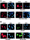
The NES-like sequence of RelA promotes cytoplasmic localization of RelA as well as p50. (A) COS cells were transfected with cDNA expression vectors encoding the RelA proteins indicated below each of the panels. The cells were stained with anti-RelA plus Texas red-conjugated rabbit IgG (αRelA), counterstained with Hoechst, and visualized as described in the legend for Fig. 1B. (B) COS cells were transfected with cDNA expression vectors encoding the proteins indicated below the panels. The cells were stained with an anti-RelA antiserum and Texas red-conjugated rabbit IgG. The expression of the RelA mutants (a and b) was visualized with a rhodamine filter (αRelA), whereas the expression of p50-GFP fusion proteins (c and d) was visualized via the autofluorescence of GFP by using an FITC filter (GFP). The nuclei of the cells were visualized by DNA staining with Hoechst (DNA).
FIG. 4
FIG. 4
Cytoplasmic expression of RelA is sensitive to a nuclear export inhibitor, LMB. (A) COS cells were transfected with cDNA expression vectors encoding the indicated RelA proteins or p50-GFP-NES. After 48 h, the cells were either not treated (NT) or treated for 6 h with 40 ng of LMB per ml, followed by immunofluorescence staining. The RelA and its mutants were stained with anti-RelA and Texas red-conjugated rabbit IgG and were visualized with a rhodamine filter, while the p50-GFP-NES proteins were directly visualized with an FITC filter. Nuclei of the cells were stained with Hoechst, and the images are shown in the lower panels. (B and C) COS cells were transfected with RelA WT, and 48 h posttransfection, the cells were incubated for 6 h with the indicated amounts of LMB. The expression of the transfected RelA and the induced endogenous IκBα was detected by a Western blot assay with anti-RelA and anti-IκBα (B). The intensity of the protein bands was quantitated by densitometry and presented as a percentage of that from the untreated cells (C).
FIG. 4
FIG. 4
Cytoplasmic expression of RelA is sensitive to a nuclear export inhibitor, LMB. (A) COS cells were transfected with cDNA expression vectors encoding the indicated RelA proteins or p50-GFP-NES. After 48 h, the cells were either not treated (NT) or treated for 6 h with 40 ng of LMB per ml, followed by immunofluorescence staining. The RelA and its mutants were stained with anti-RelA and Texas red-conjugated rabbit IgG and were visualized with a rhodamine filter, while the p50-GFP-NES proteins were directly visualized with an FITC filter. Nuclei of the cells were stained with Hoechst, and the images are shown in the lower panels. (B and C) COS cells were transfected with RelA WT, and 48 h posttransfection, the cells were incubated for 6 h with the indicated amounts of LMB. The expression of the transfected RelA and the induced endogenous IκBα was detected by a Western blot assay with anti-RelA and anti-IκBα (B). The intensity of the protein bands was quantitated by densitometry and presented as a percentage of that from the untreated cells (C).
FIG. 5
FIG. 5
p50 induces the nuclear accumulation of RelA. (A) COS cells were transfected with RelA together with cDNA expression vectors encoding either GFP (a to c) or p50-GFP (d to f). The cells were stained with the C-terminal specific anti-RelA antibody plus Texas red-conjugated rabbit IgG. The expression of RelA and GFP or p50-GFP in the same cells was visualized with rhodamine (upper panels) and FITC (middle panels) filters, respectively. The nuclei of the cells were visualized by Hoechst staining (lower panels). Note that most of the cells were cotransfected. A cell expressing only RelA is indicated by the arrow. (B) COS cells were transfected with RelA and p50-GFP-NES, and the transfected cells were subjected to immunofluorescence as described above. A cell expressing only RelA is indicated by the arrow, which shows cytoplasmic expression of RelA.
FIG. 6
FIG. 6
Free IκBα accumulates in the nucleus but is excluded from the nucleus when coexpressed with RelA and p50. (A) COS cells were transfected with an expression vector encoding the IκBα-GFP fusion protein either alone (left panels) or together with RelA (right panels). The transfected cells were subjected to immunostaining with anti-RelA, and the subcellular localization of IκBα-GFP and RelA was visualized by a fluorescence microscope with FITC (upper panels) and rhodamine (middle panels) filters, respectively. DNA staining is shown in the lower panels. (B) COS cells were transfected with p50-GFP together with RelA (left panels) or p50-GFP together with RelA and IκBα (right panels). The cells were subjected to immunofluorescence analyses as described above, and the subcellular localization of p50-GFP and RelA was visualized with FITC (upper panels) and rhodamine (middle panels) filters, respectively. DNA staining is shown in the lower panels.

Similar articles

Cited by

References

    1. Arenzana-Seisdedos F, Turpin P, Rodriguez M, Thomas D, Hay R T, Virelizier J-L, Dargemont C. Nuclear localization of IκBα promotes active transport of NF-κB from the nucleus to the cytoplasm. J Cell Sci. 1997;110:369–378. - PubMed
    1. Baeuerle P, Baichwal V R. NF-κB as a frequent target for immunosuppressive and anti-inflammatory molecules. Adv Immunol. 1997;65:111–137. - PubMed
    1. Baeuerle P A. IκB-NF-κB structures: at the interface of inflammation control. Cell. 1998;95:729–731. - PubMed
    1. Baeuerle P A, Baltimore D. A 65-kD subunit of active NF-κB is required for inhibition of NF-κB by IκB. Genes Dev. 1989;3:1689–1698. - PubMed
    1. Baldwin A S., Jr The NF-κB and IκB proteins: new discoveries and insights. Annu Rev Immunol. 1996;14:649–683. - PubMed

Publication types