

The severity of murray valley encephalitis in mice is linked to neutrophil infiltration and inducible nitric oxide synthase activity in the central nervous system
- PMID: 10482632
- PMCID: PMC112899
- DOI: 10.1128/JVI.73.10.8781-8790.1999
The severity of murray valley encephalitis in mice is linked to neutrophil infiltration and inducible nitric oxide synthase activity in the central nervous system
Abstract
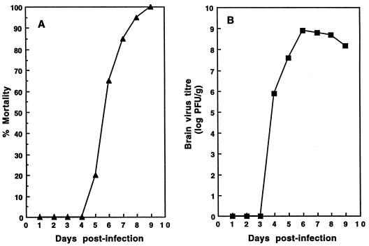
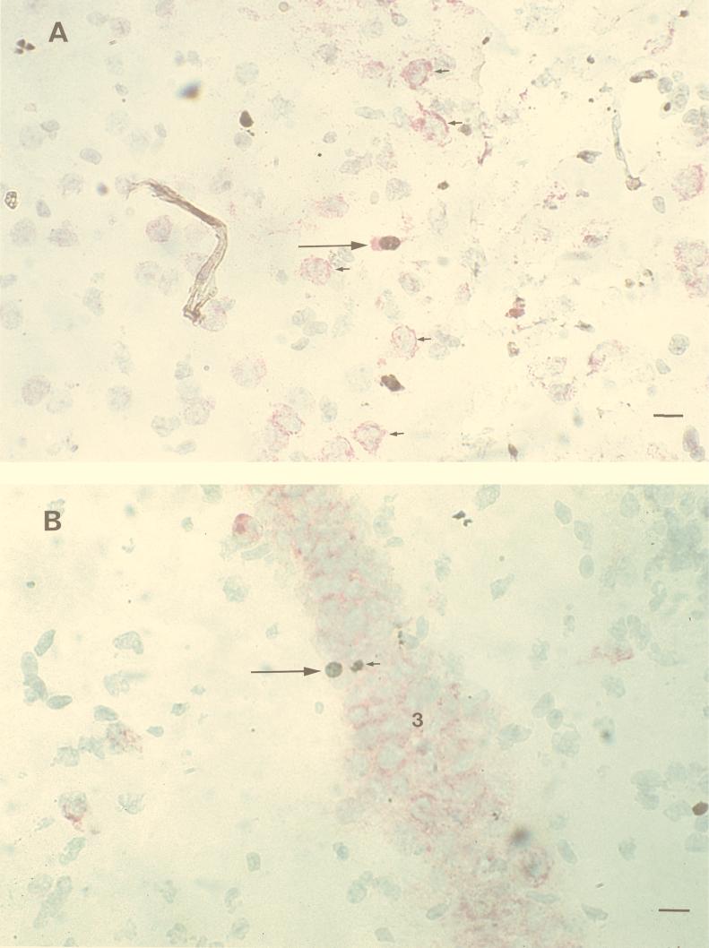
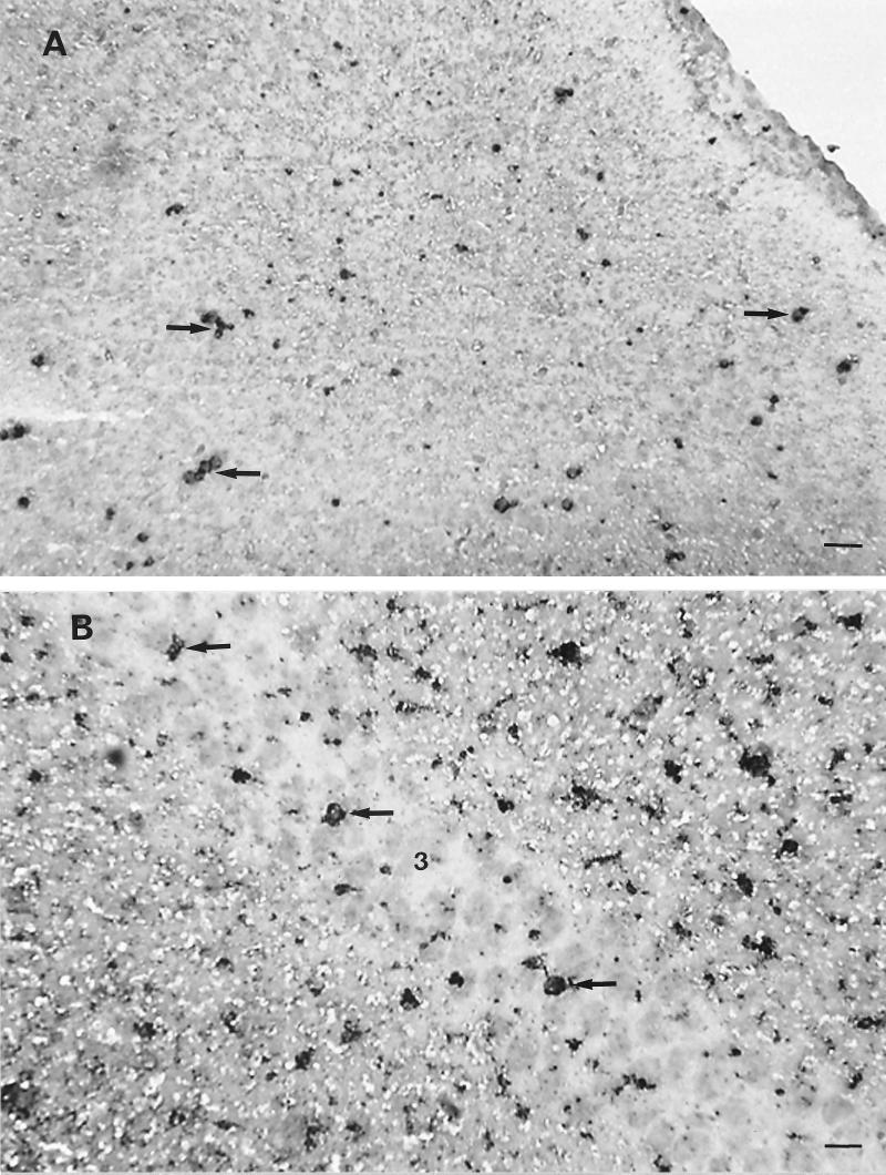
A study of immunopathology in the central nervous system (CNS) during infection with a virulent strain of Murray Valley encephalitis virus (MVE) in weanling Swiss mice following peripheral inoculation is presented. It has previously been shown that virus enters the murine CNS 4 days after peripheral inoculation, spreads to the anterior olfactory nucleus, the pyriform cortex, and the hippocampal formation at 5 days postinfection (p.i.), and then spreads throughout the cerebral cortex, caudate putamen, thalamus, and brain stem between 6 and 9 days p.i. (P. C. McMinn, L. Dalgarno, and R. C. Weir, Virology 220:414-423, 1996). Here we show that the encephalitis which develops in MVE-infected mice from 5 days p.i. is associated with the development of a neutrophil inflammatory response in perivascular regions and in the CNS parenchyma. Infiltration of neutrophils into the CNS was preceded by increased expression of tumor necrosis factor alpha and the neutrophil-attracting chemokine N51/KC within the CNS. Depletion of neutrophils with a cytotoxic monoclonal antibody (RB6-8C5) resulted in prolonged survival and decreased mortality in MVE-infected mice. In addition, neutrophil infiltration and disease onset correlated with expression of the enzyme-inducible nitric oxide synthase (iNOS) within the CNS. Inhibition of iNOS by aminoguanidine resulted in prolonged survival and decreased mortality in MVE-infected mice. This study provides strong support for the hypothesis that Murray Valley encephalitis is primarily an immunopathological disease.
Figures
Similar articles
-
Morphological features of Murray Valley encephalitis virus infection in the central nervous system of Swiss mice.Int J Exp Pathol. 2000 Feb;81(1):31-40. doi: 10.1046/j.1365-2613.2000.00135.x. Int J Exp Pathol. 2000. PMID: 10718862 Free PMC article.
-
A comparison of the spread of Murray Valley encephalitis viruses of high or low neuroinvasiveness in the tissues of Swiss mice after peripheral inoculation.Virology. 1996 Jun 15;220(2):414-23. doi: 10.1006/viro.1996.0329. Virology. 1996. PMID: 8661392
-
Protective immune responses to the E and NS1 proteins of Murray Valley encephalitis virus in hybrids of flavivirus-resistant mice.J Gen Virol. 1996 Jun;77 ( Pt 6):1287-94. doi: 10.1099/0022-1317-77-6-1287. J Gen Virol. 1996. PMID: 8683218
-
Murray Valley encephalitis: case report and review of neuroradiological features.Australas Radiol. 2003 Mar;47(1):61-3. doi: 10.1046/j.1440-1673.2003.01105.x. Australas Radiol. 2003. PMID: 12581057 Review.
-
Australian X disease, Murray Valley encephalitis and the French connection.Vet Microbiol. 1995 Sep;46(1-3):79-90. doi: 10.1016/0378-1135(95)00074-k. Vet Microbiol. 1995. PMID: 8545982 Review.
Cited by
-
Nitric oxide and virus infection.Immunology. 2000 Nov;101(3):300-8. doi: 10.1046/j.1365-2567.2000.00142.x. Immunology. 2000. PMID: 11106932 Free PMC article. Review.
-
Neuroadapted yellow fever virus 17D: genetic and biological characterization of a highly mouse-neurovirulent virus and its infectious molecular clone.J Virol. 2001 Nov;75(22):10912-22. doi: 10.1128/JVI.75.22.10912-10922.2001. J Virol. 2001. PMID: 11602731 Free PMC article.
-
Duck Tembusu virus exhibits neurovirulence in BALB/c mice.Virol J. 2013 Aug 14;10:260. doi: 10.1186/1743-422X-10-260. Virol J. 2013. PMID: 23941427 Free PMC article.
-
Neuroprotection and progenitor cell renewal in the injured adult murine retina requires healing monocyte-derived macrophages.J Exp Med. 2011 Jan 17;208(1):23-39. doi: 10.1084/jem.20101202. Epub 2011 Jan 10. J Exp Med. 2011. PMID: 21220455 Free PMC article.
-
Lack of both Fas ligand and perforin protects from flavivirus-mediated encephalitis in mice.J Virol. 2002 Apr;76(7):3202-11. doi: 10.1128/jvi.76.7.3202-3211.2002. J Virol. 2002. PMID: 11884544 Free PMC article.
References
-
- Anthony D, Dempster R, Fearn S, Clements J, Wells G, Perry V H, Walker K. CXC chemokines generate age-related increases in neutrophil-mediated brain inflammation and blood-brain barrier breakdown. Curr Biol. 1998;8:923–926. - PubMed
-
- Anthony D C, Bolton S J, Fearn S, Perry V H. Age-related effects of interleukin-1β on polymorphonuclear neutrophil-dependent increases in blood-brain barrier permeability in rats. Brain. 1997;120:435–444. - PubMed
-
- Bell M D, Taub D D, Perry V H. Overriding the brain’s intrinsic resistance to leukocyte recruitment with intraparenchymal injection of recombinant chemokines. Neuroscience. 1996;74:283–292. - PubMed
-
- Boje K M. Inhibition of nitric oxide synthase attenuates blood-brain barrier disruption during experimental meningitis. Brain Res. 1996;720:75–83. - PubMed
-
- Boyle D B. Comparative studies of two related Australian flaviviruses: Murray Valley encephalitis and Kunjin. Ph.D. thesis. Canberra, Australia: Australian National University; 1979.
Publication types
MeSH terms
Substances
LinkOut - more resources
Full Text Sources
