. 1999 Oct;73(10):8415-26.
doi: 10.1128/JVI.73.10.8415-8426.1999.

Activation of cJUN N-terminal kinase by herpes simplex virus type 1 enhances viral replication

Affiliations

Activation of cJUN N-terminal kinase by herpes simplex virus type 1 enhances viral replication

T I McLean et al. J Virol. 1999 Oct.

Abstract

Signal transduction pathways convey signals generated at the cell surface into the cell nucleus in order to initiate a program of gene expression that is characteristic for particular stimuli. Here we present evidence that infection by herpes simplex virus type 1 activated the two terminal kinases, cJUN N-terminal kinase (JNK) and p38, of stress-activated signal transduction kinase cascades. By using a solid-phase kinase assay, a phospho-specific antibody, and extracts prepared from a variety of infected cell types, we determined that activation of both kinases began 3 to 4 h postinfection (p.i.) and remained elevated out to 14 h p.i. Through the use of UV-irradiated or antibody-neutralized wild-type virus and the temperature-sensitive mutant tsB7, the high level of JNK activation was shown to be dependent on viral gene expression. Activation of JNK following infection by vi13, an ICP4 mutant virus that does not express early or late genes, suggested that only virus entry and immediate-early gene expression were necessary for JNK activation. The activation of JNK and p38 correlated with increased chloramphenicol acetyltransferase (CAT) activity in reporter assays dependent upon the activity of cJUN and ATF2 trans-activation domains. Increased CAT activity dependent on TRE and CRE promoter sites was also observed in response to herpes simplex virus infection. The activities of ERK and ERK-dependent transcription factors were unchanged or depressed following infection, showing that activation of JNK and p38 was a specific event. Finally, the activation of JNK was important for the efficiency of viral replication. The yield of virus in NIH 3T3 cells stably expressing JIP-1, an inhibitor of JNK translocation to the nucleus, was reduced 70% compared to that of control cells, in single-step growth experiments.


Figures
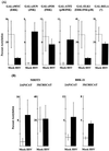
FIG. 1
FIG. 1
HSV activates the TAD of cJUN and ATF2 and stimulates AP-1 activity. (A) NIH 3T3 cells (5 × 10⁵) were transfected with 2.5 μg of a 5×GAL-CAT reporter and 150 to 500 ng of the various effectors, each of which encodes a fusion protein containing the GAL4 DNA-binding domain and the TAD of the respective transcription factors. Under each effector heading is the reported MAPK, if known, that activates the TAD of that transcription factor. (B) NIH 3T3 or BHK-21 cells (5 × 10⁵) were transfected with 2.5 μg of 2AP1CAT or 3×CRECAT reporter. 2AP1CAT contains two consensus AP-1 sites upstream of the CAT gene. 3×CRECAT contains three copies of the CRE in front of the adenovirus E1b promoter upstream of the CAT gene. For all CAT experiments, cells were refed 18 h after transfection, and 12 h later, the cells were either mock infected or infected with KOS (MOI = 5). Eighteen hours p.i., the cells were harvested. CAT assays were performed with equivalent amounts of protein from each sample. Results were quantitated on an AMBIS scanner and are the average of at least three separate experiments.
FIG. 2
FIG. 2
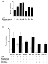
Activation of cJUN and ATF2 TADs is RAS independent. (A) NIH 3T3 cells (5 × 10⁵) were transfected with 2.5 μg of a 5×GAL-CAT reporter, 500 ng of GAL-c-JUN, and various amounts of pZIP-CDC25X, with or without 250 ng of pZIP-RAS15A. An adjusted amount of pZIP vector was added accordingly to equalize the amount of transfected DNA between the samples. (B) NIH 3T3 cells (5 × 10⁵) were transfected with 2.5 μg of a 5×GAL-CAT reporter and 500 ng of either GAL-c-JUN or GAL-ATF-2 and either 250 ng of pZIP-RAS15A, 200 ng of pZIP-CDC25X, or equivalent amounts of empty pZIP vector. For all CAT experiments, cells were refed 18 h after transfection, and 12 h later, the cells were either mock infected or infected with KOS (MOI = 5). Eighteen hours p.i., the cells were harvested. CAT assays were performed with equivalent amounts of protein from each sample. Results were quantitated on an AMBIS scanner and are the average of at least three separate experiments, except in panel A, which was performed only once.
FIG. 3
FIG. 3
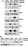
HSV activates JNK but not ERK. (A) BHK-21, U2OS, NIH 3T3, Vero, and C33-A cells were mock infected, infected with KOS (MOI = 5) for 10 h, or treated with anisomycin (20 μg/ml) for 20 min. Cells were then harvested to produce lysates. Equivalent numbers of cells were assayed in each sample. The top panel for each cell type is the result of an in vitro solid substrate kinase assay using 10 μg of GST-cJUN (aa 1 to 79) conjugated to GST-Sepharose beads, as described in Materials and Methods. Two different GST-cJUN substrates were used: a wild-type (WT) GST-cJUN and a mutant (AA) GST-cJUN. In the mutant construct, the serines that are phosphorylated to cause cJUN trans-activation (serines 63 and 73) have been mutated to alanines. The bottom panel for each cell type is an immunoblot, probed with an antibody to JNK, performed on a fraction of the pull down. (B) The same cell types used in panel A were used to compare the level of ERK activity in mock-infected cells (M) versus infected cells at 10 h p.i. (I). The top panel shows results from an in vitro immune complex kinase assay for ERK on extracts from serum-starved cells, as described in Materials and Methods. The bottom panel is an immunoblot from a fraction of the immunoprecipitation probed with an antibody against ERK.
FIG. 4
FIG. 4
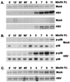
Activation of JNK occurs with early kinetics. U2OS cells were mock infected or infected with KOS (MOI = 5) and harvested to produce cellular lysates at the indicated times p.i. An equivalent number of cells was assayed in each sample. (A) The top two panels show results from an experiment using 10 μg of GST-cJUN (aa 1 to 79) as the precipitating agent and substrate in kinase reactions performed as described in Materials and Methods. The bottom two panels are the results with 6 μg of GST-ATF2 (aa 1 to 254) as the precipitating agent and substrate. (B) Cell-equivalent amounts of mock-infected and wild-type KOS-infected U2OS cellular lysates prepared for JNK kinase assays and collected at the indicated times p.i. were run on 10% polyacrylamide gels. Proteins were transferred to a PVDF membrane and probed for activated p38 (p-p38) or total p38 (p38). One-sixth the amount of protein was loaded onto gels to be used for detecting total p38 as was loaded onto gels to be used for detecting activated p38. (C) Samples of lysate were first precleared with normal rabbit serum and protein A beads. ERK1 antibody was then added to precipitate the kinase. Myelin basic protein (10 μg) was included in the kinase reaction as a substrate, as described in Materials and Methods.
FIG. 5
FIG. 5
Events prior to viral gene expression are not sufficient for JNK activation. (A) U2OS cells were mock infected, infected with KOS (MOI = 5), or infected with KOS (MOI = 5) previously incubated with an antibody specific for viral gB (ss10), gC (VII 13-7), gD (III 174-1.2), or gH (52S). Cells were harvested to produce cellular lysates at the indicated times p.i. Solid substrate kinase assays were performed with GST-cJUN as described in Materials and Methods. (B) U2OS cells were mock infected, infected with KOS (MOI = 5), or infected with KOS (MOI = 5) that had been irradiated with UV light sufficiently to reduce the effective titer by 3 logs. Cellular lysates were prepared at the indicated times p.i., and in vitro solid substrate kinase assays were performed with GST-cJUN as described in Materials and Methods. (C) U2OS cells preincubated at 33 or 39.5°C were mock infected, infected with tsB7 (MOI = 5 based on titer at 33°C), or infected with HFEM, the strain from which tsB7 was derived. Infections then proceeded at the indicated temperatures, and cellular lysates were prepared at the indicated times p.i. In vitro solid substrate kinase assays were performed with GST-cJUN as described in Materials and Methods.
FIG. 6
FIG. 6
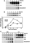
JNK activation is dependent upon viral IE gene expression. (A) Cell-equivalent amounts of wild-type KOS-infected U2OS cellular lysates prepared for JNK kinase assays were run on 12% polyacrylamide gels. Proteins were transferred to a PVDF membrane and probed for viral proteins ICP4, ICP8, and gC. (B) An in vitro solid substrate kinase assay was performed with GST-cJUN as described in Materials and Methods with lysates prepared at the indicated times p.i. from wild-type KOS- and vi13-infected cells (MOI = 5). The kinase data points were derived by scanning the autorads on an LKB laser densitometer. (C) Immunoblot analysis of wild-type and vi13 ICPs was with the same antibodies as in panel A, with the addition of ICP0.
FIG. 7
FIG. 7
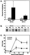
Preventing JNK from activating nuclear targets decreases the efficiency of viral replication. (A) Two stably transfected NIH 3T3 cell lines, G13.5 (control cell line) and G14.1 (JIP-1 overexpressing cell line), were isolated as described in Materials and Methods and compared for their ability to support GAL-cJUN-dependent CAT activity. Cells were transfected with Lipofectamine reagent with 2.5 μg of 5×GAL-CAT and 500 ng of GAL-cJUN and then infected with KOS (MOI = 5) 24 h later. Eighteen hours p.i., the cells were harvested, and CAT assays were performed as described in Materials and Methods. (B) G13.5 and G14.1 cells were mock infected or infected with KOS (MOI = 5) for 8 h. Cells were harvested, and fractionated extracts were prepared. Equal amounts of protein from each fraction were run on 12% polyacrylamide gels. Proteins were transferred to a PVDF membrane and probed for JNK. MC, mock-infected cytoplasm; MN, mock-infected nucleus; IC, KOS-infected cytoplasm; IN, KOS-infected nucleus. (C) The two cell lines were infected with HSV at an MOI of 5. Cells and medium were harvested at the times indicated and frozen and thawed four times. Titers of aliquots were determined on Vero cell monolayers.

Similar articles

Cited by

References

    1. Ace C I, McKee T A, Ryan J M, Cameron J M, Preston C M. Construction and characterization of a herpes simplex virus type 1 mutant unable to transinduce immediate-early gene expression. J Virol. 1989;63:2260–2269. - PMC - PubMed
    1. Aktas H, Cai H, Cooper G M. Ras links growth factor signaling to the cell cycle machinery via regulation of cyclin D1 and the Cdk inhibitor p27KIP1. Mol Cell Biol. 1997;17:3850–3857. - PMC - PubMed
    1. Alvarez E, Northwood I C, Gonzales F A, Latour D A, Seth A, Abate C, Curran T, Davis R J. Pro-Leu-Ser/Thr-Pro is a consensus primary sequence for substrate protein phosphorylation. J Biol Chem. 1991;266:15277–15285. - PubMed
    1. Angel P, Karin M. The role of Jun, Fos, and the AP-1 complex in cell-proliferation and transformation. Biochim Biophys Acta. 1991;1072:129–157. - PubMed
    1. Batterson W, Roizman B. Characterization of the herpes simplex virion-associated factor responsible for the induction of α genes. J Virol. 1983;46:371–377. - PMC - PubMed

Publication types

Substances