. 1999 Sep 15;19(18):7901-12.
doi: 10.1523/JNEUROSCI.19-18-07901.1999.

Effects of roundabout on growth cone dynamics, filopodial length, and growth cone morphology at the midline and throughout the neuropile

Affiliations

Effects of roundabout on growth cone dynamics, filopodial length, and growth cone morphology at the midline and throughout the neuropile

M J Murray et al. J Neurosci. .

Abstract

roundabout (robo) encodes an axon guidance receptor that controls midline crossing in the Drosophila CNS. In robo mutants, axons that normally project ipsilaterally can cross and recross the midline. Growth cones expressing Robo are believed to be repelled from the midline by the interaction of Robo and its ligand Slit, an extracellular protein expressed by the midline glia. To help understand the cellular basis for the midline repulsion mediated by Robo, we used time-lapse observations to compare the growth cone behavior of the ipsilaterally projecting motorneuron RP2 in robo and wild-type embyros. In wild-type embryos, filopodia can project across the midline but are quickly retracted. In robo mutants, medial filopodia can remain extended for longer periods and can develop into contralateral branches. In many cases RP2 produces both ipsilateral and contralateral branches, both of which can extend into the periphery. The growth cone also exhibits longer filopodia and more extensive branching both at the midline and throughout the neuropile. Cell injections in fixed stage 13 embryos confirmed and quantified these results for both RP2 and the interneuron pCC. The results suggest that Robo both repels growth cones at the midline and inhibits branching throughout the neuropile by promoting filopodial retraction.


Figures
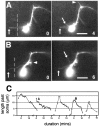
Fig. 1.
Fig. 1.
Extension of medial filopodia by RP2 in wild-type embryos. A, B, Two examples of wild-type time-lapse sequences in which RP2 extends a filopodium medially across the commissures. In this and remaining figures in this article, the midline (indicated by dotted lines) is situated to the left of RP2’s soma (see image conventions in Materials and Methods). In wild-type sequences the midline was not recorded but is estimated to be ∼5 μm from the medial edge of RP2’s soma. Filopodia are shown first at their point of maximum extension (up arrows at0 min) and then again shortly afterward, during their retraction (up arrows; A,4 min; B, 6 min). The extension of new filopodia from adjacent regions of the neuron can continue while the medial filopodium is retracted (down arrows; A, 4 min;B, 6 min). C, Time course of extension and retraction of five medial filopodia (taken from the 25 wild-type sequences) that extended ≥10 μm past the medial edge of RP2’s soma. Missing data points correspond to frames where the filopodium was out of focus. The second and fourth time courses (black) represent the filopodia from Figure 1,A and B, respectively. Typically a medial filopodium rapidly attains its maximum length, is rapidly partially retracted, and then is fully retracted more gradually. Scale bars, 10 μm.
Fig. 2.
Fig. 2.
Axonal trajectories and growth cone morphologies of RP2 in robo mutant (A–E,G, H) and wild-type (F) embryos. A, Stage 13/14robo embryo. RP2 has developed a contralateral branch that, after crossing the midline, follows the usual pathway anteriorly along the longitudinal connectives and then laterally along the ISN.B, Stage 14 robo embryo. RP2 has developed both a normal ipsilateral branch and a long thin contralateral branch (arrow). C, Stage 14/15 robo embryo. RP2 has extended a normal ipsilateral branch but has also developed a branch in the ipsilateral posterior direction (arrow). D, Stage 15robo embryo. RP2 has bifurcated into ipsilateral and contralateral branches, both of which have exited the CNS.E, Stage 16 robo embryo. RP2 has projected contralaterally, but then turned posteriorly, has exited the CNS in the next posterior ISN, has passed under the ventral muscle field, and is exploring the dorsal muscle region. F, Midstage 13 wild-type embryo. RP2 displays a typical growth cone morphology for this developmental stage consisting of a single dominant axonal branch with 13 filopodia ranging in length from ∼1 to 4 μm.G, Midstage 13 robo embryo. RP2 has developed a contralateral branch that has crossed the midline and migrated anteriorly to the contralateral ISN. Filopodia are longer than usual, ranging from ∼1 to 8 μm, and are exploring the normal ipsilateral direction and the contralateral posterior direction (arrowheads). H, Midstage 13robo embryo. RP2 has developed two ipsilateral branches and has an unusually long (9 μm) filopodium (arrowhead). Scale bars (shown in A forB, C, F–H): 10 μm.
Fig. 3.
Fig. 3.
robo sequence in which RP2 extends an ipsilateral axon. At 0 min RP2 has developed an ipsilateral branch (arrow) and extends a filopodium along a more lateral ipsilateral pathway (arrowhead). The more lateral branch continues to develop with the extension of a long filopodium (arrowheads, 9–13 min) that subsequently thickens (arrow at 28min), resulting in two ipsilateral branches (arrows at41 min). RP2 begins to extend numerous long filopodia (arrowheads at 46–47, 50,52 min), some of which thicken (right arrow, 50 min), resulting in a highly branched morphology (53 min). During this period, RP2 extends a filopodium in a lateral direction (up arrow at52 min) that thickens (up arrow,60 min), redirecting the axon laterally (up arrow, 140 min). The original ipsilateral branch continues to extend new filopodia (arrowhead at 101min) and persists to the end of recording (arrowhead at140 min). Scale bar, 10 μm.
Fig. 4.
Fig. 4.
robo sequences in which RP2 extends axons in aberrant directions. Dotted lines show position of the midline. A, RP2 develops a contralateral axon. At0 min, RP2 has extended a filopodium in the ipsilateral direction (arrowhead) and two filopodia (right arrow and up arrow) in a contralateral direction. At 17 min the ipsilateral filopodium and more posterior of the two contralateral filopodia have been retracted. The more anterior contralateral filopodium thickens but is then retracted (data not shown). At 39 min a new filopodium extends contralaterally (right arrow,39 min) and subsequently thickens into a contralateral branch (right arrows, 47–86 min). Having crossed the midline, the axon migrates anteriorly along the longitudinal connectives (arrowhead at 86min) and extends a lateral filopodium at the ISN (arrowat 154 min), which subsequently thickens, redirecting the axon along the contralateral ISN (up arrow at225 min). B, RP2 develops a contralateral branch. B1, At 0 min RP2 has a process in the ipsilateral direction (arrow) that is retracted as the contralateral branch (arrow at 105min) develops. B2, Detail of branch formation from 9 to 24 min. From 9 to 10 min, a filopodium extends in a contralateral direction (arrow at 10 min) and thickens (arrow at 12 min). In subsequent frames a second filopodium extends (up arrow at 13min) and also thickens (up arrow at 16min), resulting in two contralateral processes (arrowsat 16 min). These appear to merge as the contralateral branch matures and new filopodia extend from its tip (24min). C, RP2 develops an ipsilateral posterior branch. At 0 min RP2 is still undergoing axonogenesis and is extending filopodia in all directions. By 28 min RP2 has developed an ipsilateral branch (out of focus; right arrow at 28 min) with a filopodium extending from the tip of this branch along the ipsilateral pathway (up arrowhead at 28 min). From the posterior side of the soma, another ipsilateral filopodium extends and develops into an ipsilateral posterior branch (down arrowheads at28, 34, 43, and131 min). At 28 min another filopodium emerges from the posterior side of the soma (down arrow), extends across the midline (down arrow at 34 min), and begins to thicken (down arrow at 38 min). New filopodia continue to explore the contralateral direction (down arrow at 43 min), but the contralateral branch is eventually resorbed as the ipsilateral posterior branch matures (arrowhead at131 min). Scale bars: A,B1, C, 10 μm; B2, 5 μm.
Fig. 5.
Fig. 5.
robo sequence showing the time course of a medial filopodium. A, RP2 has extended a normal ipsilateral branch that is turning laterally along the ISN.Dotted line shows position of the midline.B, Detail of A showing extension of medial filopodium over a period of 30 min. Filopodium is first detected at a length of ∼9 μm (up arrow at 0min) and extends to a maximum length of 18 μm (18min). At 25 min the filopodium thickens (up arrow), and a new filopodium extends from this thickened region (down arrow at 26–27 min). The original filopodium is retracted, and its base appears to be translocated forward along the new filopodium (up arrow at27–28 min). The combined process is then retracted and disappears at 39 min (data not shown). C, Time course of extension of filopodium with time course of wild-type filopodium from Figure 1B overlaid. Where the wild-type filopodium is quickly retracted after reaching its maximum length, therobo filopodium repeatedly extends and retracts as it explores the contralateral side before being fully retracted.D, Histogram of the period spent extended across the midline for filopodia and any subsequent transient branch that formed (Fig. 4C, down arrow at 38min) in robo and wild-type sequences. Wild-type values were calculated using both 5 and 3 μm as the estimated position of the midline from the medial edge of RP2’s soma (see Results). In both cases the robo medial filopodia tended to remain extended past the midline for longer periods of time. Scale bars:A, 10 μm; B, 5 μm.
Fig. 6.
Fig. 6.
Filopodial length distributions for RP2 in time-lapse sequences (A, B) and fixed midstage 13 embryos (C, D).A, RP2 filopodial length distributions in time-lapse sequences. Filopodia ≥9 μm were measured in wild-type (n = 17) and robo(n = 22) time-lapse sequences, and the results for each genotype were pooled and normalized for the total duration of sequences (55.8 hr for robo; 63.4 hr for wild-type) (see Materials and Methods for details), resulting in a histogram of the number of filopodia of a given length per hour. B, RP2 cumulative filopodial length distributions in time-lapse sequences.Graph shows the number of filopodia more than or equal to a given length per hour for robo and wild-type sequences. C, D, Filopodial length distributions in fixed midstage 13 embryos. Figure2F–H shows single-plane projections of typical cells from these data sets. n values give total number of cells injected for each genotype. C, RP2 filopodial length distributions in midstage 13 embryos for four genotypes:robo/robo, robo/CyO, CyO/CyO, and wild-type. D, RP2 cumulative filopodial length distributions in midstage 13 embryos for the four genotypes. Error bars represent SEM.
Fig. 7.
Fig. 7.
Growth cone morphologies and filopodial length distributions of pCC. A, Typical pCC growth cone at midstage 13 in robo/CyO embryo. pCC has a single dominant branch extending ipsilaterally along the connectives, with filopodia ranging from ∼1 to 4 μm in length. B, pCC in midstage 13 robo embryo. pCC is in the process of crossing the midline and exhibits longer filopodia and a high degree of branching. C, pCC in stage 14 roboembryo. pCC has bifurcated into ipsilateral and contralateral branches.D, pCC filopodial length distributions in midstage 13 embryos for two genotypes: robo/robo androbo/CyO. E, pCC cumulative filopodial length distributions in midstage 13 embryos for the two genotypes. Scale bars (shown in B for A andB): 10 μm. Error bars represent SEM.

Similar articles

Cited by

References

    1. Bentley D, O’Connor TP. Cytoskeletal events in growth cone steering. Curr Opin Neurobiol. 1994;4:43–48. - PubMed
    1. Brose K, Bland KS, Wang KH, Arnott D, Henzel W, Goodman CS, Tessier-Lavigne M, Kidd T. Slit proteins bind Robo receptors and have an evolutionarily conserved role in repulsive axon guidance. Cell. 1999;96:795–806. - PubMed
    1. Fan J, Raper JA. Localized collapsing cues can steer growth cones without inducing their full collapse. Neuron. 1995;14:263–274. - PubMed
    1. Gertler FB, Comer AR, Juang J, Ahern SM, Clark MJ, Liebl EC, Hoffmann FM. enabled, a dosage-sensitive suppressor of mutations in the Drosophila Abl tyrosine kinase, encodes an Abl substrate with SH3 domain-binding properties. Genes Dev. 1995;9:521–533. - PubMed
    1. Hu S, Reichardt LF. From membrane to cytoskeleton: enabling a connection. Neuron. 1999;22:419–422. - PMC - PubMed

Publication types

LinkOut - more resources