

Serological evidence of human infection with the protozoan Neospora caninum
- PMID: 10473533
- PMCID: PMC95770
- DOI: 10.1128/CDLI.6.5.765-767.1999
Serological evidence of human infection with the protozoan Neospora caninum
Abstract
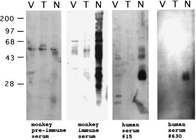
Neospora caninum is a protozoan parasite that is closely related to Toxoplasma gondii. Dogs are a definitive host. Prior to its discovery in 1988, N. caninum infection in animals was often mistakenly diagnosed as toxoplasmosis. Neosporosis in animals is characterized by encephalitis, abortion, and other conditions that clinically and pathologically resemble toxoplasmosis. The potential of N. caninum to infect humans is unknown. Therefore, evidence of human exposure to this parasite was sought by screening for antibodies in blood donors by indirect fluorescent antibody (IFA) tests and immunoblotting. Of 1,029 samples screened, 69 (6.7%) had titers of 1:100 by IFA testing. Fifty of the 69 (72%) sera that were positive for N. caninum were also negative for a closely related protozoan pathogen of humans, T. gondii. Immunoblot analysis confirmed the specificity of the positive sera for N. caninum antigens, with several sera recognizing multiple Neospora antigens with molecular masses similar to those of antigens recognized by monkey anti-N. caninum serum. An immunodominant antigen of approximately 35 kDa was observed with 12 sera. These data provide evidence of human exposure to N. caninum, although the antibody titers in healthy donors were low. The significance of human exposure to, and possible infection with, this parasite is unknown and warrants further study.
Figures

Similar articles
-
Evaluation of serological tests for the diagnosis of Neospora caninum infection in dogs: optimization of cut off titers and inhibition studies of cross-reactivity with Toxoplasma gondii.Vet Parasitol. 2007 Feb 28;143(3-4):234-44. doi: 10.1016/j.vetpar.2006.08.028. Epub 2006 Sep 12. Vet Parasitol. 2007. PMID: 16973287
-
Serological diagnosis of bovine neosporosis by Neospora caninum monoclonal antibody-based competitive inhibition enzyme-linked immunosorbent assay.J Clin Microbiol. 1996 Jun;34(6):1423-8. doi: 10.1128/jcm.34.6.1423-1428.1996. J Clin Microbiol. 1996. PMID: 8735092 Free PMC article.
-
Evaluation of Toxoplasma gondii and Neospora caninum infections in sheep from Uberlândia, Minas Gerais State, Brazil, by different serological methods.Vet Parasitol. 2011 Feb 10;175(3-4):252-9. doi: 10.1016/j.vetpar.2010.10.017. Epub 2010 Oct 16. Vet Parasitol. 2011. PMID: 21075529
-
The antigenic composition of Neospora caninum.Int J Parasitol. 1999 Aug;29(8):1175-88. doi: 10.1016/s0020-7519(99)00085-5. Int J Parasitol. 1999. PMID: 10576569 Review.
-
Cell type- and species-specific host responses to Toxoplasma gondii and its near relatives.Int J Parasitol. 2020 May;50(5):423-431. doi: 10.1016/j.ijpara.2020.05.001. Epub 2020 May 11. Int J Parasitol. 2020. PMID: 32407716 Free PMC article. Review.
Cited by
-
Transcriptome and histopathological changes in mouse brain infected with Neospora caninum.Sci Rep. 2015 Jan 21;5:7936. doi: 10.1038/srep07936. Sci Rep. 2015. PMID: 25604996 Free PMC article.
-
Extracellular Vesicles Secreted by Neospora caninum Are Recognized by Toll-Like Receptor 2 and Modulate Host Cell Innate Immunity Through the MAPK Signaling Pathway.Front Immunol. 2018 Jul 24;9:1633. doi: 10.3389/fimmu.2018.01633. eCollection 2018. Front Immunol. 2018. PMID: 30087675 Free PMC article.
-
A review of neosporosis and pathologic findings of Neospora caninum infection in wildlife.Int J Parasitol Parasites Wildl. 2015 Apr 24;4(2):216-38. doi: 10.1016/j.ijppaw.2015.04.002. eCollection 2015 Aug. Int J Parasitol Parasites Wildl. 2015. PMID: 25973393 Free PMC article. Review.
-
Are genetically modified protozoa eligible for ATMP status? Concerning the legal categorization of an oncolytic protozoan drug candidate.Gene Ther. 2024 May;31(5-6):295-303. doi: 10.1038/s41434-024-00445-1. Epub 2024 Mar 1. Gene Ther. 2024. PMID: 38429432
-
Congenital Transmission of Apicomplexan Parasites: A Review.Front Microbiol. 2021 Sep 29;12:751648. doi: 10.3389/fmicb.2021.751648. eCollection 2021. Front Microbiol. 2021. PMID: 34659187 Free PMC article. Review.
References
-
- Anderson M L, Blanchard P C, Barr B C, Dubey J P, Hoffman R L, Conrad P A. Neospora-like protozoan infection as a major cause of abortion in California dairy cattle. J Am Vet Med Assoc. 1991;198:241–244. - PubMed
-
- Barr B C, Conrad P A, Sverlow K W, Tarantal A F, Hendrickx A G. Experimental fetal and transplacental Neospora infection in the nonhuman primate. Lab Investig. 1994;71:236–242. - PubMed
-
- Conrad P A, Sverlow K, Anderson M, Rowe J, BonDurant R, Tuter G, Breitmeyer R, Palmer C, Thurmond M, Ardans A. Detection of serum antibody responses in cattle with natural or experimental Neospora infections. J Vet Diagn Investig. 1993;5:572–578. - PubMed
-
- Dubey J P, Beattie C P. Toxoplasmosis of animals and man. Boca Raton, Fla: CRC Press; 1988.
Publication types
MeSH terms
Substances
Grants and funding
LinkOut - more resources
Full Text Sources
