. 1999 Aug 9;146(3):645-58.
doi: 10.1083/jcb.146.3.645.

Insoluble gamma-tubulin-containing structures are anchored to the apical network of intermediate filaments in polarized CACO-2 epithelial cells

Affiliations

Insoluble gamma-tubulin-containing structures are anchored to the apical network of intermediate filaments in polarized CACO-2 epithelial cells

P J Salas. J Cell Biol. .

Abstract

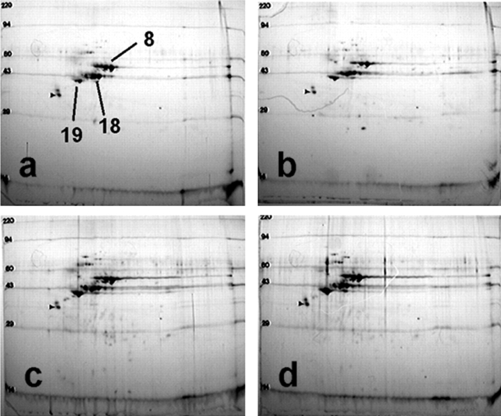
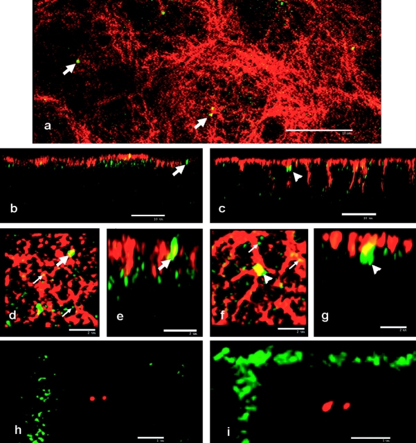
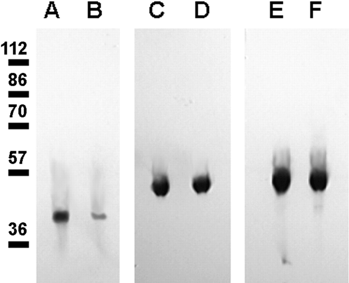
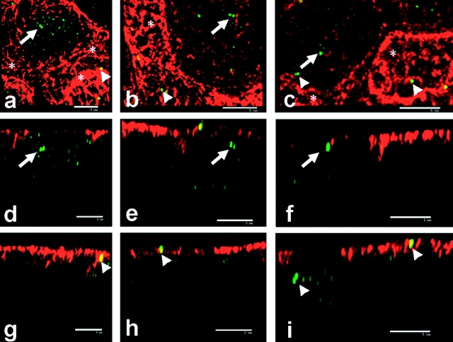
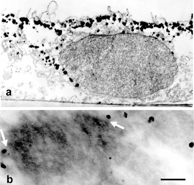
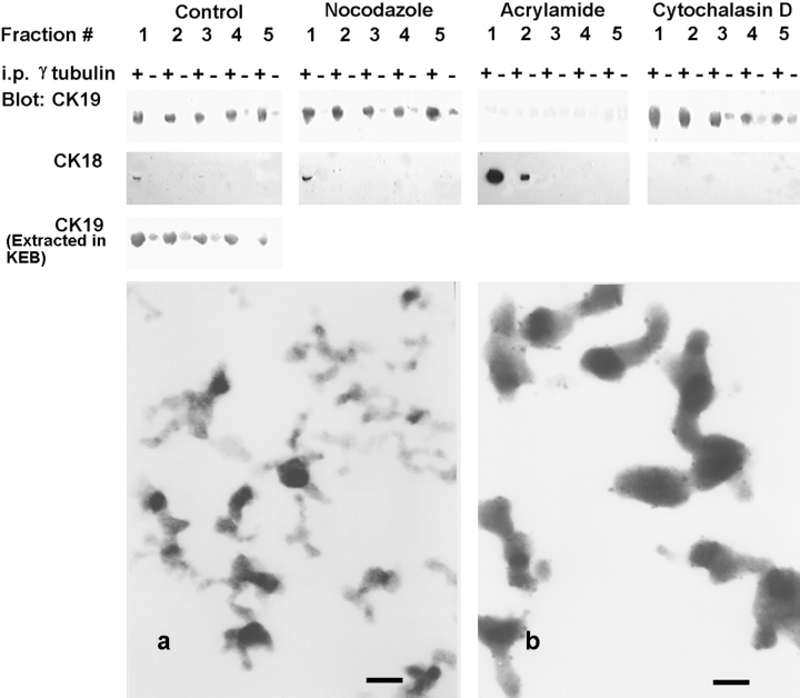
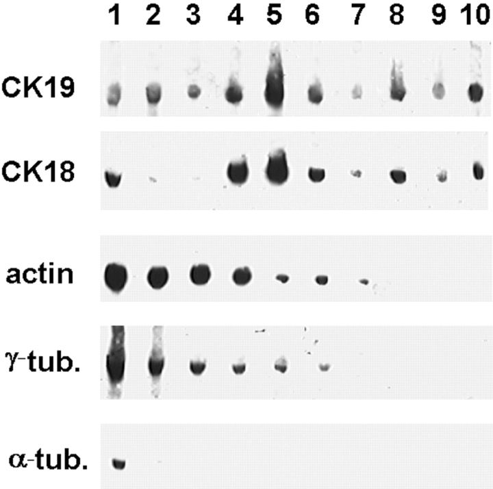
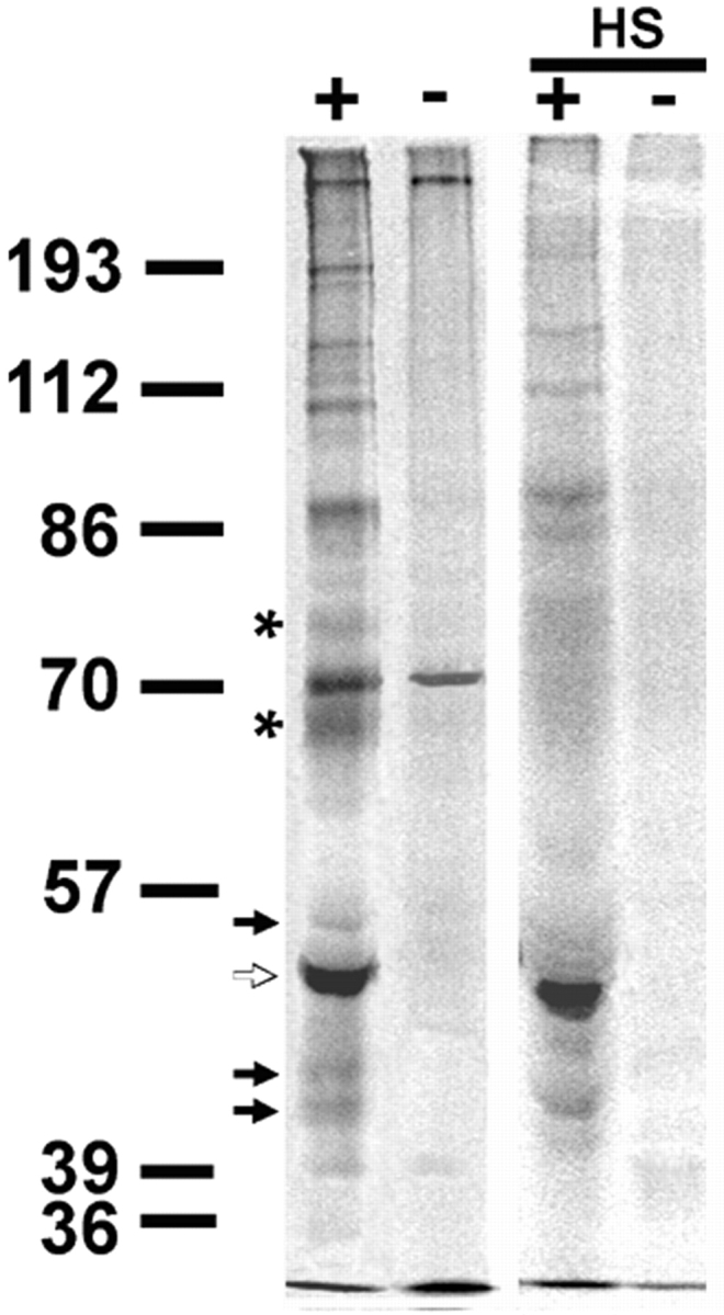
We have previously shown that a thin ( approximately 1 microm) layer of intermediate filaments located beneath the apical membrane of a variety of simple epithelial cells participates in the organization of apical microfilaments and microtubules. Here, I confirmed the apical distribution of gamma-tubulin-containing structures (potential microtubule-organizing centers) in CACO-2 cells and demonstrated perfect colocalization of centrosomes and nearly 50% of noncentrosomal gamma-tubulin with apical intermediate filaments, but not with apical F-actin. Furthermore, the antisense-oligonucleotide-mediated downregulation of cytokeratin 19, using two different antisense sequences, was more efficient than anticytoskeletal agents to delocalize centrosomes. Electron microscopy colocalization suggests that binding occurs at the outer boundary of the pericentriolar material. Type I cytokeratins 18 and 19 present in these cells specifically coimmunoprecipitated in multi-protein fragments of the cytoskeleton with gamma-tubulin. The size and shape of the fragments, visualized at the EM level, indicate that physical trapping is an unlikely explanation for this result. Drastic changes in the extraction protocol did not affect coimmunoprecipitation. These results from three independent techniques, indicate that insoluble gamma-tubulin-containing structures are attached to apical intermediate filaments.


Figures

Figure 1
Figure 1
Expression of cytokeratins in CACO-2 cells. Purified IF proteins from CACO-2 monolayers confluent for 3 d (a), 7 d (b), 9 d (c), and 15 d (d) were separated by two-dimensional electrophoresis (IEF, first dimension and SDS-PAGE in the second dimension). Notice that only cytokeratins 8, 18, and 19 were expressed at all times. Small arrowheads point to an internal standard, tropomyosin (mol wt 33,000, pI 5.2). Molecular mass standards are given in kD.
Figure 5
Figure 5
Separation of noncentrosomal γ-tubulin from CK19 IF induced by acrylamide but not by other anticytoskeletal agents. CACO-2 cells were grown on Transwell™ filters for 9 d. Before fixation, the monolayers were changed to DME supplemented with 33 μM nocodazole, 2 μM cytochalasin D (Cytoch. D), 5 mM acrylamide (Acryl.), or none (Control). The distance from noncentrosomal γ-tubulin spots to the nearest bundle of CK19 IF was measured in three-dimensional confocal deconvoluted images such as those shown in Fig. 2. The frequency of distances as percent, is shown for nine distance ranges: (1) <100 nm (below resolution), perfect colocalization as judged by this technique; (2) 100–300 nm, usually within the same confocal plane; (3) 300–600 nm; (4) 600–900 nm; (5) 900–1,200 nm; (6) 1,200–1,500 nm; (7) 1,500–1,800 nm; (8) 1,800–2,100 nm; (9) 2,100–2,400 nm. Most of the cases in the ranges 3–9 were distances measured in the z-axis. A total of 1,309 small γ-tubulin spots in 54 cells were counted, in approximately similar numbers for each treatment group. The 100% was considered as the total in each particular treatment. Notice that a large percent of the γ-tubulin colocalizes with apical IF in all treatments (∼40%, range 1) except in cells treated with acrylamide (arrow).
Figure 2
Figure 2
γ-tubulin colocalizes with apical IF but not with apical F-actin in CACO-2 cells. CACO-2 cells grown on Transwell™ filters at 9 d confluency stage were fixed and processed for double indirect immunofluorescence for γ-tubulin (green), and CK19 (b, d, and e) or CK18 (a, c, f, and g) (red). The preparations were analyzed by laser scanning confocal microscopy and the stacks of confocal sections were further subjected to three-dimensional deconvolution. a, d, and f represent confocal sections in the XY plane (parallel to the monolayer). b, c, e, and g are XZ reconstructions (perpendicular to the plane of the monolayer, apical is shown up). e is the XZ cross-section of the image in d, at the level of the arrow, while g is the XZ cross-section of the image in f, at the level of the arrowhead. Large arrows point at centrosomes and small arrows at noncentrosomal γ-tubulin spots. In h and i γ-tubulin (red) signal was colocalized with FITC-phalloidin signal (green). h represents a XY section and i represents a XZ section of the same field. Bars: (a–c) 10 μm; (d–g) 2 μm; (h and i) 1 μm.
Figure 4
Figure 4
Effect of anti-CK19 phosphorothioate antisense oligonucleotide on steady-state levels of cytokeratins in CACO-2 cells. CACO-2 cells were continuously incubated with the phosphorothioate oligodeoxy nucleotide A19, antisense against the CK19 mRNA (b, d, and f) or R, its randomized sequence (a, c, and d), as a control, for three passages. In this experiment, the cells were grown on 24-mm Transwell™ filters, and the cytoskeletal IF preparation was obtained by extraction in Triton X-100 in the presence of 1.5 M KCl. The pellets were analyzed by immunoblot using anti-CK19 (a and b), anti-CK18 (c and d), and anti-CK8 (e and f) mAbs. Molecular mass standards are expressed in kD.
Figure 3
Figure 3
Redistribution of centrosomes in CACO-2 cells depleted in CK19 by antisense oligonucleotide treatment. CACO-2 cells were continuously grown on Transwell™ filters in the presence of A19 oligonucleotide. The monolayers were fixed, processed for double immunofluorescence, and analyzed by confocal microscopy and deconvolution as described in Fig. 2. a–c show different examples of cells depleted in CK19 (red signal) with neighboring nontargeted cells (*), viewed in the XY plane (parallel to the monolayer). d–i are XZ sections (apical is shown up) of the same XY image above, at the level of the arrows (d–f) or the arrowheads (g–i). Arrows and arrowheads point at centrosomes. Bars, 5 μm.
Figure 6
Figure 6
Colocalization of γ-tubulin and CK19 IF at EM level. CACO-2 cell monolayers were grown on filters as described above and fixed in methanol. CK19 was localized with Nanogold™ gold particles followed by silver developing, while γ-tubulin was localized using a standard immunoperoxidase procedure. (a) CK19-Nanogold™ signal was developed with silver for 20 min to attain 200–400 nm particles visible at low magnification. (b) The CK19-Nanogold™ signal was developed with silver for only 5 min, so that the particles were in the range 15–20 nm, and colocalized with γ-tubulin detected by immunoperoxidase (diffuse stain) reaction confined within a matrix of cross-linked albumin. Note that the minimum distance from gold particles to peroxidase stain is 10–20 nm (arrows). Bar: (a) 1.6 μm; (b) 0.1 μm.
Figure 8
Figure 8
Coimmunoprecipitation of CK19 and CK18 with γ-tubulin from native cytoskeletal fragments obtained by sonication and separated in sucrose gradients, are sensitive to acrylamide and cytochalasin D, respectively. CACO-2 cell monolayers were grown for 9 d on Transwell™ filters, and treated with anticytoskeletal agents as described in Fig. 5 before detergent extraction. Sonication fragments of cytoskeletal preparations were obtained and separated as described in Fig. 7, with the exception of the bottom immunoblot panel, obtained from samples extracted in a physiologic (KEB) buffer, in the presence of saponin, antiproteases, and phosphatase inhibitors. Each one of the top five fractions (containing fragments in the range 0–510 S) was diluted, divided in two equal aliquots, and subjected to immunoprecipitation with anti–γ-tubulin antibody (+) or with nonimmune purified rabbit IgG at an equivalent concentration (−). After extensive washes, the protein A–agarose beads were eluted in SDS-urea, the eluates were TCA precipitated, run in SDS-PAGE, and blotted. The same nitrocellulose sheets were sequentially probed, stripped off, and reprobed with anti-CK19 mAb and with anti-CK18 mAb. (Bottom panels) Electron microscopy of the cytoskeletal fragments in fractions 1 (a) and 5 (b). Samples from the same gradients, before immunoprecipitation, were fixed in glutaraldehyde, pelleted, embedded, sectioned, and stained for TEM. Bars, 60 nm.
Figure 7
Figure 7
Distribution of cytoskeletal proteins from sonication fragments of cytoskeletal preparations of CACO-2 cells in sucrose gradients after velocity sedimentation. Cytoskeletal preparations of 9 d confluent CACO-2 monolayers were obtained by extraction in 1% Triton X-100, in EB-antiprotease cocktail. The cytoskeletons were extensively sonicated avoiding heating, and run in 20–60% sucrose gradients under rate centrifugation conditions to separate the fragments by size. Ten 1-ml fractions of the gradient were collected, diluted in PBS and pelleted (approximate sedimentation coefficient ranges per fraction: (1) 0–230S; (2) up to 430S; (3) up to 625S; (4) up to 790S; (5) up to 958S; (6) up to 1,100S; (7) up to 1,246S; (8) up to 1,376S; (9) up to 1,500S; and (10) up to 1,600S). The pellets were analyzed by SDS-PAGE and blotted onto nitrocellulose (fraction 1, top of the gradient). The same nitrocellulose sheet was sequentially probed, stripped off and reprobed with antibodies against CK19, CK18, actin, γ-tubulin (γ-tub.), and α-tubulin (α-tub). A second cycle of reprobing, and reprobing in a different sequence indicated that the differences were not due to protein loss after reprobing.
Figure 9
Figure 9
Protein composition of the complexes immunoprecipitated with anti–γ-tubulin antibody. CACO-2 cell monolayers (7.5 × 10⁷ cells) were metabolically labeled with [35S]methionine-cysteine for 24 h, extracted, sonicated, and pools of fractions 1 and 2 were immunoprecipitated (+, anti–γ-tubulin antibody; −, nonimmune rabbit IgG) as described above. An equivalent amount of immunoprecipitate still bound to the beads was washed in EB supplemented with 0.7 M NaCl (HS). The eluates were analyzed by SDS-PAGE and autoradiogram. Molecular mass standards are expressed in kD.

Similar articles

Cited by

References

    1. Apodaca G., Katz L.A., Mostov K.E. Receptor-mediated transcytosis of IgA in MDCK cells is via apical recycling endosomes. J. Cell Biol. 1994;125:67–86. - PMC - PubMed
    1. Archer J., Solomon F. Deconstructing the microtubule-organizing center. Cell. 1994;76:589–591. - PubMed
    1. Bacallao R., Antony C., Dotti C., Karsenti E., Stelzer E.H.K., Simons K. The subcellular organization of Madin-Darby Canine kidney cells during the formation of a polarized epithelium. J. Cell Biol. 1989;109:2817–2832. - PMC - PubMed
    1. Brown W.J., Farquhar M.G. The mannose-6-phosphate receptor for lysosomal enzymes is concentrated in Cis Golgi cisternae. Cell. 1984;36:295–307. - PubMed
    1. Buendia B., Bré M.H., Griffiths G., Karsenti E. Cytoskeletal control of centrioles movement during the establishment of polarity in MDCK cells. J. Cell Biol. 1990;110:1123–1135. - PMC - PubMed

Publication types

MeSH terms