R5 strains of human immunodeficiency virus type 1 from rapid progressors lacking X4 strains do not possess X4-type pathogenicity in human thymus
- PMID: 10438873
- PMCID: PMC104310
- DOI: 10.1128/JVI.73.9.7817-7822.1999
R5 strains of human immunodeficiency virus type 1 from rapid progressors lacking X4 strains do not possess X4-type pathogenicity in human thymus
Abstract
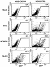
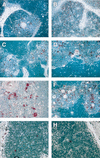
Some individuals infected with only R5 strains of human immunodeficiency virus type 1 progress to AIDS as quickly as individuals harboring X4 strains. We determined that three R5 viruses were much less pathogenic than an X4 virus in SCID-hu Thy/Liv mice, suggesting that R5 virus-mediated rapid disease progression is associated with host, not viral, factors.
Figures


Similar articles
-
HIV-1 replication and pathogenesis in the human thymus.Curr HIV Res. 2003 Jul;1(3):275-85. doi: 10.2174/1570162033485258. Curr HIV Res. 2003. PMID: 15046252 Free PMC article. Review.
-
Human immunodeficiency virus type 1 strains R5 and X4 induce different pathogenic effects in hu-PBL-SCID mice, depending on the state of activation/differentiation of human target cells at the time of primary infection.J Virol. 1999 Aug;73(8):6453-9. doi: 10.1128/JVI.73.8.6453-6459.1999. J Virol. 1999. PMID: 10400739 Free PMC article.
-
Pathogenesis of primary R5 human immunodeficiency virus type 1 clones in SCID-hu mice.J Virol. 2000 Apr;74(7):3205-16. doi: 10.1128/jvi.74.7.3205-3216.2000. J Virol. 2000. PMID: 10708437 Free PMC article.
-
Causal relationships between HIV-1 coreceptor utilization, tropism, and pathogenesis in human thymus.AIDS Res Hum Retroviruses. 2000 Jul 20;16(11):1039-45. doi: 10.1089/08892220050075291. AIDS Res Hum Retroviruses. 2000. PMID: 10933618
-
The role of viral coreceptors and enhanced macrophage tropism in human immunodeficiency virus type 1 disease progression.Sex Health. 2004;1(1):23-34. doi: 10.1071/sh03006. Sex Health. 2004. PMID: 16335478 Review.
Cited by
-
Human immunodeficiency virus bearing a disrupted central DNA flap is pathogenic in vivo.J Virol. 2007 Jun;81(11):6146-50. doi: 10.1128/JVI.00203-07. Epub 2007 Mar 28. J Virol. 2007. PMID: 17392373 Free PMC article. Review.
-
Increased mucosal transmission but not enhanced pathogenicity of the CCR5-tropic, simian AIDS-inducing simian/human immunodeficiency virus SHIV(SF162P3) maps to envelope gp120.J Virol. 2003 Jan;77(2):989-98. doi: 10.1128/jvi.77.2.989-998.2003. J Virol. 2003. PMID: 12502815 Free PMC article.
-
Cytopathicity of human immunodeficiency virus type 1 primary isolates depends on coreceptor usage and not patient disease status.J Virol. 2001 Sep;75(18):8842-7. doi: 10.1128/jvi.75.18.8842-8847.2001. J Virol. 2001. PMID: 11507229 Free PMC article.
-
HIV-1 replication and pathogenesis in the human thymus.Curr HIV Res. 2003 Jul;1(3):275-85. doi: 10.2174/1570162033485258. Curr HIV Res. 2003. PMID: 15046252 Free PMC article. Review.
-
Validation of the SCID-hu Thy/Liv mouse model with four classes of licensed antiretrovirals.PLoS One. 2007 Aug 1;2(7):e655. doi: 10.1371/journal.pone.0000655. PLoS One. 2007. PMID: 17668043 Free PMC article.
References
-
- Berger E A, Doms R W, Fenyo E M, Korber B T, Littman D R, Moore J P, Sattentau O J, Schuitemaker H, Sodroski J, Weiss R A. A new classification for HIV-1. Nature. 1998;391:240. - PubMed
-
- Berkowitz R D, Alexander S, Bare C, Lindquist-Stepps V, Bogan M, Moreno M E, Gibson L, Wieder E D, Kosek J, Soddart C A, McCune J M. CCR5- and CXCR4-utilizing strains of human immunodeficiency virus type 1 exhibit differential tropism and pathogenesis in vivo. J Virol. 1998;72:10108–10117. - PMC - PubMed
-
- Berkowitz, R. D., S. Alexander, and J. M. McCune. Causal relationships between the HIV-1 V3 loop, coreceptor utilization, tropism, and pathogenesis in vivo. Submitted for publication. - PubMed
-
- Berkowitz R D, Beckerman K P, Schall T J, McCune J M. CXCR4 and CCR5 expression delineates targets for HIV-1 disruption of T cell differentiation. J Immunol. 1998;161:3702–3710. - PubMed
Publication types
MeSH terms
Grants and funding
LinkOut - more resources
Full Text Sources
Medical

