A recombinant measles vaccine virus expressing wild-type glycoproteins: consequences for viral spread and cell tropism
- PMID: 10400788
- PMCID: PMC112775
- DOI: 10.1128/JVI.73.8.6903-6915.1999
A recombinant measles vaccine virus expressing wild-type glycoproteins: consequences for viral spread and cell tropism
Abstract
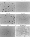
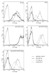
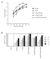
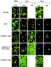
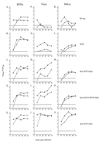
Wild-type, lymphotropic strains of measles virus (MV) and tissue culture-adapted MV vaccine strains possess different cell tropisms. This observation has led to attempts to identify the viral receptors and to characterize the functions of the MV glycoproteins. We have functionally analyzed the interactions of MV hemagglutinin (H) and fusion (F) proteins of vaccine (Edmonston) and wild-type (WTF) strains in different combinations in transfected cells. Cell-cell fusion occurs when both Edmonston F and H proteins are expressed in HeLa or Vero cells. The expression of WTF glycoproteins in HeLa cells did not result in syncytia, yet they fused efficiently with cells of lymphocytic origin. To further investigate the role of the MV glycoproteins in virus cell entry and also the role of other viral proteins in cell tropism, we generated recombinant vaccine MVs containing one or both glycoproteins from WTF. These viruses were viable and grew similarly in lymphocytic cells. Recombinant viruses expressing the WTFH protein showed a restricted spread in HeLa cells but spread efficiently in Vero cells. Parental WTF remained restricted in both cell types. Therefore, not only differential receptor usage but also other cell-specific factors are important in determining MV cell tropism.
Figures


Similar articles
-
Recombinant wild-type and edmonston strain measles viruses bearing heterologous H proteins: role of H protein in cell fusion and host cell specificity.J Virol. 2002 May;76(10):4891-900. doi: 10.1128/jvi.76.10.4891-4900.2002. J Virol. 2002. PMID: 11967306 Free PMC article.
-
Receptor usage and differential downregulation of CD46 by measles virus wild-type and vaccine strains.Proc Natl Acad Sci U S A. 1995 Apr 25;92(9):3943-7. doi: 10.1073/pnas.92.9.3943. Proc Natl Acad Sci U S A. 1995. PMID: 7732009 Free PMC article.
-
Importance of the cytoplasmic tails of the measles virus glycoproteins for fusogenic activity and the generation of recombinant measles viruses.J Virol. 2002 Jul;76(14):7174-86. doi: 10.1128/jvi.76.14.7174-7186.2002. J Virol. 2002. PMID: 12072517 Free PMC article.
-
Toward understanding the pathogenicity of wild-type measles virus by reverse genetics.Jpn J Infect Dis. 2002 Oct;55(5):143-9. Jpn J Infect Dis. 2002. PMID: 12501253 Review.
-
Measles virus interactions with cellular receptors: consequences for viral pathogenesis.J Neurovirol. 2001 Oct;7(5):391-9. doi: 10.1080/135502801753170246. J Neurovirol. 2001. PMID: 11582511 Review.
Cited by
-
Current vaccine technology with an emphasis on recombinant measles virus as a new perspective for vaccination against SARS-CoV-2.EuroMediterr J Environ Integr. 2021;6(2):61. doi: 10.1007/s41207-021-00263-6. Epub 2021 Jul 4. EuroMediterr J Environ Integr. 2021. PMID: 34250222 Free PMC article. Review.
-
Cotton rat (Sigmodon hispidus) signaling lymphocyte activation molecule (CD150) is an entry receptor for measles virus.PLoS One. 2014 Oct 8;9(10):e110120. doi: 10.1371/journal.pone.0110120. eCollection 2014. PLoS One. 2014. PMID: 25295727 Free PMC article.
-
Measles virus-induced suppression of immune responses.Immunol Rev. 2010 Jul;236:176-89. doi: 10.1111/j.1600-065X.2010.00925.x. Immunol Rev. 2010. PMID: 20636817 Free PMC article. Review.
-
Previously unrecognized amino acid substitutions in the hemagglutinin and fusion proteins of measles virus modulate cell-cell fusion, hemadsorption, virus growth, and penetration rate.J Virol. 2009 Sep;83(17):8713-21. doi: 10.1128/JVI.00741-09. Epub 2009 Jun 24. J Virol. 2009. PMID: 19553316 Free PMC article.
-
Measles viruses possessing the polymerase protein genes of the Edmonston vaccine strain exhibit attenuated gene expression and growth in cultured cells and SLAM knock-in mice.J Virol. 2008 Dec;82(23):11979-84. doi: 10.1128/JVI.00867-08. Epub 2008 Sep 17. J Virol. 2008. PMID: 18799577 Free PMC article.
References
-
- Alkhatib G, Richardson C D, Shen S-H. Intracellular processing, glycosylation, and cell-surface expression of the measles virus fusion protein (F) encoded by a recombinant adenovirus. Virology. 1990;175:262–270. - PubMed
-
- Bartz R, Brinkmann U, Dunster L M, Rima B, ter Meulen V, Schneider-Schaulies J. Mapping amino acids of the measles virus hemagglutinin responsible for receptor (CD46) downregulation. Virology. 1996;224:334–337. - PubMed
-
- Bartz R, Firsching R, ter Meulen V, Schneider-Schaulies J. Differential receptor usage by measles virus strains. J Gen Virol. 1998;79:1015–1025. - PubMed
-
- Buckland R, Malvoisin E, Beauverger P, Wild T F. A leucine zipper structure present in the measles virus fusion protein is not required for its tetramerization but is essential for fusion. J Gen Virol. 1992;73:1703–1707. - PubMed
Publication types
MeSH terms
Substances
Associated data
- Actions
Grants and funding
LinkOut - more resources
Full Text Sources
Other Literature Sources

