. 1999 Jul;10(7):2407-23.
doi: 10.1091/mbc.10.7.2407.

A role for Tlg1p in the transport of proteins within the Golgi apparatus of Saccharomyces cerevisiae

Affiliations
Free PMC article

A role for Tlg1p in the transport of proteins within the Golgi apparatus of Saccharomyces cerevisiae

J G Coe et al. Mol Biol Cell. 1999 Jul.
Free PMC article

Abstract

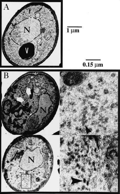
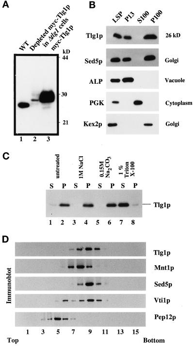
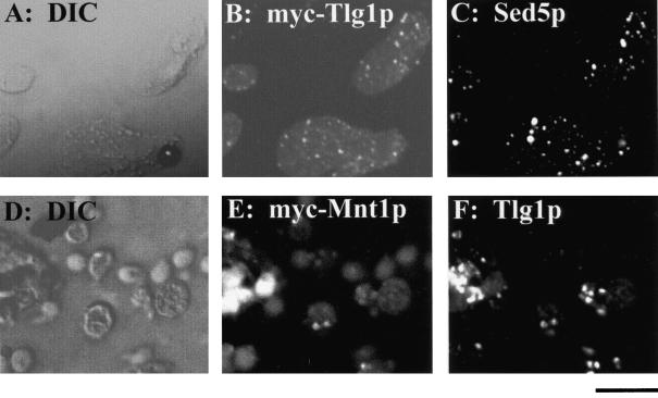
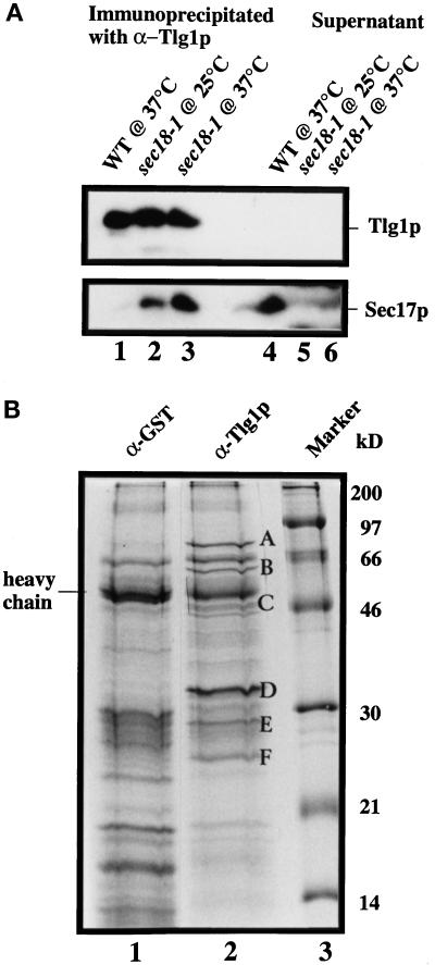
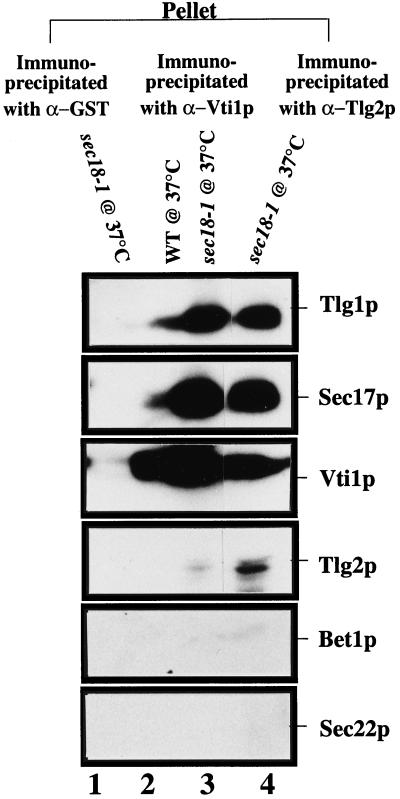
Members of the syntaxin protein family participate in the docking-fusion step of several intracellular vesicular transport events. Tlg1p has been identified as a nonessential protein required for efficient endocytosis as well as the maintenance of normal levels of trans-Golgi network proteins. In this study we independently describe Tlg1p as an essential protein required for cell viability. Depletion of Tlg1p in vivo causes a defect in the transport of the vacuolar protein carboxypeptidase Y through the early Golgi. Temperature-sensitive (ts) mutants of Tlg1p also accumulate the endoplasmic reticulum/cis-Golgi form of carboxypeptidase Y at the nonpermissive temperature (38 degrees C) and exhibit underglycosylation of secreted invertase. Overexpression of Tlg1p complements the growth defect of vti1-11 at the nonpermissive temperature, whereas incomplete complementation was observed with vti1-1, further suggesting a role for Tlg1p in the Golgi apparatus. Overexpression of Sed5p decreases the viability of tlg1 ts mutants compared with wild-type cells, suggesting that tlg1 ts mutants are more susceptible to elevated levels of Sed5p. Tlg1p is able to bind His6-tagged Sec17p (yeast alpha-SNAP) in a dose-dependent manner and enters into a SNARE complex with Vti1p, Tlg2p, and Vps45p. Morphological analyses by electron microscopy reveal that cells depleted of Tlg1p or tlg1 ts mutants incubated at the restrictive temperature accumulate 40- to 50-nm vesicles and experience fragmentation of the vacuole.


Figures

Figure 1
Figure 1
TLG1 is essential for cell growth. (A) Disruption strategy to create a null mutation of TLG1 in the yeast genome. The arrow represents the direction of transcription. (B) Dissection result of heterozygous W303 yeast diploids containing Δtlg1::LEU2. Correct integrants verified by Southern blotting were incubated on a sporulation plate for 3 d at 30°C. Asci were glusulase treated, and tetrads were segregated on a dissection microscope. Surviving progeny were wild-type for TLG1. (C) Dissection of Y100 (SEY6210 × SEY6211)-, Y98 (MATaade2-1, ade3Δ his3-11,-15 leu2-3,-112 trp1-Δ1 ura3-1 can1-100)-, and Y97 (RSY255 × SEY6210)-based asci in which two of the spores from each tetrad contain Δtlg1::LEU2. The middle and right panels of C were dissection plates incubated at 30°C for 5 d. All other plates were incubated for 3 d at 30°C. Δtlg1 cells grew poorly, as revealed by the tiny colonies, particularly in Y97 and Y98 cells. (D) Reintroduction of an episomal TLG1 under the control of an inducible GAL1 promoter in Δtlg1::LEU2 cells from W303 background restores cell growth in galactose-based media but not in glucose-based media. The growth curves of wild type (open squares), a heterozygous diploid of Δtlg1::LEU2 (open triangles), and Δtlg1::LEU2 cells (closed circles) containing the wild-type TLG1 under the control of the GAL1 promoter (pYTLG1) are shown. Cells were grown in YEP-galactose (D) to induce or in YEP-glucose (E) liquid media to deplete the Tlg1p. Cell growth was measured by optical density at 600 nm at the indicated times.
Figure 2
Figure 2
Depletion of plasmid-borne Tlg1p in Δtlg1::LEU2 cells leads to an accumulation of the ER form of CPY (p1CPY). The wild-type and Δtlg1::LEU2 cells containing the GAL1-TLG1 plasmid (JC20) were grown in 1% galactose at 30°C, pelleted, washed, and incubated in 2% glucose medium at 30°C for the indicated periods. Cells (3 OD600nm) were pulse labeled with [35S]methionine for 15 min and chased for a further 30 min at 30°C. CPY was isolated from cell extracts by immunoprecipitation with 6 μg of anti-CPY antibodies and resolved by 8% SDS-PAGE. The different forms of CPY are indicated on the left. p1CPY, p2CPY, and mCPY represent the ER/early Golgi, late Golgi, and mature vacuolar forms of CPY, respectively. After 2–4 h of incubation in glucose, processing of p1CPY to p2CPY and mCPY is inhibited.
Figure 3
Figure 3
Temperature-sensitive tlg1 mutants are defective for growth at 38°C. Δtlg1::LEU2 cells in W303 haploids containing either TLG1 or its mutant alelles under the control of its endogenous promoter were streaked on a glucose-based selective plate without trptophan and incubated for 3 d at either 23 or 38°C. The orientation of the respective cells is indicated on the left.
Figure 4
Figure 4
Transport of CPY in the early secretory pathway is affected at the restrictive temperature for tlg1 ts mutants. (A) Accumulation of p1CPY at 38°C in Δtlg1 cells bearing various tlg1 ts mutant alelles. Cells were initially grown in yeast standard minimal glucose medium (without Trp, Leu, and Met) at 23°C, after which half of each sample was shifted to 38°C for 60 min. The cultures were pulse labeled with [35S]methionine for 15 min and chased for 60 min at either 23 or 38°C. CPY was immunoprecipitated from cell lysates and analyzed by SDS-PAGE. (B) The ER/cis-Golgi form of CPY is poorly glycosylated by α-1,6-mannose-specific enzymes in tlg1 ts cells at 38°C. tlg1 ts mutants were grown at 38°C, labeled for 15 min with [35S]methionine, and chased for 20 min at 38°C. The immunoprecipitated CPY was heated at 90°C for 15 min, and the supernatants were reimmunoprecipitated with either anti-CPY or anti-α-1,6-mannose-specific antibodies. The second immunoprecipitates were analyzed by SDS-PAGE and autoradiography. The different forms of CPY are indicated on the left. p1CPY, p2CPY, and mCPY represent the ER/early Golgi, late Golgi, and mature vacuolar forms of CPY, respectively.
Figure 5
Figure 5
Secretion of the underglycosylated form and intracellular accumulation of core-glycosylated forms of invertase in tlg1 cells at 38°C. (A) Autoradiograph of invertase immunoprecipitations from Δtlg1 strains bearing the TLG1, tlg1-15 ts, or tlg1-24 ts mutant alelles. Cells expressing myc-tagged invertase were grown at 23 or 38°C for 1 h and converted to spheroplasts. The spheroplasts were further incubated for 1 h at 23 or 38°C, and the resulting spheroplast (I, intracellular fraction) and the culture media (E, extracellular fraction) were analyzed by immunoblot to detect myc-tagged invertase. Note the intracellular accumulation of the core glycosylated invertase and the underglycosylation of the secreted invertase at the 38°C in tlg1-24 cells. (B) The secreted hypoglycosylated invertase is modified by α-1,6- and α-1,3-mannose-specific linkages. The c-myc-tagged invertase was immunoprecipitated from the extracellular (E) fractions of spheroplasts that were incubated at 38°C. Immunoprecipitates were heated to 90°C for 15 min, and the supernatants were diluted in IP buffer. Samples were reimmunoprecipitated with anti-invertase, anti-α-1,6-mannose-specific antiserum, and anti-α-1,3-mannose-specific antiserum. Immunoprecipitates were analyzed on 7% SDS-PAGE and autoradiographed.
Figure 6
Figure 6
Accumulation of vesicles and fragmentation of the vacuole upon depletion of cellular Tlg1p. Wild-type (A) and Tlg1p-depleted cells containing the episomal TLG1 under the GAL1 promoter in Δtlg1::LEU2 cells (B) were incubated in YEP-glucose medium for 16 h at 30°C and then examined by electron microscopy. V, vacuole; N, nucleus. Large white arrows and the region within the box indicate the areas where 50-nm vesicles accumulated. The large dark structures of irregular size shown in left panels of B are fragmented vacuoles. Right panels of B are enlargements of the regions indicated by large arrows in the left panels. Large white arrows in the left panels indicate transport vesicles. Bars, 1 μm for the whole cell and 0.15 μm for enlarged regions shown in the right panels of B.
Figure 7
Figure 7
Tlg1p is an integral membrane protein that cofractionates mainly with the Golgi fraction. (A) Polyclonal antibodies against Tlg1p specifically recognize Tlg1p in wild-type cells (lane 1), Δtlg1 cells that have been depleted of the myc-tagged Tlg1p regulated by the episomal GAL1 promoter (lane 2), and Δtlg1 cells overexpressing myc-tagged Tlg1p (lane 3). (B) Yeast homogenates were separated into different fractions by differential centrifugation. The resulting low-speed pellet (LSP), 13,000 × g pellet (P13), 100,000 × g supernatant (S100), and 100,000 × g pellet (P100) fractions were analyzed by immunoblot to detect proteins as indicated. Tlg1p has a similar distribution as the Golgi t-SNARE Sed5p. (C) Tlg1p is an integral membrane protein. Yeast membranes were extracted with different buffers as indicated and separated into pellet (P) and supernatant (S) fractions, which were analyzed by immunoblot to detect Tlg1p. (D) The majority of Tlg1p cofractionated with Sed5p. The 13,000 × g supernatant from RSY255 cells was fractionated on a sucrose gradient as described in MATERIALS AND METHODS. Fractions were collected from the top of the gradient and analyzed for the distribution of Vti1p, Mnt1p, Tlg1p, Sed5p, and Pep12p by immunoblotting.
Figure 8
Figure 8
Golgi association of Tlg1p as revealed by imunofluorescence microscopy. Cells expressing the myc-tagged Tlg1p were double labeled with 9E10 antibody (B) and polyclonal antibodies against endogenous Sed5p (C). Similarly, cells expressing myc-tagged Mnt1p were double labeled with 9E10 (E) and polyclonal antibodies against the endogenous Tlg1p (F). Also shown are the corresponding differential interference contrast images. Bar, 4 μm.
Figure 9
Figure 9
Characterization of Tlg1p-containing SNARE complex. Spheroplasts of sec18-1 cells were incubated at either 25 or 37°C for 1 h and lysed with Triton X-100, and extracts were prepared as described (see MATERIALS AND METHODS). (A) Twenty milligrams of the extract from the indicated cells were incubated with anti-Tlg1p antibodies (200 μg), which had been covalently coupled to cyanogen bromide–activated Sepharose CL4B (400 μg). The supernatants and eluted immunoprecipitates were TCA precipitated, and formula image of the samples was analyzed by immunoblot for Tlg1p and Sec17p. Sec17p was not coimmunoprecipitated by anti-Tlg1p antibodies when extracts from wild-type cells were used (lanes 1 and 4). However, Sec17p was coimmunoprecipitated by anti-Tlg1p antibodies when sec18-1 cell extracts were used (lanes 2 and 3), particularly when spheroplasts were preincubated at 37°C (lane 3). (B) Large-scale immunoprecipitation of the Tlg1p–SNARE complex from sec18-1 cells. Proteins immunoprecipitated with anti-GST antibodies (lane 1) or anti-Tlg1p antibodies (lane 2) were resolved on a 12% SDS-PAGE gel and stained with Coomassie brilliant blue. The molecular mass markers are shown in lane 3. The heavy chain of the rabbit antibody is detectable despite the chemical cross-linking. All of the specific protein bands denoted by different letters were individually excised and sequenced for identity (refer to Table 3).
Figure 10
Figure 10
Tlg1p, Vti1p, and Tlg2p are components of the same complex. Spheroplasts of wild-type or sec18-1 yeast cells were preincubated for 1 h at 37°C. Cell extracts were then prepared as described (Sogaard et al., 1994). Extracts (20 mg of protein) from wild-type (lane 2) and sec18-1 mutant cells (lanes 1, 3, and 4) were incubated with anti-Vti1p (lanes 2 and 3), anti-Tlg2p (lane 4), and anti-GST (lane 1) antibodies, which had been covalently coupled to protein A-Sepharose CL4B. Immunoprecipitates were eluted with glycine elution buffer, and formula image of the sample was analyzed by immunoblot to detect the respective proteins.

Similar articles

Cited by

References

    1. Abeliovich H, Grote E, Novick P, Ferro-Novick S. Tlg2p, a yeast syntaxin homolog that resides on the Golgi and endocytic structures. J Biol Chem. 1998;273:11719–11727. - PubMed
    1. Banfield DK, Lewis MJ, Pelham HRB. A SNARE-like protein required for traffic through the Golgi complex. Nature. 1995;375:806–809. - PubMed
    1. Becherer KA, Rieder SE, Emr SD, Jones EW. Novel syntaxin homologue, Pep12p, required for the sorting of lumenal hydrolases to the lysosome-like vacuole in yeast. Mol Biol Cell. 1996;7:579–594. - PMC - PubMed
    1. Berber G, Dumont J, Gilliquet V, Bolle P-A, Higler F. The YDp plasmids, a uniform set of vectors bearing a versatile gene disruption cassettes for Saccharomyces cerevisiae. Yeast. 1991;7:475–477. - PubMed
    1. Bock JB, Lin RC, Scheller RH. A new syntaxin family member implicated in targeting of intracellular transport vesicles. J Biol Chem. 1996;271:17961–17965. - PubMed

MeSH terms