. 1999 Jun 28;145(7):1497-509.
doi: 10.1083/jcb.145.7.1497.

Direct involvement of ezrin/radixin/moesin (ERM)-binding membrane proteins in the organization of microvilli in collaboration with activated ERM proteins

Affiliations

Direct involvement of ezrin/radixin/moesin (ERM)-binding membrane proteins in the organization of microvilli in collaboration with activated ERM proteins

S Yonemura et al. J Cell Biol. .

Abstract

Ezrin/radixin/moesin (ERM) proteins have been thought to play a central role in the organization of cortical actin-based cytoskeletons including microvillar formation through cross-linking actin filaments and integral membrane proteins such as CD43, CD44, and ICAM-2. To examine the functions of these ERM-binding membrane proteins (ERMBMPs) in cortical morphogenesis, we overexpressed ERMBMPs (the extracellular domain of E-cadherin fused with the transmembrane/cytoplasmic domain of CD43, CD44, or ICAM-2) in various cultured cells. In cultured fibroblasts such as L and CV-1 cells, their overexpression significantly induced microvillar elongation, recruiting ERM proteins and actin filaments. When the ERM-binding domains were truncated from these molecules, their ability to induce microvillar elongation became undetectable. In contrast, in cultured epithelial cells such as MTD-1A and A431 cells, the overexpression of ERMBMPs did not elongate microvilli. However, in the presence of EGF, overexpression of ERMBMPs induced remarkable microvillar elongation in A431 cells. These results indicated that ERMBMPs function as organizing centers for cortical morphogenesis by organizing microvilli in collaboration with activated ERM proteins. Furthermore, immunodetection with a phosphorylated ERM-specific antibody and site-directed mutagenesis suggested that ERM proteins phosphorylated at their COOH-terminal threonine residue represent activated ERM proteins.


Figures
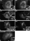
Figure 1
Figure 1
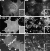
Microvillar elongation in L fibroblasts overexpressing ERMBMPs. L transfectants transiently expressing E-cadherin chimeric molecules with the transmembrane/cytoplasmic domain of CD43 (E-43, a and b), CD44 (E-44, c and d), or ICAM-2 (E-ICAM-2, e and f) were doubly stained with anti– E-cadherin mAb (a, c, and e), and anti-ERM mAb (b, d, and f). In these cells, microvilli were significantly elongated, where both ERMBMPs and ERM proteins were recruited (inset, higher magnification). Cells were transfected by microinjection. As a control, parental L cells were stained with anti-ERM mAb (g). Bar, a–g, 10 μm; insets, 2.5 μm.
Figure 2
Figure 2
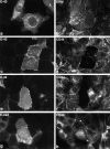
L fibroblasts overexpressing membrane proteins lacking the ERM-binding ability. L transfectants transiently expressing wild-type E-cadherin (E-cad, a and b), E-44/20-70, a mutant E-44 lacking the juxta-membrane ERM-binding region (E-44/20-70, c and d), or E-43/KRR:NGG, a mutant E-43 bearing site-directed disrupted ERM-binding region (E-43/ KRR:NGG, e and f), were doubly stained with anti–E-cadherin mAb (a, c, and e) and anti-ERM mAb (b, d, and f). No elongated microvilli were observed. Cells were transfected by microinjection (a, b, e, and f) or by lipofection (c and d). Bar, a–f, 10 μm; insets, 2.5 μm.
Figure 3
Figure 3
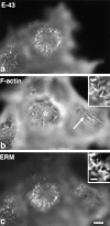
Changes in cortical actin filament organization in L fibroblasts transiently overexpressing E-43. Cells were triply stained with anti–E-cadherin mAb (E-43, a), FITC-phalloidin (F-actin, b), and anti-ERM mAb (ERM, c). Nontransfected cells (compare a with b) were characterized by dorsal stress fibers (arrow in b) and short microvilli (c), whereas in E-43–overexpressing cells long microvillar core actin bundles were induced with concomitant disruption of dorsal stress fibers (asterisk in b). The precise colocalization of F-actin and ERM proteins in elongated microvilli in E-43–expressing cells is shown at higher magnification in the insets. Cells were transfected by microinjection. Bar, a–c, 10 μm; insets, 2 μm.
Figure 4
Figure 4
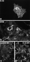
Microvillar elongation in CV-1 fibroblasts transiently overexpressing E-43. Cells were doubly stained with anti–E-cadherin mAb (E-43, a) and anti-ERM mAb (ERM, b). In E-43– expressing CV-1 cells, microvilli were significantly elongated by recruiting E-43 as well as ERM proteins as compared to surrounding nontransfected cells (a and b). E-43 overexpression did not appear to increase the density of microvilli. Microvillar elongation was more clearly demonstrated at higher magnification when nontransfected cells (c) and E-43–overexpressing cells (d) were stained with anti-ERM mAb. Cells were transfected by microinjection. Bars, a and b, 10 μm; c and d, 5 μm.
Figure 5
Figure 5
Length distribution of microvilli in E-43–expressing (open box) and nontransfected (closed box) CV-1 cells. The length of each microvillus was measured from anti-ERM mAb-stained images (see Fig. 4 b). In nontransfected cells, microvilli were relatively short (0.53 ± 0.33 μm; n = 320), whereas in E-43– overexpressing cells they were significantly elongated (3.5 ± 1.4 μm; n = 300). Cells were transfected by lipofection.
Figure 6
Figure 6
Scanning electron microscopic surface images of nontransfected (a) and E-43–overexpressing (b) CV-1 cells. E-43– expressing cells were identified by surface labeling by 0.1-μm beads (arrows) which recognized the extracellular domain of E-cadherin (see Materials and Methods in detail). Note the significant difference in microvillar length between a and b. Cells were transfected by microinjection. Bar, 1 μm.
Figure 7
Figure 7
Transient overexpression of E-43 or E-44 in MTD-1A epithelial cells. Cells were doubly stained with anti–E-cadherin mAb (a) and anti-ERM mAb (b) or with anti–E-cadherin pAb (c, e, and g) and anti-CD44 mAb (d, f, and h). Overexpression of E-43 (a–d) or E-44 (e and f) did not induce microvillar elongation (a and b), but caused exclusion of CD44 from preexisting microvilli (c–f). Overexpression of E-cadherin (g and h) did not affect the distribution of CD44. Cells were transfected by microinjection. Bar, 10 μm.
Figure 8
Figure 8
Transient overexpression of E-43 or E-43/KRR:NGG in A431 cells. Cells were doubly stained with anti–E-cadherin pAb (a and c)/anti-ezrin mAb, M11 (b and d), or anti–E-cadherin mAb, ECCD-2 (e)/anti-ERM pAb, TK89 (f) in the absence (a and b) or presence (c–f) of EGF. In this experiment, cells were stained with antibodies after Triton X-100 treatment, resulting in intense cytoplasmic staining with anti–E-cadherin (a, c, and e). M11 did not bind to ezrin in the cytoplasm strongly (b and d). When cells were exposed to EGF for 30 s, even in nontransfected cells, short ezrin-positive microvilli appeared on the cell surface, but E-43– expressing cells elongated significantly longer ezrin-positive microvilli (d). In sharp contrast, cells expressing E-43/KRR: NGG, which lacks the binding ability to ERM proteins, elongated short ERM-positive microvilli at the similar level to the surrounding nontransfected cells (f) at 30 s after EGF treatment. Cells were transfected by microinjection. Bar, 10 μm.
Figure 11
Figure 11
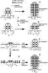
Possible involvement of ERMBMPs and activated ERM proteins in organizing microvilli. (A) Fibroblastic cells such as L cells and CV-1 cells bear short microvilli on their surface. In this model, ERM proteins were effectively activated in response to increases in amounts of ERMBMP in these cells. Overexpressed ERMBMPs such as E-43, E-44, and E-ICAM-2 then recruit these activated ERM proteins and actin filaments to elongate microvilli. (B) Epithelial cells such as MTD-1A cells bear short microvilli on their surface. The activation process of ERM proteins is postulated to be suppressed in these cells. Due to the limited amount of activated ERM proteins, overexpressed ERMBMPs do not elongate microvilli but compete out endogenous CD44 from preexisting microvilli. (C) A431 cells carry only very short microvilli in the absence of EGF. The activation process of ERM proteins is also postulated to be suppressed in these epithelial cells. EGF treatment released this suppression mechanism, resulting in an increase in the amount of activated ERM proteins, which are recruited to the cytoplasmic domain of endogenous ERMBMPs to elongate microvilli. (D) Even when E-43 is overexpressed in A431 cells in the absence of EGF, microvilli are not elongated due to the suppression of the activation process of ERM proteins. (E) When E-43 is overexpressed in A431 cells in the presence of EGF, increased amounts of activated ERM proteins are recruited to the overexpressed E-43 to cause overelongation of microvilli.
Figure 9
Figure 9
Phosphorylation of the COOH-terminal threonine of ERM proteins in EGF-treated A431 cells. (A) Serum-starved A431 cells were incubated with EGF for 30 s, then immediately they were applied onto SDS-PAGE followed by immunoblotting with anti-phosphotyrosine mAb (4G10) (upper panel, P-Y), a mAb (297S) specific for CPERM (middle panel, CP-ERM), or anti-ERM pAb (TK89) (lower panel, ERM). The same nitrocellulose membrane was repeatedly probed with each antibody, confirming that ezrin and moesin were predominant in A431 cells, that ezrin but not moesin was tyrosine-phosphorylated, and that both ezrin and moesin were phosphorylated at the COOH-terminal threonine residue. Similar immunoblotting was performed also in MTD-1A cells and L cells. EGF-R, EGF receptor; E, ezrin; R, radixin; M, moesin. Bars indicate molecular masses of 200, 116, 97, 66, 45, 31, and 21 kD from the top, respectively. (B) Immunofluorescence images of serum-starved A431 cells doubly stained with mAb 297S specific for CPERM (a and c) and TK88 specific for all ERM proteins (b and d) before (a and b) and 30 s after (c and d) EGF treatment. CPERM appeared in large amounts at microvilli and cell–cell borders only in EGF-treated cells (c). Some amount of total ERM proteins appeared to be translocated from the cytoplasm to microvilli after EGF treatment (c and d). Bar, 10 μm.
Figure 10
Figure 10
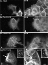
Localization of site-directed mutants of ezrin in serum-starved A431 cells. VSVG-tagged ezrin (Ez-VSVG) (a and b), Ez-VSVG mutant which T567 was mutated to alanine (Ez-T/A-VSVG) (c and d), or Ez-VSVG mutant which T567 was mutated to aspartic acid (Ez-T/D-VSVG) (e–h) was transfected to A431 cells by microinjection. Cells were doubly stained with anti-VSVG mAb (P5D4) (a, c, and e)/anti-ERM pAb (TK89) (b, d, and f), or anti-VSVG mAb (P5D4) (g)/ FITC-phalloidin (h). Ez-VSVG and Ez-T/A-VSVG did not affect cell morphology and were codistributed with endogenous ERM proteins diffusely in the cytoplasm as well as on the cell surface (a–d). In contrast, Ez-T/ D-VSVG induced microvillar elongation and was localized exclusively at the cell surface structures such as microvilli (e and f). Actin filaments were recruited to Ez-T/D-VSVG– positive elongated microvilli throughout their length (inset, g and h). Occasionally, on the cell surface or intracellular vesicles overexpressed Ez-T/D-VSVG formed large aggregates that recruited actin filaments (arrows in g and h). Bar, 10 μm; insets, 2.5 μm.

Similar articles

Cited by

References

    1. Algrain M, Turunen O, Vaheri A, Louvard D, Arpin M. Ezrin contains cytoskeleton and membrane binding domains accounting for its proposed role as a membrane-cytoskeletal linker. J Cell Biol. 1993;120:129–140. - PMC - PubMed
    1. Arpin M, Algrain M, Louvard D. Membrane-actin microfilament connections: an increasing diversity of players related to band 4.1. Curr Opin Cell Biol. 1994a;6:136–141. - PubMed
    1. Arpin M, Friederich E, Algrain M, Vernel F, Louvard D. Functional differences between L- and T-plastin isoforms. J Cell Biol. 1994b;127:1995–2008. - PMC - PubMed
    1. Berryman M, Gary R, Bretscher A. Ezrin oligomers are major cytoskeletal components of placental microvilli: a proposal for their involvement in cortical morphogenesis. J Cell Biol. 1995;131:1231–1242. - PMC - PubMed
    1. Bretscher A. Rapid phosphorylation and reorganization of ezrin and spectrin accompany morphological changes induced in A-431 cells by epidermal growth factor. J Cell Biol. 1989;108:921–930. - PMC - PubMed

Publication types

MeSH terms