. 1999 Jul;19(7):5189-202.
doi: 10.1128/MCB.19.7.5189.

Trithorax- and Polycomb-group response elements within an Ultrabithorax transcription maintenance unit consist of closely situated but separable sequences

Affiliations

Trithorax- and Polycomb-group response elements within an Ultrabithorax transcription maintenance unit consist of closely situated but separable sequences

S Tillib et al. Mol Cell Biol. 1999 Jul.

Abstract

In Drosophila, two classes of genes, the trithorax group and the Polycomb group, are required in concert to maintain gene expression by regulating chromatin structure. We have identified Trithorax protein (TRX) binding elements within the bithorax complex and have found that within the bxd/pbx regulatory region these elements are functionally relevant for normal expression patterns in embryos and confer TRX binding in vivo. TRX was localized to three closely situated sites within a 3-kb chromatin maintenance unit with a modular structure. Results of an in vivo analysis showed that these DNA fragments (each approximately 400 bp) contain both TRX- and Polycomb-group response elements (TREs and PREs) and that in the context of the endogenous Ultrabithorax gene, all of these elements are essential for proper maintenance of expression in embryos. Dissection of one of these maintenance modules showed that TRX- and Polycomb-group responsiveness is conferred by neighboring but separable DNA sequences, suggesting that independent protein complexes are formed at their respective response elements. Furthermore, we have found that the activity of this TRE requires a sequence (approximately 90 bp) which maps to within several tens of base pairs from the closest neighboring PRE and that the PRE activity in one of the elements may require a binding site for PHO, the protein product of the Polycomb-group gene pleiohomeotic. Our results show that long-range maintenance of Ultrabithorax expression requires a complex element composed of cooperating modules, each capable of interacting with both positive and negative chromatin regulators.


Figures

FIG. 1
FIG. 1
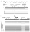
Localization of the TRX protein in the BX-C. (Top) A diagram of the molecular organization of the BX-C. Map coordinates (in kilobases) are based on the complete sequence of the BX-C (22). The Ubx, abdA, and AbdB transcription units and regulatory regions are shown above the DNA. PREs (7, 16, 32, 39) and Pc protein binding regions (43) are shown beneath the DNA as filled bars and arrows, respectively. The TRX protein binding regions are indicated by arrows above phage clones that contain TRX binding fragments, which are shown as horizontal lines. (Bottom) Phage clones are indicated by numbers. Equal amounts (∼1 μg) of each of the 27 overlapping λ Charon clones (2) were digested with EcoRI and transferred onto a nylon membrane. The membrane was hybridized with 32P-labeled PCR-amplified fragments of genomic DNA obtained after two rounds of amplification-immunoprecipitation with TRX antibody (see Materials and Methods). Positions of molecular weight markers are indicated on the left. Asterisks indicate two bands also seen in control experiments with preimmune serum that were probably due to repeats in genomic DNA (these bands were not seen in the preimmune serum control lane when cloned phage DNA was used for immunoprecipitation, as in Fig. 2).
FIG. 2
FIG. 2
Localization of TRX protein in the bxd region of Ubx. DNA fragments obtained after one round of immunoprecipitation of pMBO1253 DNA (containing a 14.4-kb SalI-HindIII fragment with the map coordinates −18.1 to 3.7 [40] with TRX antibody or with preimmune serum from the same rabbit, following incubation with nuclear extracts (see Materials and Methods) were 32P labeled and used to probe filters containing the same DNA digested with Sau3A (S), MspI (M), or both (SM). Fragment B was detected following incubation of DNA with nuclear extract in binding buffer 1 (A), while fragments C and D were detected by using binding buffer 2 (B). Lengths of binding fragments are shown on the left. Gels in panel B were run for a longer time to resolve a doublet in the S lane. The initial DNA mixture was obtained by end labeling λ1253 DNA digested with Sau3A and MspI. The coordinates of the TRX binding fragments in the complete sequence of the BX-C (accession no. U31961) are as follows: B, 217,111 to 217,626; C, 218,834 to 219,314; D, 219,701 to 220,118. (C) TRX binding to the 4-kb N transgene (Fig. 3B) on polytene chromosomes. Localization of the N18 transgene to the 100F region of 3R by in situ hybridization (top). TRX is not localized at 100F in the wild-type larva (middle). The new TRX binding site appears at 100F in the N18 transformant larva (bottom). Arrows indicate the site of insertion of the N transgene.
FIG. 2
FIG. 2
Localization of TRX protein in the bxd region of Ubx. DNA fragments obtained after one round of immunoprecipitation of pMBO1253 DNA (containing a 14.4-kb SalI-HindIII fragment with the map coordinates −18.1 to 3.7 [40] with TRX antibody or with preimmune serum from the same rabbit, following incubation with nuclear extracts (see Materials and Methods) were 32P labeled and used to probe filters containing the same DNA digested with Sau3A (S), MspI (M), or both (SM). Fragment B was detected following incubation of DNA with nuclear extract in binding buffer 1 (A), while fragments C and D were detected by using binding buffer 2 (B). Lengths of binding fragments are shown on the left. Gels in panel B were run for a longer time to resolve a doublet in the S lane. The initial DNA mixture was obtained by end labeling λ1253 DNA digested with Sau3A and MspI. The coordinates of the TRX binding fragments in the complete sequence of the BX-C (accession no. U31961) are as follows: B, 217,111 to 217,626; C, 218,834 to 219,314; D, 219,701 to 220,118. (C) TRX binding to the 4-kb N transgene (Fig. 3B) on polytene chromosomes. Localization of the N18 transgene to the 100F region of 3R by in situ hybridization (top). TRX is not localized at 100F in the wild-type larva (middle). The new TRX binding site appears at 100F in the N18 transformant larva (bottom). Arrows indicate the site of insertion of the N transgene.
FIG. 3
FIG. 3
Map of constructs used to detect TREs and PREs in the bxd region of Ubx. (A) Partial map of the BX-C including the bxd/pbx regulatory region. Map coordinates are as in Fig. 1. A basal 13-kb construct is shown as a solid bar beneath the DNA line. Deletions within TRX binding elements used to generate transgenic lines are indicated as open boxes beneath the basal construct. The coordinates of the deleted regions in the constructs are as follows: ΔA, HindIII-BamHI (nt 214,875 to 216,285); ΔB, MspI-MspI (nt 217,111 to 217,626); ΔC, MspI-Sau3A (nt 218,835 to 219,249); ΔD, Sau3A-MspI (nt 219,700 to 220,118); ΔC1 (nt 218,835 to 218,959). (B) Map of the multiple TRE-PRE-containing expression maintenance modules. The basal 4-kb construct with a mini-white reporter gene contains the C and D TRX binding elements, approximately a 1-kb fragment which separates the B and C elements, and approximately 0.8 kb of flanking sequences. The coordinates of the deletions in the constructs are as follows: ΔB and ΔC1 are as in panel A; ΔC2 (nt 218,960 to 219,088); ΔC3 (nt 219,089 to 219,249). ΔBC1-A, ΔBC1-B, and ΔBC1-C are deletions of nt 1 to 27, 28 to 61, and 86 to 122, respectively, in the C1 fragment indicated above. The mutation AC to TG in the C1-D fragment and the deletion of the PHO binding site in the C3 fragment are indicated by stars. Consensus binding sites for PHO (filled circles) and GAGA (open circles) in the C fragment are indicated above the map. H, HindIII; E, EcoRI; B, BamHI; P, PstI; K, KpnI; S, Sau3A; M, MspI.
FIG. 4
FIG. 4
Expression of lacZ in transformant lines in wild-type and mutant trx embryos. (A) lacZ RNA, detected by in situ hybridization to whole-mount embryos, in the N, ΔB (upper row), and all the other tested lines (not shown) is expressed in PS6 to 13 at embryonic stage 10. At embryonic stages 15 and 16, expression of the N construct is still restricted to the region of the embryo posterior to PS6, while in ΔB, ΔC, and ΔD embryos, lacZ is strongly expressed in the anterior neuromeres and in the supraesophageal ganglia (right). At this stage in ΔBC embryos expression of lacZ in the anterior is strongly decreased compared to expression in ΔB, ΔC, and ΔD embryos. In all embryos with the deletion constructs, expression of lacZ in the posterior parasegments is weaker than in N embryos. The anterior region is to the left. (B) Expression of lacZ RNA in the CNS of N, ΔB, ΔC, and ΔD lines. Expression in neuromeres posterior to PS5 is visibly reduced in the deletion lines. The extent and pattern of anteriorly expressed lacZ are different in each of the ΔB, ΔC, and ΔD lines. The anterior end is at the top. (C) The effect of trxB11 mutation on the expression of N, ΔC, and ΔD transgenes. Expression of lacZ RNA in the CNS of N, ΔC, and ΔD transgenes in wild-type and mutant trx embryos. Embryos were doubly stained with antibodies against β-galactosidase (brown) and lightly stained by in situ hybridization with a probe specific to endogenous Ubx RNA (blue). trxB11 mutant embryos were identified by a decrease in Ubx expression primarily in PS6. Expression in neuromeres posterior to PS5 is visibly reduced in the trx mutant in the N line. In the ΔC and ΔD lines, no reduction of β-galactosidase expression in the trxB11 mutant is seen in cells at the periphery of the CNS, where expression of endogenous Ubx is strongly affected by trxB11 mutations (8, 24). In the trxB11 mutant embryos, expression is severely reduced in the anterior neuromeres of the ΔC and ΔD embryos. In the ΔC and ΔD lines, trxB11 mutation also causes an increase of expression in the cells along the midline of the CNS posterior to PS6. This effect is likely to be indirect, due to decreased repression by Ubx and abd-A proteins, since in a trxB11 mutant, expression of all BX-C genes is decreased. Consistent with this interpretation, it has been shown previously that several transgenes, including one similar to the N construct, contain elements that mediate partial repression by the Ubx and abd-A genes (39). Arrowheads indicate PS6.
FIG. 4
FIG. 4
Expression of lacZ in transformant lines in wild-type and mutant trx embryos. (A) lacZ RNA, detected by in situ hybridization to whole-mount embryos, in the N, ΔB (upper row), and all the other tested lines (not shown) is expressed in PS6 to 13 at embryonic stage 10. At embryonic stages 15 and 16, expression of the N construct is still restricted to the region of the embryo posterior to PS6, while in ΔB, ΔC, and ΔD embryos, lacZ is strongly expressed in the anterior neuromeres and in the supraesophageal ganglia (right). At this stage in ΔBC embryos expression of lacZ in the anterior is strongly decreased compared to expression in ΔB, ΔC, and ΔD embryos. In all embryos with the deletion constructs, expression of lacZ in the posterior parasegments is weaker than in N embryos. The anterior region is to the left. (B) Expression of lacZ RNA in the CNS of N, ΔB, ΔC, and ΔD lines. Expression in neuromeres posterior to PS5 is visibly reduced in the deletion lines. The extent and pattern of anteriorly expressed lacZ are different in each of the ΔB, ΔC, and ΔD lines. The anterior end is at the top. (C) The effect of trxB11 mutation on the expression of N, ΔC, and ΔD transgenes. Expression of lacZ RNA in the CNS of N, ΔC, and ΔD transgenes in wild-type and mutant trx embryos. Embryos were doubly stained with antibodies against β-galactosidase (brown) and lightly stained by in situ hybridization with a probe specific to endogenous Ubx RNA (blue). trxB11 mutant embryos were identified by a decrease in Ubx expression primarily in PS6. Expression in neuromeres posterior to PS5 is visibly reduced in the trx mutant in the N line. In the ΔC and ΔD lines, no reduction of β-galactosidase expression in the trxB11 mutant is seen in cells at the periphery of the CNS, where expression of endogenous Ubx is strongly affected by trxB11 mutations (8, 24). In the trxB11 mutant embryos, expression is severely reduced in the anterior neuromeres of the ΔC and ΔD embryos. In the ΔC and ΔD lines, trxB11 mutation also causes an increase of expression in the cells along the midline of the CNS posterior to PS6. This effect is likely to be indirect, due to decreased repression by Ubx and abd-A proteins, since in a trxB11 mutant, expression of all BX-C genes is decreased. Consistent with this interpretation, it has been shown previously that several transgenes, including one similar to the N construct, contain elements that mediate partial repression by the Ubx and abd-A genes (39). Arrowheads indicate PS6.
FIG. 5
FIG. 5
Expression of lacZ in ΔC1 and ΔC3 transformant lines. lacZ RNA in the N, ΔC1, and ΔC3 embryos (left column) is expressed in PS6 to 13 at embryonic stage 10. In ΔC1 embryos, at embryonic stage 15 and 16 (right column), there is no expression anterior to PS6. At this stage in ΔC1 embryos, expression of lacZ is decreased compared to expression in N. In ΔC3 embryos, the anterior boundary is shifted to PS5 (small arrowhead). Large arrowheads indicate PS6.
FIG. 6
FIG. 6
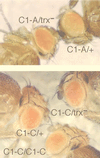
The effect of the trxB11 mutation in heterozygotes on expression of the white gene, and pairing-sensitive repression in ΔBC1-A and ΔBC1-C transgenic flies. Expression of white in the eyes of the ΔBC1-A heterozygous line is strongly decreased in trxB11 heterozygotes (top) (C1-A/+ appears to the left of the label, and C1-A/trx is shown to the right of the label), but expression of white in the ΔBC1-C heterozygous line is unaffected by the trxB11 mutation (bottom). Expression of white is significantly decreased in the eyes of homozygotes compared to that in heterozygotes in the ΔBC1-C line.
FIG. 7
FIG. 7
Binding of the TRX protein is affected by mutations in the C TRE. DNA fragments obtained after one round of immunoprecipitation of pCaSpeR3 DNA (containing either intact 4-kb BamHI-KpnI fragment, N, or the same fragment with the ΔC1-B, ΔC1-C, and ΔC1-D deletions) with TRX (T) antibody or with preimmune serum (P), following incubation with nuclear extracts (see Materials and Methods), were run on the agarose gel and transferred onto a nylon membrane. The filters were probed separately with the 32P-labeled C (upper panel) and D (lower panel) DNA fragments.

Similar articles

Cited by

References

    1. Adamson A L, Shearn A. Molecular genetic analysis of Drosophila ash2, a member of the trithorax group required for imaginal disc pattern formation. Genetics. 1996;144:621–633. - PMC - PubMed
    1. Bender W, Akam M, Karch F, Beachy P A, Peifer M, Spierer P, Lewis I B, Hogness D S. Molecular genetics of the bithorax complex in Drosophila melanogaster. Science. 1983;221:23–29. - PubMed
    1. Bienz M, Müller J. Transcriptional silencing of homeotic genes in Drosophila. BioEssays. 1995;17:775–784. - PubMed
    1. Breen T R, Harte P J. trithorax regulates multiple homeotic genes in the bithorax and Antennapedia complexes and exerts different tissue-specific, parasegment-specific and promoter-specific effects on each. Development. 1993;117:119–134. - PubMed
    1. Brown J L, Mucci D, Whiteley M, Dirksen M L, Kassis J A. The Drosophila Polycomb group gene pleiohomeotic encodes a DNA binding protein with homology to the transcription factor YY1. Mol Cell. 1998;1:1057–1064. - PubMed

Publication types

LinkOut - more resources