. 1999 May 3;145(3):619-31.
doi: 10.1083/jcb.145.3.619.

Laminin polymerization induces a receptor-cytoskeleton network

Affiliations

Laminin polymerization induces a receptor-cytoskeleton network

H Colognato et al. J Cell Biol. .

Abstract

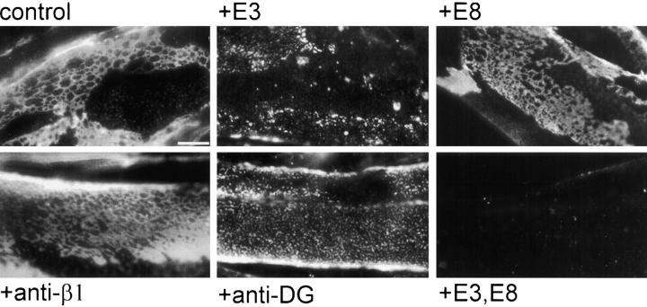
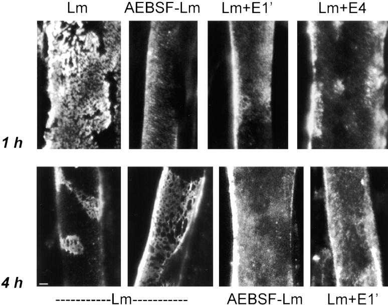
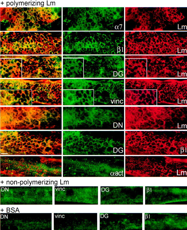
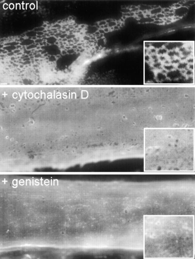
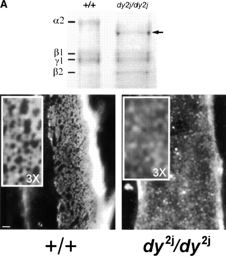
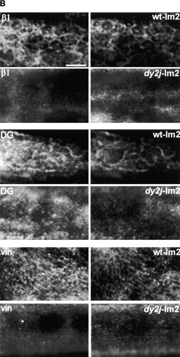
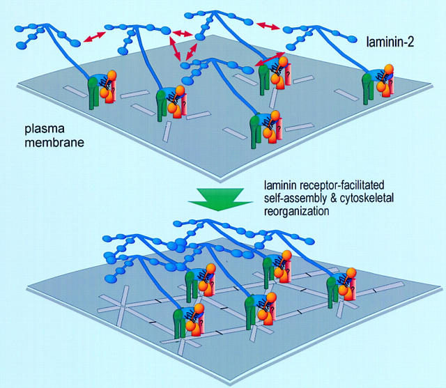
The transition of laminin from a monomeric to a polymerized state is thought to be a crucial step in the development of basement membranes and in the case of skeletal muscle, mutations in laminin can result in severe muscular dystrophies with basement membrane defects. We have evaluated laminin polymer and receptor interactions to determine the requirements for laminin assembly on a cell surface and investigated what cellular responses might be mediated by this transition. We found that on muscle cell surfaces, laminins preferentially polymerize while bound to receptors that included dystroglycan and alpha7beta1 integrin. These receptor interactions are mediated through laminin COOH-terminal domains that are spatially and functionally distinct from NH2-terminal polymer binding sites. This receptor-facilitated self-assembly drives rearrangement of laminin into a cell-associated polygonal network, a process that also requires actin reorganization and tyrosine phosphorylation. As a result, dystroglycan and integrin redistribute into a reciprocal network as do cortical cytoskeleton components vinculin and dystrophin. Cytoskeletal and receptor reorganization is dependent on laminin polymerization and fails in response to receptor occupancy alone (nonpolymerizing laminin). Preferential polymerization of laminin on cell surfaces, and the resulting induction of cortical architecture, is a cooperative process requiring laminin- receptor ligation, receptor-facilitated self-assembly, actin reorganization, and signaling events.


Figures

Figure 4
Figure 4
Generation of nonpolymerizing laminin using AEBSF. (A) Binding of [14C]AEBSF to laminin proteolytic fragments. Laminin incubated with [14C]AEBSF was treated with elastase to generate standard laminin elastase fragments (Fig. 2 A, fragment map of laminin). Total laminin digest was separated using SDS-PAGE and carbon-14 incorporation was determined by phosphoanalysis, shown here relative to fragment migration positions. Most [14C]AEBSF bound to E1′, a fragment shown to contain sites essential for polymerization. (B) AEBSF-treated laminin (open circles) and untreated laminin (closed circles) were evaluated for their ability to form a polymer in solution. Laminin was incubated for 3 h at 37°C and the polymer fraction was separated by centrifugation (see Materials and Methods). Untreated laminin showed concentration-dependent polymerization, whereas AEBSF-treated laminin remained in solution. (C) AEBSF-treated laminin (open circles) and untreated laminin (closed circles) were compared for their ability to support adhesion of C2C12 myoblasts. Both treated and untreated laminin supported adhesion and showed a similar dependence on substrate concentration. In addition, laminin fragments (E3, E8, and C8 and 9) treated with AEBSF bound to fused myotubes (not shown).
Figure 2
Figure 2
Laminin uses its COOH-terminal long arm to bind to the myotube surface. (A) Structure/function map of laminin proteolytic fragments and recombinant proteins, shown as a composite of features common to laminin-1 (α1β1γ1) and laminin-2 (α2β1γ1). Proteolytic fragments include: E1′, E4, E8, and E3. Recombinant proteins include: α1(VI-IVb), α2(VI-IVb), and α2(G). Receptor binding sites are: α1β1, α2β1, α7β1 integrin, and α-dystroglycan (αDG). Binding sites for extracellular matrix molecules: laminin polymer– forming regions (domains V and VI of α-, β-, and γ-short arms), entactin/nidogen (En/Nd), and agrin. Mutations used include the following: 57–amino acid region deleted in α2 chain of dystrophic dy2J mouse (Δdy2J). (B) Direct binding of laminin COOH-terminal long arm proteins to the myotube surface. Laminin-1 and laminin-2 bound myotubes and formed a reticular pattern after 1 h (insets). Recombinant laminin α-subunit proteins from the NH2-terminal short arm region (α1- and α2[VI-IVb]′) showed no detectable binding. COOH-terminal proteins, including proteolytic fragments E8 and E3, and recombinant α2-G domain protein showed widespread attachment to the myotube surface, but remained in a diffuse, punctate distribution. Insets show regions at two times the magnification.
Figure 2
Figure 2
Laminin uses its COOH-terminal long arm to bind to the myotube surface. (A) Structure/function map of laminin proteolytic fragments and recombinant proteins, shown as a composite of features common to laminin-1 (α1β1γ1) and laminin-2 (α2β1γ1). Proteolytic fragments include: E1′, E4, E8, and E3. Recombinant proteins include: α1(VI-IVb), α2(VI-IVb), and α2(G). Receptor binding sites are: α1β1, α2β1, α7β1 integrin, and α-dystroglycan (αDG). Binding sites for extracellular matrix molecules: laminin polymer– forming regions (domains V and VI of α-, β-, and γ-short arms), entactin/nidogen (En/Nd), and agrin. Mutations used include the following: 57–amino acid region deleted in α2 chain of dystrophic dy2J mouse (Δdy2J). (B) Direct binding of laminin COOH-terminal long arm proteins to the myotube surface. Laminin-1 and laminin-2 bound myotubes and formed a reticular pattern after 1 h (insets). Recombinant laminin α-subunit proteins from the NH2-terminal short arm region (α1- and α2[VI-IVb]′) showed no detectable binding. COOH-terminal proteins, including proteolytic fragments E8 and E3, and recombinant α2-G domain protein showed widespread attachment to the myotube surface, but remained in a diffuse, punctate distribution. Insets show regions at two times the magnification.
Figure 1
Figure 1
Laminin binds to the myotube surface and forms a network. Laminin attached to the surface of C2C12 myotubes was visualized using polyclonal antibodies against specific laminin subunits followed by rhodamine-conjugated secondary antibodies. After incubation with 1 μg/ml laminin-1 for 15 min, laminin was seen on the dorsal myotube surface in a diffuse punctate pattern (a and a′). In 1 h, laminin was organized in a more aggregated, reticular pattern (b and b′). Myotubes incubated with 10 μg/ml laminin-1 (c, c′, and d) for 4 h showed an extensive repeating polygonal network of laminin on their surface (see c′ for a higher magnification view of this network, arrow depicts polygonal unit). At 4 h, many myotubes also contain regions cleared of laminin, with surface networks appearing in more compact clusters (d). Control myotubes incubated with BSA show little laminin staining (e). Fibronectin (10 μg/ml for 4 h) forms fibrillar structures primarily in regions containing myoblasts, not myotubes (f, phase micrograph, f′, antifibronectin). Bar, 5 μm.
Figure 3
Figure 3
Blocking receptor interactions disrupts laminin surface networks. Laminin COOH-terminal proteins and/or receptor-blocking antibodies were used to compete for laminin binding sites. Myotubes were preincubated with the following proteins for 1 h: BSA (control), fragment E3, fragment E8, anti–β1 integrin blocking antibody, antidystroglycan (anti-DG), or a combination of fragments E3 and E8. Laminin incubated thereafter (5 μg/ml for 1 h) was visualized using an antibody specific for the NH2-terminal region of the laminin β1 subunit (anti-E4, which does not recognize COOH-terminal fragments E3 and E8). Blockade using fragment E3 alone resulted in a large decrease of laminin binding and network formation on the cell surface, whereas blockade of the long arm integrin binding sites using E8 or integrin blocking antibodies had minimal effect. Treatment with antibodies against α-dystroglycan disrupted laminin surface networks, to a lesser degree than blockade with fragment E3. Blockade of both E3- and E8-mediated cell recognition domains completely inhibited laminin binding to the surface. Bar, 10 μm.
Figure 5
Figure 5
Laminin polymer bonds are required for network formation on the cell surface. Laminins were treated with reagents that block laminin polymerization, but not cell adhesion activities. Myotubes were incubated with these polymer-deficient laminins for 1 h at 1 μg/ml (top) or for 4 h at 10 μg/ml (bottom) then evaluated for the presence of laminin aggregates. In the presence of short arm fragments E1′ and E4, laminin was unable to organize into reticular clusters in 1 h (untreated laminin shown at left). Similarly, AEBSF-treated laminin (Fig. 4) did not reorganize on the cell surface into aggregates. Formation of ordered laminin networks (at 4 h) was also blocked by polymer-disrupting reagents E1′ and AEBSF. Bar, 5 μm.
Figure 6
Figure 6
Laminin polymerization induces a selective reorganization of cell surface receptors and components of the cortical cytoskeleton. Myotubes were incubated with 10 μg/ml laminin-1 for 4 h, followed by double indirect immunofluorescence to visualize sets of bound laminin and various sarcolemmal proteins: Lm, laminin; α7, α7 integrin subunit; β1, β1 integrin subunit; DG, dystroglycan; vinc, vinculin; DN, dystrophin; α-act, α-actinin. Merged images of paired sets are shown to the far left in yellow. Sarcolemmal proteins in myotubes treated with BSA or with nonpolymerizing (AEBSF-treated) laminin are shown in the bottom two rows of images. In the presence of laminin that can polymerize, receptors and cytoskeletal proteins reorganize into a similar polygonal pattern, colocalizing with each other and with laminin. Bar, 5 μm; insets are twice the magnification.
Figure 7
Figure 7
Inhibition of actin reorganization or tyrosine phosphorylation prevents formation of laminin surface networks. Myotubes were preincubated with either cytochalasin, an agent that interferes with assembly and disassembly of actin filaments, or genistein, an agent that blocks tyrosine phosphorylation. Treated and untreated myotubes were incubated with 5 μg/ml laminin and evaluated for the presence of laminin surface networks after 4 h. Cytochalasin and genistein both prevented characteristic laminin surface networks from forming and disrupted laminin reorganization and clearance. Bar, 5 μm; insets are twice the magnification.
Figure 8
Figure 8
Polymer-defective laminin from dy2J/dy2J mice does not form surface networks and does not induce sarcolemmal reorganization. (A) The top frame shows an SDS-PAGE of laminin from skeletal muscle of +/+ and dy2J/dy2J mice. Proteins extracted from mouse skeletal muscle were separated by SDS-PAGE under reducing conditions and visualized with Coomassie blue. The arrow depicts dy2J laminin α2 subunit, roughly 25 kD smaller than native α2. Bottom frame: dy2J–α2-laminin binds to the myotube surface but fails to form networks. On myotubes incubated for 4 h, α2-laminin from wild-type mice formed networks (+/+), whereas dy2J–α2-laminin remained in a punctate, diffusely distributed pattern (dy2J/dy2J). Bar, 5 μm; insets, three times the magnification. (B) dy2J–α2-laminin does not induce reorganization of sarcolemmal proteins into a cortical network. Myotubes were incubated for 4 h with laminin from either wild-type (wt-lm2) or dy2J/dy2J mice (dy2j-lm2) and costained to visualize laminin α2 subunit and b1 integrin subunit (β1), dystroglycan (DG), or vinculin (vin). α2-Laminin induces a reorganization of integrin, dystroglycan, and vinculin, whereas dy2J–α2-laminin does not. Bar, 10 μm.
Figure 8
Figure 8
Polymer-defective laminin from dy2J/dy2J mice does not form surface networks and does not induce sarcolemmal reorganization. (A) The top frame shows an SDS-PAGE of laminin from skeletal muscle of +/+ and dy2J/dy2J mice. Proteins extracted from mouse skeletal muscle were separated by SDS-PAGE under reducing conditions and visualized with Coomassie blue. The arrow depicts dy2J laminin α2 subunit, roughly 25 kD smaller than native α2. Bottom frame: dy2J–α2-laminin binds to the myotube surface but fails to form networks. On myotubes incubated for 4 h, α2-laminin from wild-type mice formed networks (+/+), whereas dy2J–α2-laminin remained in a punctate, diffusely distributed pattern (dy2J/dy2J). Bar, 5 μm; insets, three times the magnification. (B) dy2J–α2-laminin does not induce reorganization of sarcolemmal proteins into a cortical network. Myotubes were incubated for 4 h with laminin from either wild-type (wt-lm2) or dy2J/dy2J mice (dy2j-lm2) and costained to visualize laminin α2 subunit and b1 integrin subunit (β1), dystroglycan (DG), or vinculin (vin). α2-Laminin induces a reorganization of integrin, dystroglycan, and vinculin, whereas dy2J–α2-laminin does not. Bar, 10 μm.
Figure 9
Figure 9
A model for receptor-facilitated laminin polymerization on the sarcolemmal plasma membrane. Receptor-facilitated self-assembly of laminin occurs through synergistic laminin–laminin polymer interactions and laminin–receptor interactions. On the muscle sarcolemmal membrane, the three NH2-terminal short arms of laminin mediate polymer interactions, whereas the COOH-terminal long arm of laminin mediates receptor binding. This basement membrane assembly process in turn mediates an organizational signaling, rearranging the cortical cellular architecture and assembling matrix, receptor, and cytoskeletal elements into a polygonal meshwork. We find that formation of this organized network is an actively driven cooperative process that requires laminin receptor–facilitated self-assembly, actin reorganization, and tyrosine phosphorylation.

Similar articles

Cited by

References

    1. Allamand V, Sunada Y, Salih MA, Straub V, Ozo CO, Al-Turaiki MH, Akbar M, Kolo T, Colognato H, Zhang X, Sorokin LM, Yurchenco PD, et al. Mild congenital muscular dystrophy in two patients with an internally deleted laminin alpha2-chain. Hum Mol Genet. 1997;6:747–752. - PubMed
    1. Bloch W, Forsberg E, Lentini S, Brakebusch C, Martin K, Krell HW, Weidle UH, Addicks K, Faessler R. β1 integrin is essential for teratoma growth and angiogenesis. J Cell Biol. 1997;139:265–278. - PMC - PubMed
    1. Boni-Schnetzler M, Pilch PF. Mechanism of epidermal growth factor receptor autophosphorylation and high-affinity binding. Proc Natl Acad Sci USA. 1987;84:7832–7836. - PMC - PubMed
    1. Brown JC, Wiedemann H, Timpl R. Protein binding and cell adhesion properties of two laminin isoforms (AmB1eB2e, AmB1sB2e) from human placenta. J Cell Sci. 1994;107:329–338. - PubMed
    1. Calof AL, Campanero MR, O'Rear JJ, Yurchenco PD, Lander AD. Domain-specific activation of neuronal migration and neurite outgrowth-promoting activities of laminin. Neuron. 1994;13:117–130. - PubMed

Publication types

MeSH terms