. 1999 Apr 19;145(2):347-61.
doi: 10.1083/jcb.145.2.347.

Phosphorylation of adducin by Rho-kinase plays a crucial role in cell motility

Affiliations

Phosphorylation of adducin by Rho-kinase plays a crucial role in cell motility

Y Fukata et al. J Cell Biol. .

Abstract

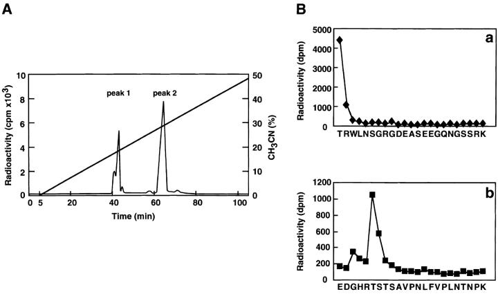
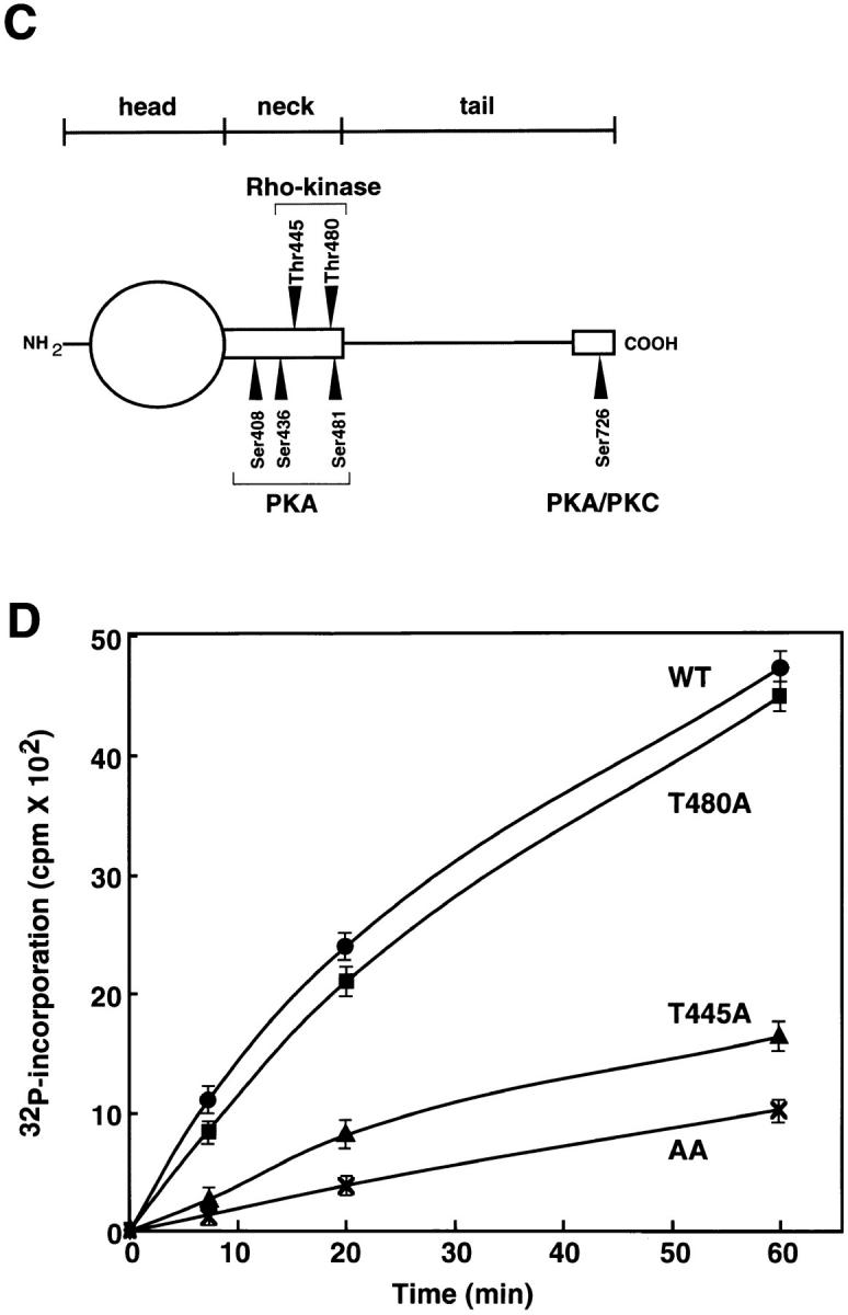
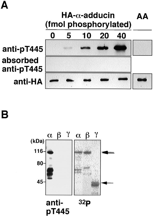
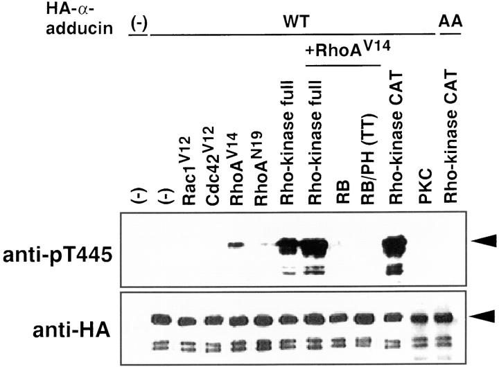
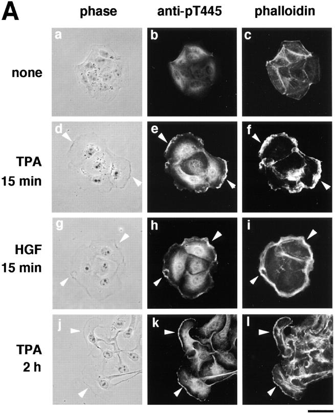
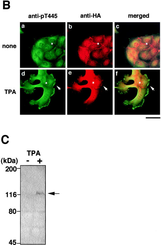
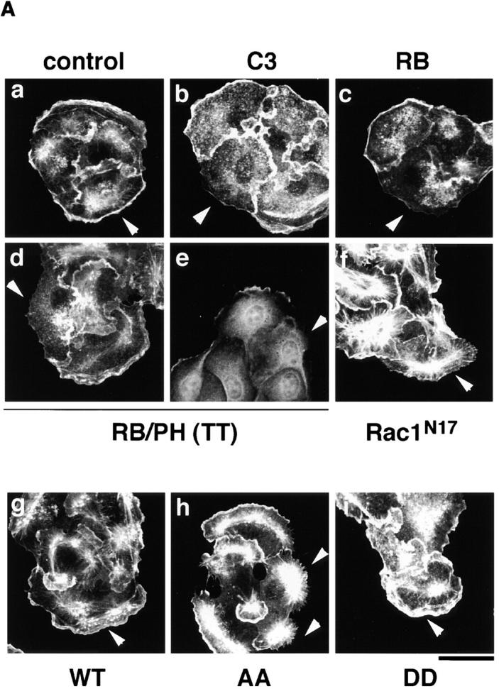
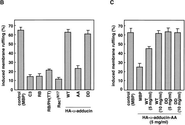
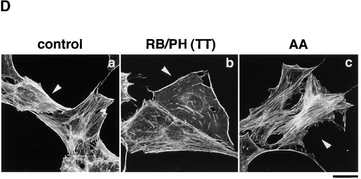
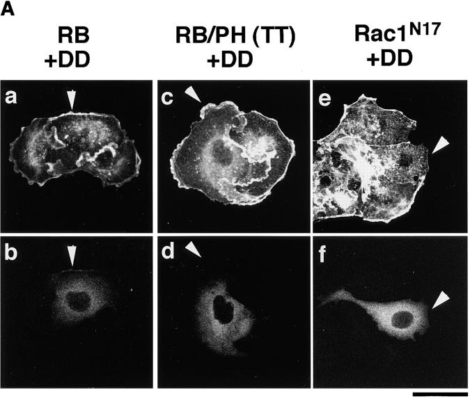
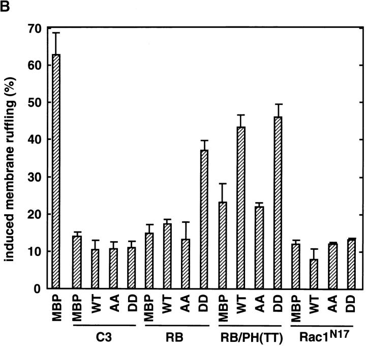
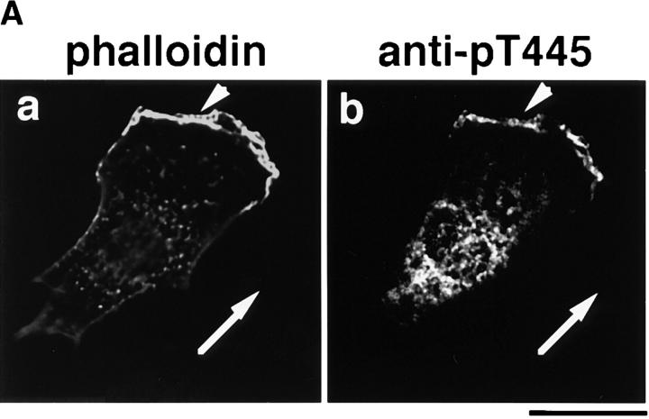
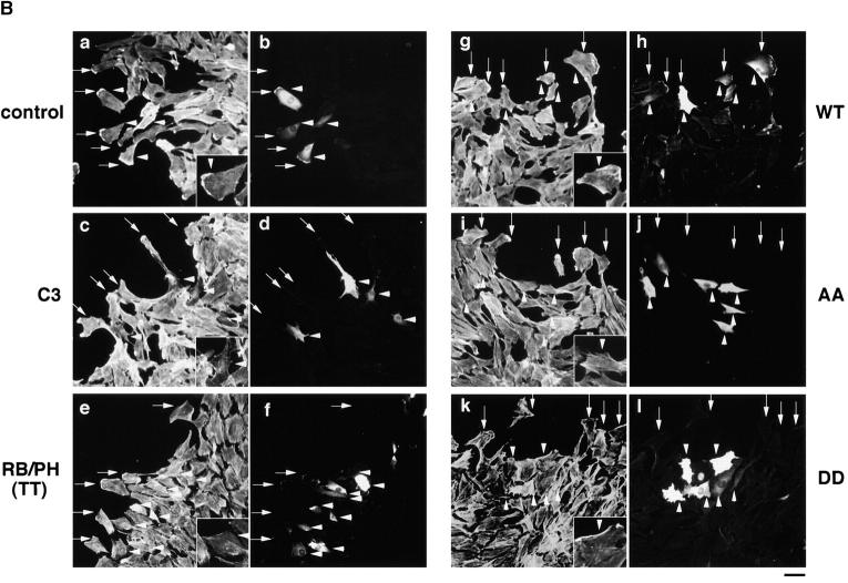
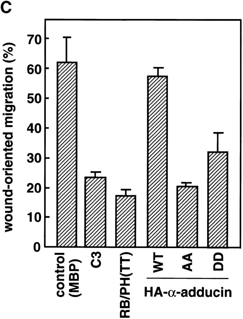
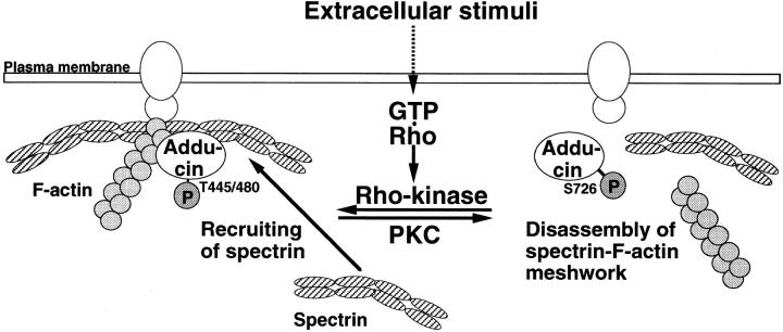
Adducin is a membrane skeletal protein that binds to actin filaments (F-actin) and thereby promotes the association of spectrin with F-actin to form a spectrin-actin meshwork beneath plasma membranes such as ruffling membranes. Rho-associated kinase (Rho- kinase), which is activated by the small guanosine triphosphatase Rho, phosphorylates alpha-adducin and thereby enhances the F-actin-binding activity of alpha-adducin in vitro. Here we identified the sites of phosphorylation of alpha-adducin by Rho-kinase as Thr445 and Thr480. We prepared antibody that specifically recognized alpha-adducin phosphorylated at Thr445, and found by use of this antibody that Rho-kinase phosphorylated alpha-adducin at Thr445 in COS7 cells in a Rho-dependent manner. Phosphorylated alpha-adducin accumulated in the membrane ruffling area of Madin-Darby canine kidney (MDCK) epithelial cells and the leading edge of scattering cells during the action of tetradecanoylphorbol-13-acetate (TPA) or hepatocyte growth factor (HGF). The microinjection of Botulinum C3 ADP-ribosyl-transferase, dominant negative Rho-kinase, or alpha-adducinT445A,T480A (substitution of Thr445 and Thr480 by Ala) inhibited the TPA-induced membrane ruffling in MDCK cells and wound-induced migration in NRK49F cells. alpha-AdducinT445D,T480D (substitution of Thr445 and Thr480 by Asp), but not alpha-adducinT445A,T480A, counteracted the inhibitory effect of the dominant negative Rho-kinase on the TPA-induced membrane ruffling in MDCK cells. Taken together, these results indicate that Rho-kinase phosphorylates alpha-adducin downstream of Rho in vivo, and that the phosphorylation of adducin by Rho-kinase plays a crucial role in the regulation of membrane ruffling and cell motility.


Figures

Figure 1
Figure 1
Determination of sites of phosphorylation of α-adducin by Rho-kinase. (A) Reverse-phase HPLC for AP-1 fragments of α-adducin phosphorylated by Rho-kinase was carried out under the conditions described in Materials and Methods. CH3CN, acetonitrile. (B) Amino acid sequences of phosphopeptides derived from α-adducin phosphorylated by Rho-kinase. The radioactive peaks 1 (panel a) and 2 (panel b) were separated. Phosphoamino acid sequencing of each peak was carried out with a peptide sequencer, and the fractions obtained from each Edoman degradation cycle were measured for 32P in a liquid scintillation counter. (C) Structure and phosphorylation sites of α-adducin. A model for α-adducin based on Matsuoka et al. (1996) is presented. The phosphorylation sites of α-adducin for PKA, PKC, and Rho-kinase are presented. (D) Preferential phosphorylation of α-adducin at Thr445 by Rho-kinase in vitro. GST-α-adducin (WT, circles), GST-α-adducin-AA (AA, X), GST-α-adducinT445A (T445A, triangles), and GST-α-adducinT480A (T480A, squares) were phosphorylated by GST-CAT for various periods. Data are means ± SEM of triplicate determinations.
Figure 1
Figure 1
Determination of sites of phosphorylation of α-adducin by Rho-kinase. (A) Reverse-phase HPLC for AP-1 fragments of α-adducin phosphorylated by Rho-kinase was carried out under the conditions described in Materials and Methods. CH3CN, acetonitrile. (B) Amino acid sequences of phosphopeptides derived from α-adducin phosphorylated by Rho-kinase. The radioactive peaks 1 (panel a) and 2 (panel b) were separated. Phosphoamino acid sequencing of each peak was carried out with a peptide sequencer, and the fractions obtained from each Edoman degradation cycle were measured for 32P in a liquid scintillation counter. (C) Structure and phosphorylation sites of α-adducin. A model for α-adducin based on Matsuoka et al. (1996) is presented. The phosphorylation sites of α-adducin for PKA, PKC, and Rho-kinase are presented. (D) Preferential phosphorylation of α-adducin at Thr445 by Rho-kinase in vitro. GST-α-adducin (WT, circles), GST-α-adducin-AA (AA, X), GST-α-adducinT445A (T445A, triangles), and GST-α-adducinT480A (T480A, squares) were phosphorylated by GST-CAT for various periods. Data are means ± SEM of triplicate determinations.
Figure 2
Figure 2
Specificity of anti-pT445. (A) HA-α-adducin (40 fmol), containing the indicated amounts of HA-α-adducin phosphorylated by Rho-kinase, and HA-α-adducin-AA (40 fmol) phosphorylated by Rho-kinase were subjected to immunoblot analysis with anti-pT445, anti-pT445 preincubated with a 100-fold amount of antigen-phosphopeptide, and anti-HA antibody. The amount of phosphates incorporated into HA-α-adducin was simultaneously determined by [γ-32P]ATP. (B) Anti-pT445 reacts with only α-adducin phosphorylated by Rho-kinase. HA-α-adducin, HA-β-adducin, or 6 X His-γ-adducin fragment (319–671 aa) was separately phosphorylated with GST-CAT, resolved by SDS-PAGE, and visualized by an image analyzer (right). 40 fmol (as 32P-incorporated amount) of phosphorylated proteins was subjected to immunoblot analysis with anti-pT445 (left). From the top, arrows indicate the positions of the intact HA-α-adducin, HA-β-adducin, or 6 X His-γ-adducin, respectively. Anti-pT445 also bound to the degradation products of HA-α-adducin. The results are representative of three independent experiments.
Figure 3
Figure 3
The phosphorylation of α-adducin via the Rho/Rho-kinase pathway in vivo. pEF-BOS-HA-α-adducin (WT) (3 μg) was cotransfected into COS7 cells with pEF-BOS vector (6 μg), pEF-BOS encoding HA-Rac1V12 (3 μg), HA-Cdc42V12 (3 μg), HA-RhoAV14 (3 μg), HA-RhoAN19 (3 μg), myc-Rho-kinase (full-length) (3 μg), HA-RhoAV14 (3 μg) and myc-Rho-kinase (full-length) (3 μg), HA-RhoAV14 (3 μg) and myc-RB (6 μg), HA-RhoAV14 (3 μg) and myc-RB/PH (TT) (6 μg), and myc-CAT (3 μg) or pcDSRα-PKC (6 μg). pEF-BOS-HA-α-adducin-AA (3 μg) was also cotransfected with myc-CAT (3 μg). As a negative control, pEF-BOS vector (12 μg) alone was transfected. After a 24-h incubation, the transfected cells were incubated in serum-free medium for 24 h. The lysates of the cells were subjected to immunoblot analysis with indicated antibodies. The amounts of the sample in lanes of HA-α-adducin or HA-α-adducin-AA cotransfected with CAT of the upper panel were 30% of the others. Arrowheads indicate the positions of the intact band of HA-α-adducin. The anti-pT445–immunoreactive bands just below the intact HA-α-adducin correspond to endogenous α-adducin. The additional bands which anti-pT445 recognized were the degradation products, because these proteins were also detected by anti-HA antibody. The ratios of phosphorylation at Thr445 to expressed HA-α-adducin were quantitated, and the proportions relative to the ratio of CAT-induced phosphorylation of HA-α-adducin were indicated in brackets as follows: HA-α-adducin alone, or with Rac1V12 or Cdc42V12 (0%); with RhoAV14 (4%); with RhoAN19 (0.3%); with Rho-kinase (20%); with RhoAV14 and Rho-kinase (32%); with RhoAV14 and RB (0.1%); with RhoAV14 and RB/PH (TT) (0%); with CAT (100%); with PKC (0%); and HA-α-adducin-AA with CAT (0%). The results are representative of three independent experiments.
Figure 4
Figure 4
The localization of Thr445-phosphorylated α-adducin in the TPA- or HGF-induced membrane ruffling. (A) The serum-deprived MDCK cells were stimulated with 200 nM TPA (panels d–f) or 50 pM HGF (panels g–i) for 15 min or with 200 nM TPA for 2 h (panels j–l). Nonstimulated (panels a–c) and stimulated cells were doubly labeled with anti-pT445 (panels b, e, h, and k) and TRITC-phalloidin (panels c, f, i, and l). Phase-contrast images are shown (panels a, d, g, and j). Arrowheads indicate the induced membrane ruffling areas or the leading edges. Bar, 25 μm. (B) The enrichment of phosphorylated α-adducin in the membrane ruffling areas. MDCK cells stably expressing HA-α-adducin were established. Nonstimulated (panels a–c) or TPA-stimulated (panels d–f) MDCK cells stably expressing HA-α-adducin were doubly stained with anti-pT445 (green; panels a and d) and anti-HA (red; panels b and e). c and f show merged images of a and b, and d and e, respectively. Arrows indicate the induced membrane ruffling areas. Asterisks indicate the cell–cell contact sites. Bar, 25 μm. (C) The TPA-induced Thr445 phosphorylation in α-adducin. MDCK cells stably expressing HA-α-adducin were stimulated by vehicle (−) or TPA (+) for 15 min followed by incubation in the presence of calyculin A, and then lysed and subjected to immunoblot analysis with anti-pT445. An arrow indicates the phosphorylated HA-α-adducin. These results are representative of three independent experiments.
Figure 4
Figure 4
The localization of Thr445-phosphorylated α-adducin in the TPA- or HGF-induced membrane ruffling. (A) The serum-deprived MDCK cells were stimulated with 200 nM TPA (panels d–f) or 50 pM HGF (panels g–i) for 15 min or with 200 nM TPA for 2 h (panels j–l). Nonstimulated (panels a–c) and stimulated cells were doubly labeled with anti-pT445 (panels b, e, h, and k) and TRITC-phalloidin (panels c, f, i, and l). Phase-contrast images are shown (panels a, d, g, and j). Arrowheads indicate the induced membrane ruffling areas or the leading edges. Bar, 25 μm. (B) The enrichment of phosphorylated α-adducin in the membrane ruffling areas. MDCK cells stably expressing HA-α-adducin were established. Nonstimulated (panels a–c) or TPA-stimulated (panels d–f) MDCK cells stably expressing HA-α-adducin were doubly stained with anti-pT445 (green; panels a and d) and anti-HA (red; panels b and e). c and f show merged images of a and b, and d and e, respectively. Arrows indicate the induced membrane ruffling areas. Asterisks indicate the cell–cell contact sites. Bar, 25 μm. (C) The TPA-induced Thr445 phosphorylation in α-adducin. MDCK cells stably expressing HA-α-adducin were stimulated by vehicle (−) or TPA (+) for 15 min followed by incubation in the presence of calyculin A, and then lysed and subjected to immunoblot analysis with anti-pT445. An arrow indicates the phosphorylated HA-α-adducin. These results are representative of three independent experiments.
Figure 5
Figure 5
The inhibition of the TPA-induced membrane ruffling by the mutants of Rho-kinase and α-adducin. (A) The serum-deprived MDCK cells were microinjected with MBP (2 mg/ ml; panel a), C3 (0.1 mg/ml; panel b), GST-RB (2 mg/ml; panel c), MBP-RB/ PH (TT) (2 mg/ml; panels d and e), or Rac1N17 (0.2 mg/ml; panel f), HA-α-adducin (WT) (5 mg/ml; panel g), HA-α-adducin-AA (5 mg/ml; panel h), or HA-α-adducin-DD (5 mg/ml; panel i) along with a marker protein (rabbit or mouse IgG, 0.5 mg/ml). After a 30-min incubation, the cells were stimulated with 200 nM TPA for 15 min. F-actin (panels a–d and f–i) and Thr445-phosphorylated α-adducin (panel e) were visualized. Arrowheads indicate the microinjected cells. Bar, 25 μm. (B) The ratios of the membrane ruffling–induced cells to the cells injected with proteins described in A are indicated. Data are means ± SEM of triplicate determinations. (C) Specificity of the effect of HA-α-adducin-AA on the TPA-induced membrane ruffling. HA-α-adducin-AA (5 mg/ml) was microinjected along with indicated proteins. The ratios of the membrane ruffling–induced cells to the injected cells are indicated. Data are means ± SEM of triplicate determinations. (D) HA-α-adducin-AA had no effect on stress fiber formation. The serum-deprived NIH3T3 cells were microinjected with MBP (2 mg/ml; panel a), MBP-RB/PH (TT) (2 mg/ml; panel b), or HA-α-adducin-AA (5 mg/ml; panel c). After a 30-min incubation, the cells were stimulated with 50 ng/ml LPA for 20 min. F-actin was visualized. Arrowheads indicate the microinjected cells. Bar, 10 μm. These results are representative of three independent experiments.
Figure 5
Figure 5
The inhibition of the TPA-induced membrane ruffling by the mutants of Rho-kinase and α-adducin. (A) The serum-deprived MDCK cells were microinjected with MBP (2 mg/ ml; panel a), C3 (0.1 mg/ml; panel b), GST-RB (2 mg/ml; panel c), MBP-RB/ PH (TT) (2 mg/ml; panels d and e), or Rac1N17 (0.2 mg/ml; panel f), HA-α-adducin (WT) (5 mg/ml; panel g), HA-α-adducin-AA (5 mg/ml; panel h), or HA-α-adducin-DD (5 mg/ml; panel i) along with a marker protein (rabbit or mouse IgG, 0.5 mg/ml). After a 30-min incubation, the cells were stimulated with 200 nM TPA for 15 min. F-actin (panels a–d and f–i) and Thr445-phosphorylated α-adducin (panel e) were visualized. Arrowheads indicate the microinjected cells. Bar, 25 μm. (B) The ratios of the membrane ruffling–induced cells to the cells injected with proteins described in A are indicated. Data are means ± SEM of triplicate determinations. (C) Specificity of the effect of HA-α-adducin-AA on the TPA-induced membrane ruffling. HA-α-adducin-AA (5 mg/ml) was microinjected along with indicated proteins. The ratios of the membrane ruffling–induced cells to the injected cells are indicated. Data are means ± SEM of triplicate determinations. (D) HA-α-adducin-AA had no effect on stress fiber formation. The serum-deprived NIH3T3 cells were microinjected with MBP (2 mg/ml; panel a), MBP-RB/PH (TT) (2 mg/ml; panel b), or HA-α-adducin-AA (5 mg/ml; panel c). After a 30-min incubation, the cells were stimulated with 50 ng/ml LPA for 20 min. F-actin was visualized. Arrowheads indicate the microinjected cells. Bar, 10 μm. These results are representative of three independent experiments.
Figure 6
Figure 6
Effects of α-adducin mutants on the membrane ruffling in the cells introduced with the dominant negative Rho-kinase. MBP, HA-α-adducin (WT), HA-α-adducin-AA, or HA-α-adducin-DD (10 mg/ml) was comicroinjected with C3 (0.1 mg/ml), GST-RB (2 mg/ ml), MBP-RB/PH (TT) (2 mg/ml), or Rac1N17 (0.2 mg/ml) along with a marker protein (rabbit IgG, 0.5 mg/ml) into the serum-deprived MDCK cells. After a 30-min incubation, the cells were stimulated with 200 nM TPA for 15 min. (A) The cells into which HA-α-adducin-DD was comicroinjected with GST-RB (panels a and b), MBP-RB/PH (TT) (panels c and d), and Rac1N17 (panels e and f) are shown. The cells were doubly labeled with TRITC-phalloidin (panels a, c, and e) and FITC anti–rabbit Ig Ab (panels b, d, and f). Arrowheads indicate the microinjected cells. Bar, 25 μm. These results are representative of three independent experiments. (B) The ratios of the membrane ruffling–induced cells to injected cells are indicated. Data are means ± SEM of triplicate determinations.
Figure 6
Figure 6
Effects of α-adducin mutants on the membrane ruffling in the cells introduced with the dominant negative Rho-kinase. MBP, HA-α-adducin (WT), HA-α-adducin-AA, or HA-α-adducin-DD (10 mg/ml) was comicroinjected with C3 (0.1 mg/ml), GST-RB (2 mg/ ml), MBP-RB/PH (TT) (2 mg/ml), or Rac1N17 (0.2 mg/ml) along with a marker protein (rabbit IgG, 0.5 mg/ml) into the serum-deprived MDCK cells. After a 30-min incubation, the cells were stimulated with 200 nM TPA for 15 min. (A) The cells into which HA-α-adducin-DD was comicroinjected with GST-RB (panels a and b), MBP-RB/PH (TT) (panels c and d), and Rac1N17 (panels e and f) are shown. The cells were doubly labeled with TRITC-phalloidin (panels a, c, and e) and FITC anti–rabbit Ig Ab (panels b, d, and f). Arrowheads indicate the microinjected cells. Bar, 25 μm. These results are representative of three independent experiments. (B) The ratios of the membrane ruffling–induced cells to injected cells are indicated. Data are means ± SEM of triplicate determinations.
Figure 7
Figure 7
Effects of the mutants of Rho-kinase and α-adducin on cell migration in a wound healing assay. (A) The localization of Thr445-phosphorylated α-adducin in the migrating NRK49F cell. NRK49F cells which migrated in response to the wounding were stained with TRITC-phalloidin (panel a) and anti-pT445 (panel b). Arrows indicate the direction of migration. Arrowheads indicate the membrane ruffling in the leading edge. Bar, 20 μm. (B) A confluent monolayer of NRK49F cells was linearly wounded with a white chip. Soon after wounding, MBP (5 mg/ml; panels a and b), C3 (0.1 mg/ml; panels c and d), MBP-RB/PH (TT) (5 mg/ml; panels e and f), HA-α-adducin (WT) (5 mg/ml; panels g and h), HA-α-adducin-AA (5 mg/ml; panels i and j), or HA-α-adducin-DD (5 mg/ml; panels k and l) was microinjected along with a marker protein (rabbit IgG, 1.0 mg/ml) into the cytoplasm of only the cells along the wound edge. The original wound edge was determined by taking photographs soon after the cells were wounded and microinjected, and then the cells that moved (>20 μm) from the original wound edge were counted by taking photographs of the same fields after 6 h. The cells were doubly labeled by staining with TRITC-phalloidin (panels a, c, e, g, i, and k) and FITC anti–rabbit Ig Ab (panels b, d, f, h, j, and l). Arrows indicate the positions of the migrating wound edge cells. Arrowheads indicate the injected cells. Morphologies of the injected cells that showed the typical phenotypes were shown in insets. Bar, 20 μm. These results are representative of three independent experiments. (C) The ratios of migrating cells to the total injected cells are indicated. Data are means ± SEM of triplicate determinations.
Figure 7
Figure 7
Effects of the mutants of Rho-kinase and α-adducin on cell migration in a wound healing assay. (A) The localization of Thr445-phosphorylated α-adducin in the migrating NRK49F cell. NRK49F cells which migrated in response to the wounding were stained with TRITC-phalloidin (panel a) and anti-pT445 (panel b). Arrows indicate the direction of migration. Arrowheads indicate the membrane ruffling in the leading edge. Bar, 20 μm. (B) A confluent monolayer of NRK49F cells was linearly wounded with a white chip. Soon after wounding, MBP (5 mg/ml; panels a and b), C3 (0.1 mg/ml; panels c and d), MBP-RB/PH (TT) (5 mg/ml; panels e and f), HA-α-adducin (WT) (5 mg/ml; panels g and h), HA-α-adducin-AA (5 mg/ml; panels i and j), or HA-α-adducin-DD (5 mg/ml; panels k and l) was microinjected along with a marker protein (rabbit IgG, 1.0 mg/ml) into the cytoplasm of only the cells along the wound edge. The original wound edge was determined by taking photographs soon after the cells were wounded and microinjected, and then the cells that moved (>20 μm) from the original wound edge were counted by taking photographs of the same fields after 6 h. The cells were doubly labeled by staining with TRITC-phalloidin (panels a, c, e, g, i, and k) and FITC anti–rabbit Ig Ab (panels b, d, f, h, j, and l). Arrows indicate the positions of the migrating wound edge cells. Arrowheads indicate the injected cells. Morphologies of the injected cells that showed the typical phenotypes were shown in insets. Bar, 20 μm. These results are representative of three independent experiments. (C) The ratios of migrating cells to the total injected cells are indicated. Data are means ± SEM of triplicate determinations.
Figure 8
Figure 8
Model for the role of adducin phosphorylated by the Rho/Rho-kinase pathway in cell motility. A spectrin-F-actin meshwork is linked to the plasma membrane by association with membrane accessory proteins such as band 4.1. Upon the activation of Rho-kinase by Rho during the action of extracellular stimuli, α-adducin is phosphorylated at Thr445 and Thr480. Phosphorylated α-adducin binds to F-actin, and then facilitates the recruitment of spectrin to F-actin. This may promote the formation of a spectrin-F-actin meshwork beneath plasma membrane for membrane ruffling. On the other hand, PKC phosphorylates α-adducin at Ser726, and then inhibits the activity of adducin in recruiting spectrin to F-actin. This may in turn induce the disassembly of a spectrin-F-actin meshwork. Transition between the phosphorylation states of α-adducin by Rho-kinase and PKC may regulate the remodeling of membrane ruffling, which is important for cell migration.

Similar articles

Cited by

References

    1. Amano M, Ito M, Kimura K, Fukata Y, Chihara K, Nakano T, Matsuura Y, Kaibuchi K. Phosphorylation and activation of myosin by Rho-associated kinase (Rho-kinase) J Biol Chem. 1996a;271:20246–20249. - PubMed
    1. Amano M, Mukai H, Ono Y, Chihara K, Matsui T, Hamajima Y, Okawa K, Iwamatsu A, Kaibuchi K. Identification of a putative target for Rho as a serine-threonine kinase, PKN. Science. 1996b;271:648–650. - PubMed
    1. Amano M, Chihara K, Kimura K, Fukata Y, Nakamura N, Matsuura Y, Kaibuchi K. Formation of actin stress fibers and focal adhesions enhanced by Rho-kinase. Science. 1997;275:1308–1311. - PubMed
    1. Amano M, Chihara K, Nakamura N, Fukata Y, Yano T, Shibata M, Ikebe M, Kaibuchi K. Myosin II activation promotes neurite retraction during the action of Rho and Rho-kinase. Genes Cells. 1998;3:177–188. - PubMed
    1. Bennett V, Gardner K, Steiner JP. Brain adducin: a protein kinase C substrate that may mediate site-directed assembly at the spectrin-actin junction. J Biol Chem. 1988;263:5860–5869. - PubMed

Publication types

MeSH terms