Comparative Study
. 1999 Apr 5;145(1):69-81.
doi: 10.1083/jcb.145.1.69.

Golgi structure correlates with transitional endoplasmic reticulum organization in Pichia pastoris and Saccharomyces cerevisiae

Affiliations
Comparative Study

Golgi structure correlates with transitional endoplasmic reticulum organization in Pichia pastoris and Saccharomyces cerevisiae

O W Rossanese et al. J Cell Biol. .

Abstract

Golgi stacks are often located near sites of "transitional ER" (tER), where COPII transport vesicles are produced. This juxtaposition may indicate that Golgi cisternae form at tER sites. To explore this idea, we examined two budding yeasts: Pichia pastoris, which has coherent Golgi stacks, and Saccharomyces cerevisiae, which has a dispersed Golgi. tER structures in the two yeasts were visualized using fusions between green fluorescent protein and COPII coat proteins. We also determined the localization of Sec12p, an ER membrane protein that initiates the COPII vesicle assembly pathway. In P. pastoris, Golgi stacks are adjacent to discrete tER sites that contain COPII coat proteins as well as Sec12p. This arrangement of the tER-Golgi system is independent of microtubules. In S. cerevisiae, COPII vesicles appear to be present throughout the cytoplasm and Sec12p is distributed throughout the ER, indicating that COPII vesicles bud from the entire ER network. We propose that P. pastoris has discrete tER sites and therefore generates coherent Golgi stacks, whereas S. cerevisiae has a delocalized tER and therefore generates a dispersed Golgi. These findings open the way for a molecular genetic analysis of tER sites.


Figures
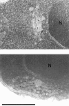
Figure 3
Figure 3
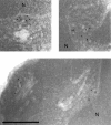
Immunoelectron microscopy of a Golgi marker in P. pastoris. P. pastoris cells of strain PPY12-OH were fixed and cryosectioned, and then incubated with a polyclonal anti–HA antibody followed by protein A-gold to detect Och1p-HA. Gold particles are consistently observed over Golgi stacks. In cells of the parental PPY12 strain, which does not express Och1p-HA, no specific labeling is seen with the anti–HA antibody (not shown). Bar, 0.5 μm. N, nuclei.
Figure 7
Figure 7
Immunoelectron microscopy of a tER marker in P. pastoris. P. pastoris cells of strain PPY12-S13G were fixed and cryosectioned. The localization of Sec13p-GFP was determined by incubating with a polyclonal anti–GFP antibody followed by protein A-gold. Gold particles are consistently observed over tER regions, which are often associated with indentations of the nuclear envelope. In cells of the parental PPY12 strain, which expresses wild-type Sec13p, no specific labeling is seen with the anti–GFP antibody (not shown). Bar, 0.5 μm. N, nuclei.
Figure 2
Figure 2
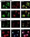
Immunofluorescence staining of Golgi and ER structures. (A) Golgi labeling in S. cerevisiae. Fixed cells of strain DBY1034/pOH were incubated with a monoclonal anti–HA antibody to label Och1p-HA, followed by Oregon green–conjugated anti–mouse antibody (green). The same cells were also incubated with a polyclonal antibody against Sec7p, followed by Texas red–conjugated anti–rabbit antibody (red). The merged image shows very little overlap between the two Golgi markers. (B) Golgi labeling in a sec14 mutant of S. cerevisiae. Strain CTY214 was grown at the permissive temperature of 23°C, and then shifted to the nonpermissive temperature of 37°C for 1 h before fixation. Antibody labeling was as in A. After the temperature shift, Sec7p-containing structures reorganize into large clusters, but the staining pattern of Och1p-HA is not significantly altered. Surprisingly, as illustrated by the cell on the right, budded sec14 cells often partition the Sec7p clusters into the bud. (C) Golgi labeling in P. pastoris. Fixed cells of strain PPY12-OH were incubated with the same primary and secondary antibodies as in A. Och1p-HA staining overlaps strongly with Sec7p staining, as shown in the merged image. As in some strains of S. cerevisiae (O.W. Rossanese, unpublished observations), Och1p-HA often gives a faint staining of the general ER in P. pastoris; for example, the cell on the lower left shows weak nuclear envelope staining, which reveals that two of the three labeled Golgi spots adjoin the nucleus. (D) General ER labeling in P. pastoris. Fixed cells of strain PPY12 were incubated with a polyclonal antibody against S. cerevisiae Pdi1p, followed by Texas red–conjugated anti–rabbit antibody (red). The same cells were also incubated with Hoechst dye to stain DNA (blue). As shown in the merged image, Pdi1p is present in the nuclear envelope and peripheral ER structures. Bar, 2 μm.
Figure 1
Figure 1
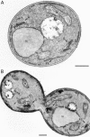
Thin-section electron microscopy of Golgi and ER structures in P. pastoris. (A) A representative P. pastoris cell. Golgi stacks are found adjacent to ER membranes, including both the nuclear envelope and peripheral ER elements. (B) A representative P. pastoris cell after treatment with nocodazole for 2.5 h. The nucleus has failed to divide. However, Golgi morphology is unaffected by the drug treatment. This experiment was performed with strain PPY1. Bars, 0.5 μm. G, Golgi stack; N, nucleus; ER, peripheral ER membranes; M, mitochondrion; V, vacuole.
Figure 4
Figure 4
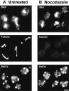
Immunofluorescence analysis of nocodazole-treated P. pastoris cells. (A) DNA, tubulin, and Sec7p distributions in untreated P. pastoris cells. Fixed cells of strain PPY12 were incubated with Hoechst dye to stain DNA. Microtubules were visualized with a monoclonal anti–tubulin antibody followed by Oregon green–conjugated anti–mouse antibody. The Golgi marker Sec7p was visualized as in Fig. 2. (B) DNA, tubulin, and Sec7p distributions in P. pastoris cells treated with nocodazole for 2.5 h. Fixed cells were labeled as in A. As previously described for S. cerevisiae (Jacobs et al., 1988), microtubules are virtually undetectable within 1 h after nocodazole addition (not shown). Bar, 2 μm.
Figure 5
Figure 5
Visualization of GFP-labeled COPII coat proteins in intact S. cerevisiae cells. (A) Sec13p-GFP fluorescence in S. cerevisiae. Cells of strain DBY1034-S13G were fixed and viewed directly. The same cells were imaged in DIC and fluorescence modes. (B) Sec23p-GFP fluorescence in S. cerevisiae strain DBY1034-S23G. The experiment was performed as in A. Bar, 2 μm.
Figure 6
Figure 6
Visualization of Sec13p-GFP in P. pastoris. (A) Sec13p-GFP fluorescence in intact cells. P. pastoris cells of strain PPY12-S13G were fixed and viewed directly. DIC and fluorescence images were collected separately, and then combined to generate the merged image. (B) Immunofluorescence localization of Sec13p-GFP and Sec7p. Fixed P. pastoris cells of strain PPY12-S13G were incubated with a monoclonal anti– GFP antibody followed by Oregon green–conjugated anti–mouse antibody (green). The same cells were also stained for Sec7p (red) as in Fig. 2. The merged image shows that Sec13-GFP– and Sec7p-containing structures are closely apposed, but distinct. Bar, 2 μm.
Figure 8
Figure 8
Immunofluorescence localization of tagged Sec12p in the two yeasts. (A) Colocalization of Sec12p-myc with Pdi1p in S. cerevisiae. Fixed cells of strain DBY1034-S12m were incubated with a monoclonal anti– myc antibody followed by Oregon green–conjugated anti–mouse antibody to visualize Sec12p-myc (green). The same cells were also stained for Pdi1p (red) as in Fig. 2. As shown in the merged image, the staining patterns for the two proteins largely overlap. (B) Localization of Sec12p-myc in P. pastoris. Fixed cells of strain PPY12-S12m were stained as in A to visualize Sec12p-myc (green); Sec7p (red) was visualized as in Fig. 2. The merged image shows that the two staining patterns are closely apposed, but distinct. Bar, 2 μm.
Figure 9
Figure 9
Summary and interpretation of the experimental data. P. pastoris contains discrete tER sites that produce coherent Golgi stacks. In S. cerevisiae, COPII vesicles bud throughout the ER network, resulting in a dispersed Golgi. The shaded portions of the ER are the regions that can function as tER. See text for details.

Similar articles

Cited by

References

    1. Acharya U, Mallabiabarrena A, Acharya JK, Malhotra V. Signaling via mitogen-activated protein kinase (MEK1) is required for Golgi fragmentation during mitosis. Cell. 1998;92:183–192. - PubMed
    1. Antebi A, Fink GR. The yeast Ca2+-ATPase homologue, PMR1, is required for normal Golgi function and localizes in a novel Golgi-like distribution. Mol Biol Cell. 1992;3:633–654. - PMC - PubMed
    1. Ayscough K, Hajibagheri NMA, Watson R, Warren G. Stacking of Golgi cisternae in Schizosaccharomyces pomberequires intact microtubules. J Cell Sci. 1993;106:1227–1237. - PubMed
    1. Bannykh SI, Balch WE. Membrane dynamics at the endoplasmic reticulum-Golgi interface. J Cell Biol. 1997;138:1–4. - PMC - PubMed
    1. Barlowe C, Schekman R. SEC12encodes a guanine-nucleotide-exchange factor essential for transport vesicle budding from the ER. Nature. 1993;365:347–349. - PubMed

Publication types

MeSH terms