

Human granulocytic ehrlichiosis agent and Ehrlichia chaffeensis reside in different cytoplasmic compartments in HL-60 cells
- PMID: 10024584
- PMCID: PMC96470
- DOI: 10.1128/IAI.67.3.1368-1378.1999
Human granulocytic ehrlichiosis agent and Ehrlichia chaffeensis reside in different cytoplasmic compartments in HL-60 cells
Abstract
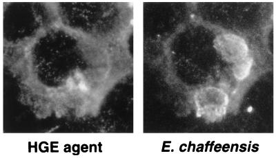
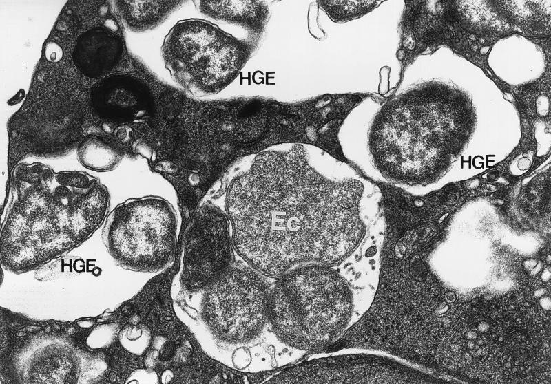
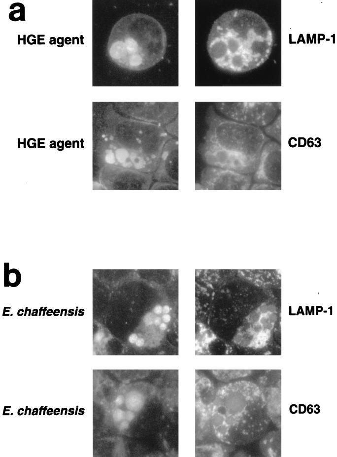
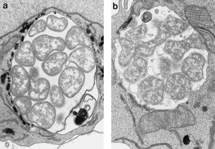
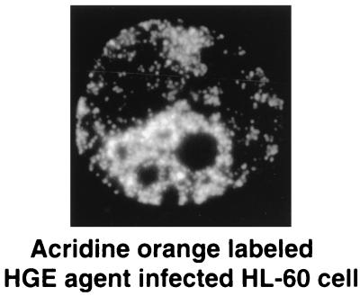
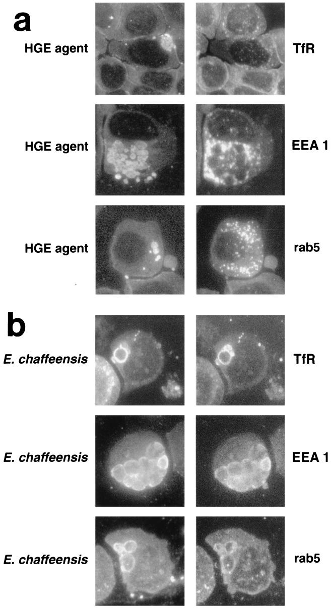
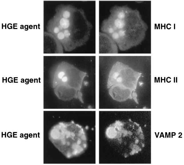
The human granulocytic ehrlichiosis (HGE) agent resides and multiplies exclusively in cytoplasmic vacuoles of granulocytes. Double immunofluorescence labeling was used to characterize the nature of the HGE agent replicative inclusions and to compare them with inclusions containing the human monocytic ehrlichia, Ehrlichia chaffeensis, in HL-60 cells. Although both Ehrlichia spp. can coinfect HL-60 cells, they resided in separate inclusions. Inclusions of both Ehrlichia spp. were not labeled with either anti-lysosome-associated membrane protein 1 or anti-CD63. Accumulation of myeloperoxidase-positive granules were seen around HGE agent inclusions but not around E. chaffeensis inclusions. 3-(2, 4-Dinitroanilino)-3'-amino-N-methyldipropylamine and acridine orange were not localized to either inclusion type. Vacuolar-type H+-ATPase was not colocalized with HGE agent inclusions but was weakly colocalized with E. chaffeensis inclusions. E. chaffeensis inclusions were labeled with the transferrin receptor, early endosomal antigen 1, and rab5, but HGE agent inclusions were not. Some HGE agent and E. chaffeensis inclusions colocalized with major histocompatibility complex class I and II antigens. These two inclusions were not labeled for annexins I, II, IV, and VI; alpha-adaptin; clathrin heavy chain; or beta-coatomer protein. Vesicle-associated membrane protein 2 colocalized to both inclusions. The cation-independent mannose 6-phosphate receptor was not colocalized with either inclusion type. Endogenously synthesized sphingomyelin, from C6-NBD-ceramide, was not incorporated into either inclusion type. Brefeldin A did not affect the growth of either Ehrlichia sp. in HL-60 cells. These results suggest that the HGE agent resides in inclusions which are neither early nor late endosomes and does not fuse with lysosomes or Golgi-derived vesicles, while E. chaffeensis resides in an early endosomal compartment which accumulates the transferrin receptor.
Figures
Similar articles
-
Ehrlichia chaffeensis and E. sennetsu, but not the human granulocytic ehrlichiosis agent, colocalize with transferrin receptor and up-regulate transferrin receptor mRNA by activating iron-responsive protein 1.Infect Immun. 1999 May;67(5):2258-65. doi: 10.1128/IAI.67.5.2258-2265.1999. Infect Immun. 1999. PMID: 10225882 Free PMC article.
-
The agent of Human Granulocytic Ehrlichiosis resides in an endosomal compartment.J Clin Invest. 1998 May 1;101(9):1932-41. doi: 10.1172/JCI1544. J Clin Invest. 1998. PMID: 9576758 Free PMC article.
-
Western blot analysis of sera reactive to human monocytic ehrlichiosis and human granulocytic ehrlichiosis agents.J Clin Microbiol. 2001 Nov;39(11):3982-6. doi: 10.1128/JCM.39.11.3982-3986.2001. J Clin Microbiol. 2001. PMID: 11682518 Free PMC article.
-
Subversion of RAB5-regulated autophagy by the intracellular pathogen Ehrlichia chaffeensis.Small GTPases. 2019 Sep;10(5):343-349. doi: 10.1080/21541248.2017.1332506. Epub 2017 Jul 5. Small GTPases. 2019. PMID: 28650718 Free PMC article. Review.
-
Tick-borne ehrlichiosis infection in human beings.J Vector Borne Dis. 2008 Dec;45(4):273-80. J Vector Borne Dis. 2008. PMID: 19248653 Review.
Cited by
-
Early transcriptional response of human neutrophils to Anaplasma phagocytophilum infection.Infect Immun. 2005 Dec;73(12):8089-99. doi: 10.1128/IAI.73.12.8089-8099.2005. Infect Immun. 2005. PMID: 16299303 Free PMC article.
-
The Anaplasma phagocytophilum-occupied vacuole selectively recruits Rab-GTPases that are predominantly associated with recycling endosomes.Cell Microbiol. 2010 Sep 1;12(9):1292-307. doi: 10.1111/j.1462-5822.2010.01468.x. Epub 2010 Mar 25. Cell Microbiol. 2010. PMID: 20345488 Free PMC article.
-
Ehrlichia chaffeensis: a prototypical emerging pathogen.Clin Microbiol Rev. 2003 Jan;16(1):37-64. doi: 10.1128/CMR.16.1.37-64.2003. Clin Microbiol Rev. 2003. PMID: 12525424 Free PMC article. Review.
-
Host membrane lipids are trafficked to membranes of intravacuolar bacterium Ehrlichia chaffeensis.Proc Natl Acad Sci U S A. 2020 Apr 7;117(14):8032-8043. doi: 10.1073/pnas.1921619117. Epub 2020 Mar 19. Proc Natl Acad Sci U S A. 2020. PMID: 32193339 Free PMC article.
-
Ehrlichia type IV secretion effector ECH0825 is translocated to mitochondria and curbs ROS and apoptosis by upregulating host MnSOD.Cell Microbiol. 2012 Jul;14(7):1037-50. doi: 10.1111/j.1462-5822.2012.01775.x. Epub 2012 Mar 12. Cell Microbiol. 2012. PMID: 22348527 Free PMC article.
References
-
- Amigorena S, Drake J R, Webster P, Mellman I. Transient accumulation of new class II MHC in a novel endocytic compartment in B lymphocytes. Nature. 1994;369:113–120. - PubMed
-
- Bakken J S, Krueth J, Tilden R L, Dumler J S, Kristansen R F. Serologic evidence of human granulocytic ehrlichiosis in Norway. Eur J Clin Microbiol Infect Dis. 1997;15:829–832. - PubMed
-
- Bakken J S, Dumler J S, Chen S M, Eckman M R, Van Etta L L, Walker D H. Human granulocytic ehrlichiosis in the upper midwest United States. a new species emerging? JAMA. 1994;272:212–218. - PubMed
Publication types
MeSH terms
Substances
Grants and funding
LinkOut - more resources
Full Text Sources
Other Literature Sources
Research Materials
Miscellaneous
