. 1999 Mar;19(3):1784-99.
doi: 10.1128/MCB.19.3.1784.

The Gab1 PH domain is required for localization of Gab1 at sites of cell-cell contact and epithelial morphogenesis downstream from the met receptor tyrosine kinase

Affiliations

The Gab1 PH domain is required for localization of Gab1 at sites of cell-cell contact and epithelial morphogenesis downstream from the met receptor tyrosine kinase

C R Maroun et al. Mol Cell Biol. 1999 Mar.

Abstract

Stimulation of the hepatocyte growth factor (HGF) receptor tyrosine kinase, Met, induces mitogenesis, motility, invasion, and branching tubulogenesis of epithelial and endothelial cell lines in culture. We have previously shown that Gab1 is the major phosphorylated protein following stimulation of the Met receptor in epithelial cells that undergo a morphogenic program in response to HGF. Gab1 is a member of the family of IRS-1-like multisubstrate docking proteins and, like IRS-1, contains an amino-terminal pleckstrin homology domain, in addition to multiple tyrosine residues that are potential binding sites for proteins that contain SH2 or PTB domains. Following stimulation of epithelial cells with HGF, Gab1 associates with phosphatidylinositol 3-kinase and the tyrosine phosphatase SHP2. Met receptor mutants that are impaired in their association with Gab1 fail to induce branching tubulogenesis. Overexpression of Gab1 rescues the Met-dependent tubulogenic response in these cell lines. The ability of Gab1 to promote tubulogenesis is dependent on its pleckstrin homology domain. Whereas the wild-type Gab1 protein is localized to areas of cell-cell contact, a Gab1 protein lacking the pleckstrin homology domain is localized predominantly in the cytoplasm. Localization of Gab1 to areas of cell-cell contact is inhibited by LY294002, demonstrating that phosphatidylinositol 3-kinase activity is required. These data show that Gab1 is an important mediator of branching tubulogenesis downstream from the Met receptor and identify phosphatidylinositol 3-kinase and the Gab1 pleckstrin homology domain as crucial for subcellular localization of Gab1 and biological responses.


Figures
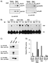
FIG. 1
FIG. 1
Association of Gab1 with cellular substrates is dependent on Met-mediated tyrosine phosphorylation of Gab1. (A) Stable MDCK cell lines expressing HA-Gab1 or vector control were serum starved in 0.02% FBS for 24 h and subsequently stimulated with HGF (100 U/ml) for 15 min. Cell lysates were subjected to immunoprecipitation (ip) with anti-HA followed by blotting with anti-PY. Products of parallel precipitations were blotted with anti-p85, anti-SHP2, or anti-HA. (B) 293T cells were transiently transfected with plasmids encoding epitope-tagged wild-type Gab1, with or without a Met chimera composed of the extracellular domain from CSF receptor fused to the Met transmembrane and intracellular domains. Cells were serum starved 24 h prior to harvest. Lysates were immunoprecipitated with anti-Met, anti-PY (PY20), or anti-HA. PI3K activity was determined as described in Materials and Methods. Phosphatidylinositol phosphates were separated by thin-layer chromatography on silica gel plates and revealed by autoradiography. The position of phosphatidylinositol 3-phosphate is shown (PIP), as is the origin (Ori). (C) Lysates from 293T cells transfected as described in panel B were subjected to immunoprecipitation with anti-HA or anti-Met and blotted with anti-HA, anti-Met, or anti-PY as indicated. (D) The incorporated radioactivity in the phosphatidylinositol 3-phosphate was quantitated with a Fujix BAS 1000 image analyzer, and the results are plotted on the bar graph as relative PI3K activity.
FIG. 2
FIG. 2
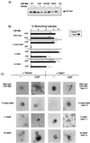
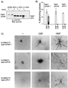
Gab1 rescues branching tubulogenesis in MDCK cells expressing mutant CSF-Met receptors that fail to bind Grb2. MDCK cell lines expressing either wild-type (WT) CSF-Met or the CSF-Met mutants Y1349/1356F, Y1356F, or N1358H were stably transfected with vector or wild-type HA-tagged Gab1. (A) Lysates from two representative lines of each experimental group were subjected to immunoprecipitation with anti-HA, and proteins resolved by SDS-PAGE were transferred to a nitrocellulose membrane and immunoblotted with anti-HA. (B) Quantitation of the tubulogenic response following stimulation with HGF and CSF in cell lines expressing either CSF-Met or mutants thereof alone (open bars) or together with Gab1 (solid bars) was undertaken as described in Materials and Methods. The responses are plotted as the percentage of cysts that have undergone branching tubulogenesis. The values were derived from three independent experiments done in duplicate. None of the cysts formed tubules in the absence of stimulation. (C) Cell lines were grown in collagen for 5 days, during which they formed cysts. rhCSF-1 (5 U/ml) was added, and 14 days later branching tubules were visualized at a magnification of ×10 and photographs taken with Kodak TMY400 film. A representative cell line for each group is shown.
FIG. 3
FIG. 3
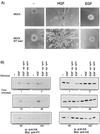
Overexpression of Gab1 does not mediate branching tubulogenesis downstream from EGF. (A) Control MDCK cells or MDCK cells overexpressing Gab1 were plated in a collagen matrix for 5 days. HGF (5 U/ml) or EGF (20 ng/ml) was added to the cultures, and photographs were taken 14 days later at a magnification of ×10. (B) MDCK cells overexpressing Gab1 were serum starved for 24 h prior to stimulation with either 100 U of HGF per ml or 100 400 ng of EGF per ml for the indicated time. Gab1 was immunoprecipitated (ip) with anti-HA. Proteins were resolved by SDS-PAGE, transferred to nitrocellulose, and probed with anti-PY or anti-HA. WT, wild type.
FIG. 4
FIG. 4
The PH domain of Gab1 is essential for Met-mediated branching tubulogenesis. (A) MDCK cells expressing the N1358H CSF-Met mutant protein [N1358H(17)] were transfected with plasmids encoding for Gab1 ΔPI3K (ΔPI3K, clones 6 and 1). Two independent N1358H CSF-Met mutant expressing lines [N1358H(17) and N1358H(1)] were transfected with Gab1 ΔPH-encoding plasmids [ΔPH(1) clones 8 and 6, ΔPH(17) clones 1 and 2]. Lysates were subjected to immunoprecipitation (ip) and blotting with anti-HA. (B) The tubulogenic response was quantitated in cells expressing Gab1ΔPI3K and ΔPH, and results from representative clones are plotted as the percentage of cysts that have formed branching tubules in response to HGF or CSF-1. Solid bars represent results from cysts that have undergone a complete tubulogenic response; i.e., the tubule length is at least five times the size of the width; the hatched bar represents a partial response. (C) Cells expressing Gab1 mutants were plated in a collagen matrix and allowed to form cysts for 5 days. rh-CSF or HGF (5 U/ml) was added, and 14 days later branching tubules were visualized by light microscopy at a magnification of ×10. Representative lines are shown.
FIG. 5
FIG. 5
Gab1 mutant proteins associate with Met and are phosphorylated following Met activation. (A) 293T cells were transiently transfected with wild-type (WT) CSF-Met or the N1358H CSF-Met mutant, together with wild-type Gab1, Gab1ΔPI3K, or Gab1ΔPH. Lysates were subjected to immunoprecipitation (ip) with anti-HA, and proteins were resolved by SDS-PAGE (8% polyacrylamide), transferred to a nitrocellulose membrane, and blotted with anti-Met. The blot was stripped and reprobed with anti-HA. (B) MDCK cells expressing N1358H CSF-Met and either Gab1ΔPI3K or Gab1ΔPH were stimulated with 2 μg of CSF-1 per ml for 15 min at 37°C. Lysates were subjected to immunoprecipitation with anti-HA and blotting with anti-PY. The blots were stripped and reprobed with anti-p85 and then with anti-SHP2. A parallel blot was probed with anti-HA. (C) MDCK cells expressing Gab1ΔPH protein were stimulated for the indicated time, and lysates were subjected to immunoprecipitation with anti-HA. Proteins were resolved by SDS-PAGE and, following transfer to nitrocellulose, were blotted with either anti-PY or anti-HA as indicated.
FIG. 6
FIG. 6
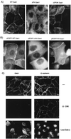
The PH domain of Gab1 is required for the localization of Gab1 to sites of cell-cell contact. (A) MDCK cells stably transfected with wild-type Gab1, Gab1ΔPH, or Gab1ΔPI3K were grown on glass coverslips in DMEM containing 10% FBS. The cells were fixed in 2% paraformaldehyde and then labeled with anti-HA followed by CY3-conjugated anti-mouse antiserum. (B) Plasmids encoding pEGFP-Gab1, pEGFP-Gab1ΔPH, or pEGFP-Gab1PH were microinjected into nuclei of MDCK cells grown in DMEM plus 10% FBS. At 2 h following the microinjections, cells were visualized. (C) MDCK cells overexpressing Gab1 were either fixed in 2% paraformaldehyde (−), treated for 10 min in CSK buffer prior to fixation (+CSK), or treated for 2 h in 5 mM EGTA-containing media (Low [Ca2+]). The cells were subsequently labeled with anti-HA or anti-E-cadherin as indicated on the figure. Photographs were taken at a magnification of ×100.
FIG. 7
FIG. 7
The localization of Gab1 is serum and PI3K dependent. (A) MDCK cells (104) stably expressing HA-Gab1 were grown in DMEM plus 10% FBS for 72 or 18 h as indicated. For serum starvation experiments, cells were first grown for 48 h in 10% FBS and then transferred for 24 h to medium containing 0.02% FBS. The cells were fixed in 2% paraformaldehyde, and the localization of Gab1 was determined following indirect immunofluorescence labeling with anti-HA followed by CY3-conjugated anti-mouse antiserum. (B) HA-Gab1-expressing MDCK cells were treated for 2 h at 37°C either with 50 μM LY294002 or with 2 μM U73122. The cells were subsequently fixed in 2% paraformaldehyde and labeled with anti-HA followed by CY3 anti-mouse antiserum. (C) MDCK cells were serum starved for 24 h prior to microinjection with plasmids encoding the p85 and p110 subunits of PI3K, together with pEGFP-Gab1 or control pEGFP plasmid. Photographs were taken 4 h following the injections. DMSO, dimethyl sulfoxide.
FIG. 7
FIG. 7
The localization of Gab1 is serum and PI3K dependent. (A) MDCK cells (104) stably expressing HA-Gab1 were grown in DMEM plus 10% FBS for 72 or 18 h as indicated. For serum starvation experiments, cells were first grown for 48 h in 10% FBS and then transferred for 24 h to medium containing 0.02% FBS. The cells were fixed in 2% paraformaldehyde, and the localization of Gab1 was determined following indirect immunofluorescence labeling with anti-HA followed by CY3-conjugated anti-mouse antiserum. (B) HA-Gab1-expressing MDCK cells were treated for 2 h at 37°C either with 50 μM LY294002 or with 2 μM U73122. The cells were subsequently fixed in 2% paraformaldehyde and labeled with anti-HA followed by CY3 anti-mouse antiserum. (C) MDCK cells were serum starved for 24 h prior to microinjection with plasmids encoding the p85 and p110 subunits of PI3K, together with pEGFP-Gab1 or control pEGFP plasmid. Photographs were taken 4 h following the injections. DMSO, dimethyl sulfoxide.
FIG. 8
FIG. 8
Gab1 is recruited to the membrane folowing Met activation. MDCK cells (104) expressing wild-type HA-Gab1 or HA-Gab1ΔPH mutant proteins were grown overnight in medium containing 10% FBS. They were then stimulated with 50 U of HGF per ml at 37°C for 15 min. Following fixation, the cells were labeled with anti-HA and photographs were taken at a magnification of ×100.
FIG. 9
FIG. 9
Gab1 is recruited to the cell surface by two distinct mechanisms. (A) One mechanism depends on the Gab1 PH domain and serum and requires PI3K activity. (B) The second mechanism is based on Met activation and is independent of the Gab1 PH domain (see Discussion for details). (C) Once recruited to and phosphorylated by Met, Met-associated or Gab1-associated PI3K activity is predicted to stabilize Gab1 association with the membrane.

Similar articles

Cited by

References

    1. Auger K R, Serunian L A, Soltoff S P, Libby P, Cantley L C. PDGF-dependent tyrosine phosphorylation stimulates production of novel polyphosphoinositides in intact cells. Cell. 1989;57:167–175. - PubMed
    1. Backer J M, Myers M J, Jr, Shoelson S E, Chin D J, Sun X J, Miralpeix M, Hu P, Margolis B, Skolnik E Y, Schlessinger J. Phosphatidylinositol 3′-kinase is activated by association with IRS-1 during insulin stimulation. EMBO J. 1992;11:3469–3479. - PMC - PubMed
    1. Bardelli A, Longati P, Gramaglia D, Stella M C, Comoglio P M. Gab1 coupling to the HGF/Met receptor multifunctional docking site requires binding of Grb2 and correlates with the transforming potential. Oncogene. 1997;15:3103–3111. - PubMed
    1. Bellusci S, Moens G, Gaudino G, Comoglio P, Nakamura T, Thiery J P, Jouanneau J. Creation of an hepatocyte growth factor/scatter factor autocrine loop in carcinoma cells induces invasive properties associated with increased tumorigenicity. Oncogene. 1994;9:1091–1099. - PubMed
    1. Burks D J, Pons S, Towery H, Smith-Hall J, Myers M G, Jr, Yenush L, White M F. Heterologous pleckstrin homology domains do not couple IRS-1 to the insulin receptor. J Biol Chem. 1997;272:27716–27721. - PubMed

Publication types

MeSH terms

LinkOut - more resources