

Bone resorption induced by parathyroid hormone is strikingly diminished in collagenase-resistant mutant mice
- PMID: 10021460
- PMCID: PMC408105
- DOI: 10.1172/JCI5481
Bone resorption induced by parathyroid hormone is strikingly diminished in collagenase-resistant mutant mice
Abstract
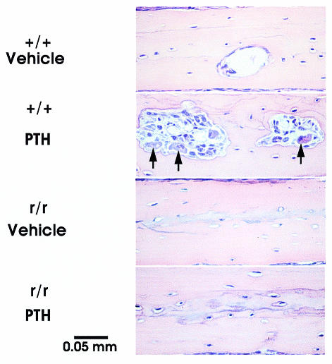
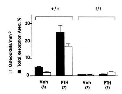
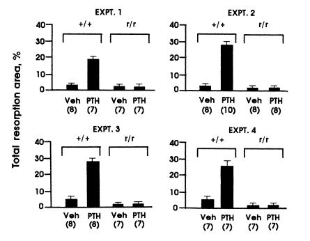
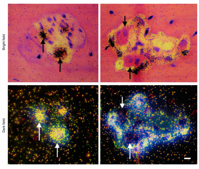
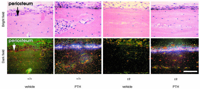
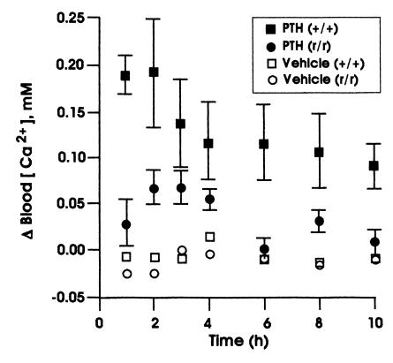
Parathyroid hormone (PTH) stimulates bone resorption by acting directly on osteoblasts/stromal cells and then indirectly to increase differentiation and function of osteoclasts. PTH acting on osteoblasts/stromal cells increases collagenase gene transcription and synthesis. To assess the role of collagenase in the bone resorptive actions of PTH, we used mice homozygous (r/r) for a targeted mutation (r) in Col1a1 that are resistant to collagenase cleavage of type I collagen. Human PTH(1-34) was injected subcutaneously over the hemicalvariae in wild-type (+/+) or r/r mice four times daily for three days. Osteoclast numbers, the size of the bone marrow spaces and periosteal proliferation were increased in calvariae from PTH-treated +/+ mice, whereas in r/r mice, PTH-induced bone resorption responses were minimal. The r/r mice were not resistant to other skeletal effects of PTH because abundant interstitial collagenase mRNA was detected in the calvarial periosteum of PTH-treated, but not vehicle-treated, r/r and +/+ mice. Calcemic responses, 0.5-10 hours after intraperitoneal injection of PTH, were blunted in r/r mice versus +/+ mice. Thus, collagenase cleavage of type I collagen is necessary for PTH induction of osteoclastic bone resorption.
Figures

Similar articles
-
Collagenase cleavage of type I collagen is essential for both basal and parathyroid hormone (PTH)/PTH-related peptide receptor-induced osteoclast activation and has differential effects on discrete bone compartments.Endocrinology. 2003 Sep;144(9):4106-16. doi: 10.1210/en.2003-0254. Endocrinology. 2003. PMID: 12933685
-
Human PTH-(7-84) inhibits bone resorption in vitro via actions independent of the type 1 PTH/PTHrP receptor.Endocrinology. 2002 Jan;143(1):171-6. doi: 10.1210/endo.143.1.8575. Endocrinology. 2002. PMID: 11751606
-
Osteocyte and osteoblast apoptosis and excessive bone deposition accompany failure of collagenase cleavage of collagen.J Clin Invest. 2000 Oct;106(8):941-9. doi: 10.1172/JCI10158. J Clin Invest. 2000. PMID: 11032854 Free PMC article.
-
Relative roles of collagenase and lysosomal cysteine-proteinases in bone resorption.Matrix Suppl. 1992;1:383-8. Matrix Suppl. 1992. PMID: 1480065 Review.
-
The regulation and regulatory role of collagenase in bone.Crit Rev Eukaryot Gene Expr. 1996;6(1):15-27. doi: 10.1615/critreveukargeneexpr.v6.i1.20. Crit Rev Eukaryot Gene Expr. 1996. PMID: 8882305 Review.
Cited by
-
Stemming bone loss by suppressing apoptosis.J Clin Invest. 1999 Aug;104(4):371-3. doi: 10.1172/JCI7991. J Clin Invest. 1999. PMID: 10449428 Free PMC article. Review. No abstract available.
-
Critical roles for collagenase-3 (Mmp13) in development of growth plate cartilage and in endochondral ossification.Proc Natl Acad Sci U S A. 2004 Dec 7;101(49):17192-7. doi: 10.1073/pnas.0407788101. Epub 2004 Nov 24. Proc Natl Acad Sci U S A. 2004. PMID: 15563592 Free PMC article.
-
Parathyroid hormone regulates the distribution and osteoclastogenic potential of hematopoietic progenitors in the bone marrow.J Bone Miner Res. 2011 Jun;26(6):1207-16. doi: 10.1002/jbmr.324. J Bone Miner Res. 2011. PMID: 21611963 Free PMC article.
-
A new mouse mutant with cleavage-resistant versican and isoform-specific versican mutants demonstrate that proteolysis at the Glu441-Ala442 peptide bond in the V1 isoform is essential for interdigital web regression.Matrix Biol Plus. 2021 May 14;10:100064. doi: 10.1016/j.mbplus.2021.100064. eCollection 2021 Jun. Matrix Biol Plus. 2021. PMID: 34195596 Free PMC article.
-
The role of collagen in extralobar pulmonary artery stiffening in response to hypoxia-induced pulmonary hypertension.Am J Physiol Heart Circ Physiol. 2010 Dec;299(6):H1823-31. doi: 10.1152/ajpheart.00493.2009. Epub 2010 Sep 17. Am J Physiol Heart Circ Physiol. 2010. PMID: 20852040 Free PMC article.
References
-
- Potts, J.T., Jr., and Jüppner, H. 1997. Parathyroid hormone and parathyroid hormone-related peptide in calcium homeostasis, bone metabolism, and bone development: the proteins, their genes, and receptors. In Metabolic bone disease and clinically related disorders. L.V. Avioli and S.M. Krane, editors. Academic Press. San Diego, CA. 51–94.
-
- Suda T, Takahashi N, Martin TJ. Modulation of osteoclast differentiation. Endocr Rev. 1992;13:66–80. - PubMed
-
- Walker DG, Lapière CM, Gross J. A collagenolytic factor in rat bone promoted by parathyroid extract. Biochem Biophys Res Commun. 1964;15:397–402. - PubMed
Publication types
MeSH terms
Substances
Grants and funding
LinkOut - more resources
Full Text Sources
Other Literature Sources
Molecular Biology Databases
Miscellaneous
