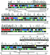
The complete nucleotide sequence of the human immunoglobulin heavy chain variable region locus
- PMID: 9841928
- PMCID: PMC2212390
- DOI: 10.1084/jem.188.11.2151
The complete nucleotide sequence of the human immunoglobulin heavy chain variable region locus
Abstract
The complete nucleotide sequence of the 957-kb DNA of the human immunoglobulin heavy chain variable (VH) region locus was determined and 43 novel VH segments were identified. The region contains 123 VH segments classifiable into seven different families, of which 79 are pseudogenes. Of the 44 VH segments with an open reading frame, 39 are expressed as heavy chain proteins and 1 as mRNA, while the remaining 4 are not found in immunoglobulin cDNAs. Combinatorial diversity of VH region was calculated to be approximately 6,000. Conservation of the promoter and recombination signal sequences was observed to be higher in functional VH segments than in pseudogenes. Phylogenetic analysis of 114 VH segments clearly showed clustering of the VH segments of each family. However, an independent branch in the tree contained a single VH, V4-44.1P, sharing similar levels of homology to human VH families and to those of other vertebrates. Comparison between different copies of homologous units that appear repeatedly across the locus clearly demonstrates that dynamic DNA reorganization of the locus took place at least eight times between 133 and 10 million years ago. One nonimmunoglobulin gene of unknown function was identified in the intergenic region.
Figures


Comment in
-
The super-information age of immunoglobulin genetics.J Exp Med. 1998 Dec 7;188(11):1973-5. doi: 10.1084/jem.188.11.1973. J Exp Med. 1998. PMID: 9841911 Free PMC article. No abstract available.
Similar articles
-
A map of the human immunoglobulin VH locus completed by analysis of the telomeric region of chromosome 14q.Nat Genet. 1994 Jun;7(2):162-8. doi: 10.1038/ng0694-162. Nat Genet. 1994. PMID: 7920635
-
Comparison and evolution of human immunoglobulin VH segments located in the 3' 0.8-megabase region. Evidence for unidirectional transfer of segmental gene sequences.J Biol Chem. 1994 Jan 28;269(4):2619-26. J Biol Chem. 1994. PMID: 8300591
-
Organization of variable region segments of the human immunoglobulin heavy chain: duplication of the D5 cluster within the locus and interchromosomal translocation of variable region segments.EMBO J. 1990 Aug;9(8):2501-6. doi: 10.1002/j.1460-2075.1990.tb07429.x. EMBO J. 1990. PMID: 2114977 Free PMC article.
-
[Structure and function of the immunoglobulin Vh gene in man].Allerg Immunol (Leipz). 1990;36(4):225-32. Allerg Immunol (Leipz). 1990. PMID: 2129098 Review. German.
-
Genetic diversity of the human immunoglobulin heavy chain VH region.Immunol Rev. 2002 Dec;190:53-68. doi: 10.1034/j.1600-065x.2002.19005.x. Immunol Rev. 2002. PMID: 12493006 Review.
Cited by
-
Addressing Technical Pitfalls in Pursuit of Molecular Factors That Mediate Immunoglobulin Gene Regulation.J Immunol. 2024 Sep 1;213(5):651-662. doi: 10.4049/jimmunol.2400131. J Immunol. 2024. PMID: 39007649
-
AIRR-C IG Reference Sets: curated sets of immunoglobulin heavy and light chain germline genes.Front Immunol. 2024 Feb 9;14:1330153. doi: 10.3389/fimmu.2023.1330153. eCollection 2023. Front Immunol. 2024. PMID: 38406579 Free PMC article.
-
IGHV allele similarity clustering improves genotype inference from adaptive immune receptor repertoire sequencing data.Nucleic Acids Res. 2023 Sep 8;51(16):e86. doi: 10.1093/nar/gkad603. Nucleic Acids Res. 2023. PMID: 37548401 Free PMC article.
-
Adaptive immune receptor genotyping using the corecount program.Front Immunol. 2023 Apr 11;14:1125884. doi: 10.3389/fimmu.2023.1125884. eCollection 2023. Front Immunol. 2023. PMID: 37114042 Free PMC article.
-
Frequent use of IGHV3-30-3 in SARS-CoV-2 neutralizing antibody responses.Front Virol. 2023 Mar 1;3:1128253. doi: 10.3389/fviro.2023.1128253. Front Virol. 2023. PMID: 37041983 Free PMC article.
References
-
- Tonegawa S. Somatic generation of antibody diversity. Nature. 1983;302:505–581. - PubMed
-
- Honjo T, Habu S. Origin of immune diversity: genetic variation and selection. Annu Rev Biochem. 1985;54:803–830. - PubMed
-
- Davis MM. T cell receptor gene diversity and selection. Annu Rev Biochem. 1990;59:475–496. - PubMed
Publication types
MeSH terms
Substances
Associated data
- Actions
- Actions
- Actions
- Actions
- Actions
LinkOut - more resources
Full Text Sources
Other Literature Sources

