Abstract
Background:
Dentinal hypersensitivity (DH) is a common dental problem and is treated non-invasively using topical application of various desensitizing agents. When there is loss of tooth structure or tooth preparation is to be followed by a bonded restoration, it requires application of dentin bonding agent. However, the effect of desensitizers on bond strength is still controversial.
Aim and Objective:
To evaluate the influence of different desensitizing agents on the bond strength of the dentin bonding agents.
Methods and Materials:
PICO strategy was used to formulate the research question. In-vitro studies conducted on human teeth to evaluate the bond strength of dentin bonding agent following the application of desensitizing agent were included. Electronic databases PubMed and Cochrane and google scholar were searched using search terms alone or in combination from the year 2010 till 2020. Search was conducted using the key words and MeSH terms (hypersensitivity, bond strength, dental adhesives, dentin bonding agents). The title and abstract were read to verify the inclusion and exclusion criteria and if further any details required, full article was accessed to check the criteria and then included or excluded. Data extraction was done using a customized data extraction form. The risk of bias was evaluated using modified Cochrane Collaboration Quality Assessment tool.
Results:
Total no of studies identified in the search were 146, after scrutiny 23 articles were eligible to be included in this study. Out of 23 articles, 17 articles were of medium bias and 6 articles were of high risk after risk of bias assessment.
Conclusion:
According to the various articles included in this study, GLUMA and 8.0%Arginine and CaCO3 when used as desensitizing agents along with different bonding agents, were found to be highly compatible without interfering with the bond strength of the dentin adhesives.
Keywords: Bonding agent, Bond strength, desensitiser, hypersensitivity
INTRODUCTION
Dentinal hypersensitivity (DH) is a common dental problem that occurs as a result of caries, noncarious lesions, or following freshly cut dentin during tooth preparations. The incidence of occurrence of DH varies from 10% to 30% in various populations with the age group varying from 20 to 50 years. The commonly accepted theory is the hydrodynamic theory which states that any stimuli that cause fluid movement within the dentinal tubules, in turn, can stimulate the nerve fiber resulting in a painful response.[1]
Hypersensitivity is treated noninvasively using topical application of various desensitizing agents. The most commonly used agents are potassium nitrate, fluorides, oxalates, GLUMA-containing agents, bonding agents, calcium phosphate/calcium carbonate, bioactive glass, strontium acetate, and casein phosphopeptide-amorphous calcium phosphate (CPP-ACP). Different lasers are also used in treating DH. These act by plugging the dentinal tubules directly or after the chemical reaction. According to Pashley et al., dentinal permeability and sensitivity are reduced when the dentinal tubules are occluded.[2] However, when there is a loss of tooth structure or tooth preparation is to be followed by a bonded restoration, it requires the application of dentin bonding agents (DBA).
In superficial dentin, which contains fewer tubules, the permeation of resin in the DBA is into intertubular dentin, which will be responsible for most of the bond strength. The dentinal tubules are present in more numbers in deep dentin and the bond strength is increased because of the intratubular resin permeability.[3] However, the effect of desensitizers on bond strength is still controversial. Some studies have demonstrated negative effects[4,5,6] and some either positive or no effect.[4,5,6,7,8]
STRUCTURED QUESTION
Does the application of dentin desensitizers influence the bond strength of DBAs?
PICO ANALYSIS
Population is extracted teeth. Intervention is the application of dentin desensitizing agents, Comparison is without application of desensitizing agent (studies with control), Outcome increases or decrease in bond strength of DBA. Study design is In-vitro study.
METHODOLOGY
Protocol and registration
PRISMA 2020 guidelines were followed in this study and the study protocol was registered with PROSPERO (Registration no-CRD42020218931).
SEARCH STRATEGY
The electronic databases PubMed, Cochrane, and Google Scholar websites were searched. The keywords used were desensitizer OR desensitizers OR dentin desensitizer OR dentin desensitizers OR desensitizing product OR desensitizing products OR desensitizing agent OR desensitizing agents OR desensitizing paste OR desensitizing pastes AND dentin bond OR dentin bond strength OR bond strength for studies in the English language from the year 2010–2020.
STUDY SELECTION
Inclusion criteria were in-vitro studies conducted on the coronal dentin surface of human extracted teeth. Exclusion criteria were studies conducted on bovine teeth, on enamel or radicular dentine of human teeth, and reviews.
DATA EXTRACTION PROCESS
Both the reviewers screened the abstract of the selected articles based on the inclusion and exclusion criteria. Both the reviewers read the articles and extracted the data such as author, year, DSA used, DBA used, type of bond strength and type of fracture that occurred and the effect of DSA on the bond strength. If there is any disagreement, the common consensus was arrived at after discussion.
QUALITY ASSESSMENT
The assessment was carried out using the criteria such as teeth free of caries, similar size sample dimension, sample size calculation, teeth randomization, blinding of the evaluator, storage, thermocycling, thermomechanical aging, fracture investigation following bond strength test. If these criteria were present it was scored “Yes” and not present was marked “No.” Both the reviewers carried out the quality assessment of the studies independently. Overall risk assessment was done with the Cochrane Risk of Bias tool using software (REVMAN 5.4.1).
RESULTS
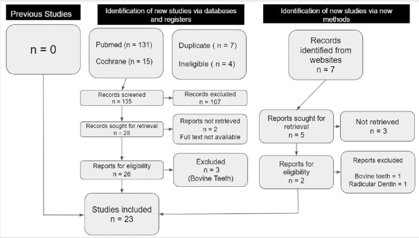
Total articles identified through database search were 146, after removal of duplicates and ineligible articles, records screened were 135, after applying inclusion and exclusion criteria 28 records were retrieved, two full texts could not be retrieved.[9,10] 26 full-text articles were screened and three articles[11,12,13] on bovine teeth were excluded. Out of seven articles identified from Google Scholar, after screening the full text, no article was eligible. A total of 23 articles were included in the quality analysis. The search process is shown in Figure 1. The data extraction done is shown in Table 1. Then, full-text articles were assessed independently by both the authors for the risk of bias of the included studies individually [Figure 2] and the overall quality of the included studies are given in Figure 3. There is an agreement of 83% between the two authors, arrived using Cohen's Kappa coefficient.
Figure 1.
PRISMA flow chart of literature search
Table 1.
Data extraction from the included studies
| Author/year | Desensitizing agents | Etch and rinse DBA | Self etch DBA | Type of bond strength/type of bond failure | Conclusion |
|---|---|---|---|---|---|
| SMA Silva et al./2010[14] | Potassium oxalate DSA | E and R DBA Adper Single Bond, One-Step and Scotchbond Multi-Purpose |
MTBS/equal among mixed and cohesive failure | BS↓with DSA | |
| Can-Karabulut DC/2011[15] | Diode laser Smart protect - Glutaraldehyde and triclosan based DSA |
Clearfil Tri S Bond | SBS/- | Short term use of diode laser and smart protect didn’t interfere with the bond strength | |
| Yahya Orcun Zorba et al./2010[16] | CPP-ACP (Tooth mousse), KNo3 (Ultra-Eze), Cervitec plus (Chlorhexedene) DSA | One E and R- XP Bond | 3 SEA AdheSE, Adper Prompt L-pop, GBond | SBS/equal of adhesive and mixed | DSA’S doesn’t affect the SBS |
| HD Arisu et al./2011[17] | Resin, glutaraldehyde, K fluoride and oxalate, bonding agent with Nd-YAG laser | SEA - Clearfil SE primer and bond | MTBS/more of adhesive | BS was reduced except Bonding agent with Nd:YAG laser | |
| Shekar Bhatiya et al./2012[18] | Ca, sodium phosphosilicate containing Novamin and K No3 | 2 E and R Prime N Bond NT and Single Bond | SBS/adhesive | Sensodent increased the BS of Prime N Bond NT where - As with Novamin no change in BS | |
| Yousry M/2012[19] | Oxalate desensitizer-D/Sense crystal | Single bottle E and R adhesive (single bond and Optibond S) | MSBS/mostly adhesive and mixed | Compromises bond strength in re-etching after oxalate treatment | |
| Yake Wang, et al./2012[20] | 8% Arginine and calcium carbonate (sensitive pro-relief) | SEA single G-Bond and two step SEA Fl Bond 2 | MTBS/- | No adverse effect of the DSA on the bonding performance to dentin when using SEA containing functional monomer such as 4 MET like G-Bond | |
| Hongye yang et al./2013[21] | 8% arginine and calcium carbonate - (sensitive pro-relief) | E and R adhesives Adper singlebond2 Adper scotchbond multipurpose |
MTBS/- | 8% arginine and calcium carbonate didn’t affect the MTBS of E and R adhesives to dentin | |
| Dandan pei et al./2013[22] | 8% Arginine CaCo3 - Sensitive pro-relief ACP-CPP - Tooth mousse, hydroxyapatite paste |
Mild SEA’S G-Bond and Clearfil SE bond |
MTBS/mostly adhesive failure | CPP-ACP didn’t influence BS of both SEA’S. ARG-CaCo3 and Hydroxy apatite paste↑BS of G-Bond and variable result for S bond | |
| Yang H and Pei D/2014[23] | Arginine - Ca Co3 Sensodyne repair and protect with Novamin |
Two etch and rinse DBA Adper single bond 2 and Adper Scotchbond | MTBS/mostly adhesive failure | Didn’t affect the bond strength | |
| Sameer makkar et al./2014[24] | Thermokind F gel, Er:YAG LASER | Selfetch adhesive (3 m ESPE Adper Single Bond 2) | TBS/- | Er-YAG laser and Fl dentrifice lowered the BS | |
| Meng Ding et al./2014[25] | Gluma, Co2 laser | SEA-Adper single bond2 | MTBS/adhesive and mixed | Gluma↑BS for eroded and sound dentin Co2 laser↓BS for sound dentin but not on eroded dentin |
|
| Luciene Santana et al./2014[26] | Sensitive pro-relief, Aqueous biosilicate | E and R- Scotchbond multipurpose 2s SEA-clearfil SE bond |
MSBS/mixed | Arginine didn’t influence but biosilicate increased BS | |
| Erhan Dilber et al./2014[27] | Gluma, Nd-YAG, Gluma+Nd-YAG | Two-step adhesive procedure (Clearfil® SE Bond, Kuraray Co. Ltd, Osaka, Japan) | SBS/mixed | Nd-YAG laser RX following Gluma DSA could be an effective RX for hypersensitivity and doesn’t affect the BS | |
| C Sabatini Z Wu et al./2015[28] | Gluma, Micro prime B (HEMA and Benzalkonium chloride) and pulpdent desensitizer (Glutaraldehyde and sodium fluoride) | SEA’S - Optibond XTR, IBond, Xeno IV | SBS/adhesive | Desensitizing agents can be used in combination with self-etching adhesives to control hypersensitivity without adversely affecting their bond strength to dentin | |
| Cortiano FM et al./2016[29] | Bisblock (oxalate), Desensiblize Nano P (capo4) | 2 step E and R Adper Single Bond Plus (SB) and OSP |
MTBS/predominantly mixed | Oxalate DSA↓BS of one of the adhesive whereas Capo4 containing didn’t affect the dentin bonding | |
| Shafiei F et al./2017[30] | CPP-ACP tooth paste | Resin modified GIC and self-adhering composites | SBS/adhesive | Improved bond strength | |
| Siso et al./2017[31] | TMD | Clearfil Universal Bond (self-etch/etch and rinse) | MTBS/mostly adhesive | TMD group showed lower MTBS than control group | |
| Gupta et al./2017[32] | BG, hydroxyapatite, and diode laser | One step self-etch adhesive (Bond Force) | SBS/- | BG and hydroxyapatite increased SBS, wheras Diode laser decreased the bond strength. | |
| Mushtaq et al./2017[33] | Systemp (poly ethylene glycol dimethacrylate) | E and R adhesive (Prime and Bond NT), 2 SEA (Xeno V+, and Futurabond DC) | SBS/- | Systemp increased the BS for Prime and Bond NT DBA and decreased it for Xeno V+and Futurabond DC |
|
| Hongye Yang 1/2018[34] | CPP-ACP and Novamin | E&R adhesive - Adper ScotchBond Multi-Purpose | MTBS/adhesive followed by mixed | DSA were compatible with adhesives | |
| Pei D et al./2019[35] | Pure nano hydroxyl apatite and Bio-repair Dentodont HAP mixture | SEA-G-Bond and Clearfil S Bond | MTBS/mixed followed by adhesive | Pure nano hydroxyl apatite resulted in comparable bond strength and other agent decreased bond strength | |
| Jung JH/2019[36] | Ag-Bioglass Nano @Mesoporous Silica Nanoparticles | Clearfil SE Bond | MTBS/- | DSA didn’t interfere with the bond strength |
BG-Bio-active Glass, DBA-Dentin Bonding Agent, MTBS-Micro- tensile Bond Strength, BS- Bond Strength, MSBS-Micro-shear Bond Strength, Nd:YAG- Neodymium doped Yttrium Aluminium Garnet, CPP-ACP - Caesin Phosphopeptide, HAp- Hydroxy Apatite, TMD- Teethmate desensitizer, GIC- Glass Ionomer Cements, DC- Dual Curing, SEA- Self -etch Adhesive, Er:YAG-Erbium doped Yttrium aluminium garnet
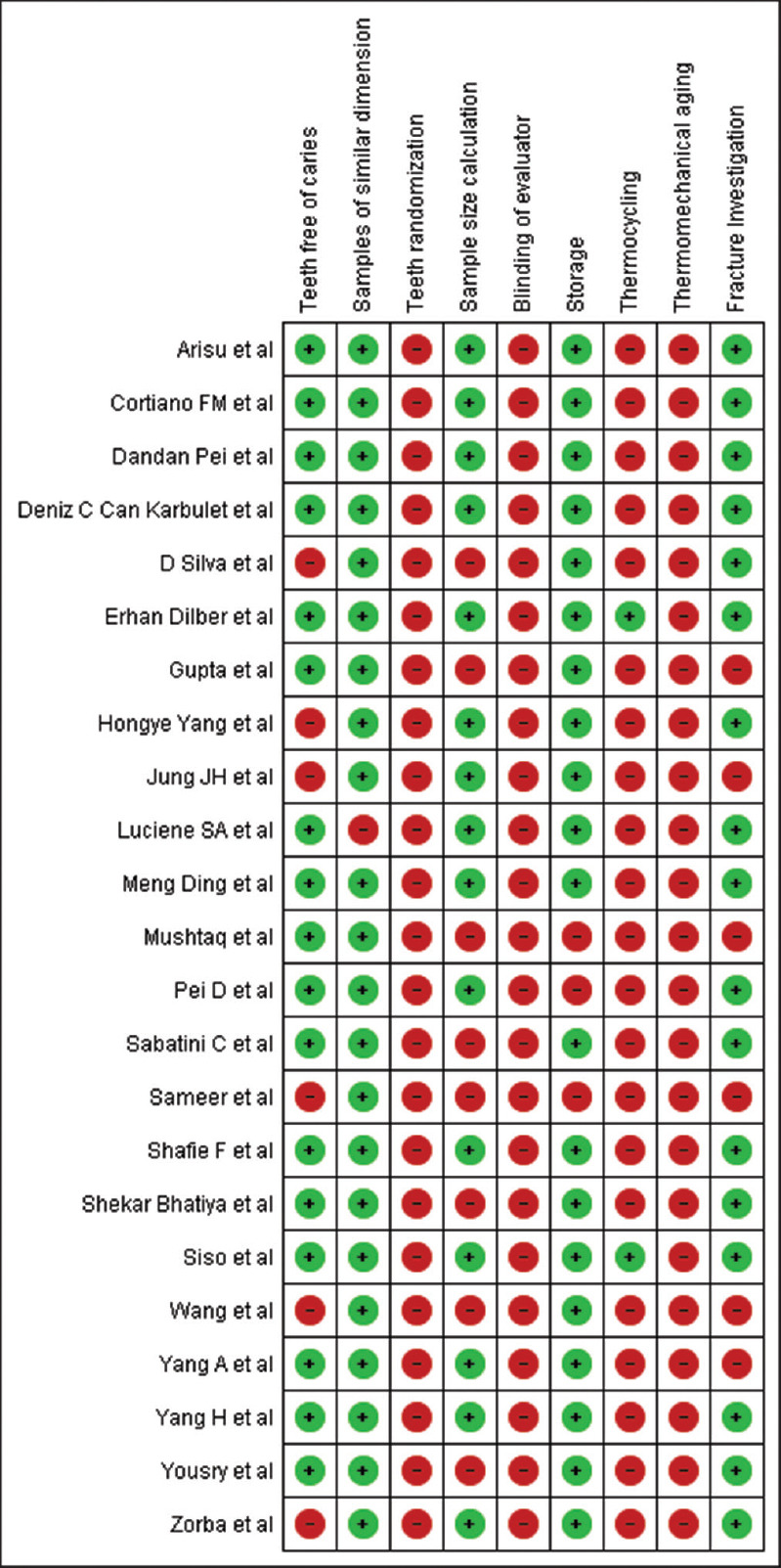
Figure 2.

Quality assessment of individual articles
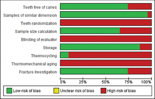
Figure 3.

Risk of Bias assessment of overall studies included in the study (REVMAN 5.4.1)
ANALYSIS OF RISK OF BIAS
Following the assessment of different items for the analysis of Risk of Bias, it was found that there was high risk of bias for sample size calculation, blinding of the evaluator, thermomechanical aging, for which none of the studies had scored Yes, where-as for thermocycling 21 studies have scored No and only two studies had scored Yes. Incomplete outcome data wherein 7 studies had not reported fracture investigation following bond strength tests. 1–3 Yes were considered high risk, 4–6 Yes considered as medium risk, and 7–9 risk as low risk. Out of 23 articles, 17 articles were of medium risk and 6 articles were of high risk after the risk of bias assessment. Since different desensitizing agents, different bonding agents used, and different bond strengths were evaluated in the included studies, meta-analysis could not be performed, thus quantification of heterogenicity by using I2 statistics was not possible.
DISCUSSION
Since bond strength studies could not be performed clinically, in-vitro studies are at the most important for evaluating the bond strength of different materials even though the same clinical conditions as in the oral cavity could not be mimicked in in-vitro studies. Of the 23 articles, nine have studied the macro-strength of which, eight were shear bond strength studies[15,16,18,27,28,30,32,33] and one was tensile bond Strength.[24] Macro tests resulted in cohesive failure and overestimation of bond strength, however, it could be considered because of its simplicity.[24,32,33,38] Fracture investigation was not done in the three studies. Of the rest of the five studies, adhesive failure was prominent in three studies[18,28,30] mixed failure was prominently reported in one study[27] and one study reported equal adhesive and mixed failures.[16] In one study, it is mentioned in material and methodology that fracture investigation was done, but the type of failure was not mentioned in the results.
14 studies have performed the micro-strength tests of which micro-tensile bond strength tests were done in 12 studies[14,17,20,21,22,23,25,29,31,34,35,36] and micro-shear bond strength bond tests were done in two studies.[19,26] More of adhesive failure was reported in eleven studies[16,17,18,19,22,23,25,28,31,34,35] and mixed failure were reported in seven studies[14,16,19,25,29,34,35] and fracture investigation was not done in seven studies.[15,20,21,24,32,33,36] Adhesive area reduction influenced the bond strength and reduced the cohesive failures in micro-tensile bond strength tests thus advantageous over macro tests to evaluate the adhesive interface.[39]
In seven studies dentin hypersensitivity models were created on the extracted teeth, by using 1% citric acid for 20 s[21,23,35,36,37] and by using 0.5 M of EDTA for 2 min[28] and 15% EDTA for 90 s.[31]
POTASSIUM NITRATE
Potassium nitrate-containing desensitizing agents, apart from interfering with the nerve conduction, also blocks the dentinal tubules following repeated application, thus reducing the bond strength[33] but according to authors[16,18] there is no interference with the bonding.
OXALATES
Oxalates application on the dentin results in the formation of calcium oxalate crystals in the dentinal tubules, which in turn interferes with the resin infiltration.[39] Oxalates are applied after acid etching since the oxalates like Bisblock have a pH of 1.5–1.8, causes an additional etching effect and thus leading to the extent of demineralization and penetration depth of the resin mismatch.[40] Compared to ethanol/water-based adhesives, acetone-based adhesives are more sensitive to moist bonding techniques, and in only one study[41] oxalates are compatible with the adhesive, i.e., Adper Single Bond.
Gluma (Glutaraldehyde and HEMA].
In eroded dentin, the glutaraldehyde in GLUMA fixes the collagen fibrils[42] exposed and HEMA helps in the infiltration of resin monomers into the collagen[43] thus increasing the bond strength.[25] When the bonding agent containing HEMA follows the application of GLUMA, the acidic effect of HEMA is repeated, thus favoring better penetration of the resin monomer achieving a greater bond strength.[44]
LASERS
Lasers work by the mechanism of thermal energy absorption,[45] thus, occluding and narrowing of dentinal tubules and melting of hydroxyapatite in the dentin.[46] These morphological changes interfere with the penetration of resin thus compromising the bond strength. Nd:YAG laser decreased the bond strength by creating morphological changes in the dentin.[47]
According to Yazici et al. Nd:YAG lasers did not interfere with the bonding.[48] Rolla JN reported that Nd-YAG laser irradiation promoted the micro-mechanical retention for self-etch adhesives, whereas did not interfere with the bonding of Universal adhesive such as Single Bond. Co2, Er-YAG, and Diode lasers decreased the bond strength of the adhesives studied.[46,32] Short-term use of red wavelength diode laser did not interfere with the bond strength when used with Clearfil Tri S Bond.[15] When Carbon dioxide laser was used, the adhesive could not infiltrate the dentin substrate that is denatured with CO2 laser adequately, hence decrease in bond strength[34] Er:YAG laser decreased the bond strength,[35] since the adhesives micro-mechanical retention to the irradiated dentin is affected.[49]
8.0% ARGININE AND CA-CO3
Calcium and phosphate ions precipitate in the alkaline environment created by the Ar-Ca-Co3 and block the dentinal tubules thus exhibiting desensitizing action.[50,51] The acidity of 4MET in adhesives is likely to solubilize the precipitate, thus reopening the tubules to permit the formation of resin tags to improve the bond strength.[52] Acidic pH of the G Bond yielded favorable bond strength whereas the S3 bond decreased the bond strength since it has a higher pH than G Bond.[22]
CASEIN PHOSPHOPEPTIDE- AMORPHOUS CALCIUM PHOSPHATE
CPP-ACP favors re-mineralization over demineralization which might resist the conditioning of the dentin during dentin bonding, reducing the bond strength.[53] On the contrary, CPP-ACP yielded favorable bonding with self-etch adhesives.[16,54] The application of CPP-ACP makes the surface more wet and reduces contact angle, thus favoring mechanical interlocking and adhesion.[55] All the studies concluded either CPP-ACP did not interfere with the bonding or increased the bond strength.
CALCIUM PHOSPHATE/BIO-SILICATE
The bond strength results are variable with different adhesives, acetone-based adhesives like Prime and Bond NT achieved favorable bond strength with Denshield because of the high vapor pressure and better penetration of acidic monomers.[10] Ethanol/water-based adhesives decrease the bond strength because of the water tree phenomenon[56] which in turn leads to incomplete polymerization[57] of the adhesive following the application of Denshield. Calcium phosphate-containing DSA did not interfere with the bonding of Etch and Rinse adhesives.[58] Clearfil Universal bond in Etch and Rinse mode achieved reduced bond strength with Teeth Mate Desensitizer[31] G Bond resulted in higher bond strength since 4MET can interact with calcium that is present in the hydroxylapatite that is formed following the application of calcium-containing DSA, whereas in Clearfil S3 bond, the higher pH gets neutralized by the DSA.[36]
DENTIN ADHESIVES
Systemp desensitizing agent increased the bond strength, when followed by the application of Prime N Bond NT, because the system acted as rewetting agent,[59] again with the adhesive may yield optimal wetting, thus dual wetting contributing to improved better strength. Xeno V+ was used, Systemp re-wetting combined with the acrylic acid present in the adhesives wetting resulted in phase separation of hydrophobic components leading to resin globule formation resulting in unfavorable bond strength.[60] Futurabond DC also decreased the bond strength, since it contains ethanol which leads to over wetting,[61] leading to the weakening of the resin dentin interface thus unfavorable bond strength.
ANTIBACTERIAL DSA
Chlorhexidine varnish reduced the bond strength,[62] whereas chlorhexidine in gel form did not have any adverse effect on the self-etch and etch and rinse adhesives[63,64] Cervitec did not interfere with the bonding of the different adhesives.[16] This review has included most of the studies evaluating the commonly used DSA and DBA and different bond strength tests, however, the limitation is data extraction limited to studies from 2010 to 2020 and studies in the English language only. In future, these types of studies have to be conducted simulating closer to the clinical conditions as far as possible, by creating hypersensitivity models, subjecting the specimens to thermocycling, thermomechanical aging. Blinding of the evaluators has to be performed to reduce the detection and performance bias.
CONCLUSION
According to the various articles included in this study, GLUMA and 8.0% arginine and Ca-C03 when used as desensitizing agents along with different bonding agents, were found to be highly compatible without interfering with the bond strength of the dentin adhesives. For other desensitizing agents, compatibility with the different DBAs should be checked before the clinical use for successful bonding.
Financial support and sponsorship
Nil.
Conflicts of interest
There are no conflicts of interest.
REFERENCES
- 1.Brannstrom M. Sensitivity of dentine. Oral Surg Oral Med Oral Path. 1996;21:517–26. doi: 10.1016/0030-4220(66)90411-7. [DOI] [PubMed] [Google Scholar]
- 2.Pashley DH, Livingston MJ, Reeder OW, Horner J. Effects of the degree of tubule occlusion on the permeability of human dentin in-vitro. Arch Oral Biol. 1978;20:1127–33. doi: 10.1016/0003-9969(78)90119-x. [DOI] [PubMed] [Google Scholar]
- 3.Susin AH, Vasconcellos WA, Saad JR, Oliveira Junior OB. Tensile bond strength of self-etching versus total-etching adhesive systems under different dentinal substrate conditions. Braz Oral Res. 2007;21:81–6. doi: 10.1590/s1806-83242007000100014. [DOI] [PubMed] [Google Scholar]
- 4.Kobler A, Schaller HG, Gernhardt CR. Effects of the desensitizing agents Gluma and Hyposen on the tensile bond strength of dentin adhesives Am J Dent. 2008;21:388–92. [PubMed] [Google Scholar]
- 5.Aranha AC, Siqueira Junior Ade S, Cavalcante LM, Pimenta LA, Marchi GM. Microtensile bond strengths of composite to dentin treated with desensitizer products. J Adhes Dent. 2006;8:85–90. [PubMed] [Google Scholar]
- 6.Kulunk S, Sarac D, Kulunk T, Karakas O. The effects of different desensitizing agents on the shear bond strength of adhesive resin cement to dentin J Esthet Restor Dent. 2011;23:380–7. doi: 10.1111/j.1708-8240.2011.00415.x. [DOI] [PubMed] [Google Scholar]
- 7.Ravikumar N, Shankar P, Indira R. Shear bond strengths of two dentin bonding agents with two desensitizers: An in vitro study. J Conserv Dent. 2011;14:247–51. doi: 10.4103/0972-0707.85802. [DOI] [PMC free article] [PubMed] [Google Scholar]
- 8.Sailer I, Tettamanti S, Stawarczyk B, Fischer J, Hämmerle CH. In vitro study of the influence of dentin desensitizing and sealing on the shear bond strength of two universal resin cements. J Adhes Dent. 2010;12:381–92. doi: 10.3290/j.jad.a17714. [DOI] [PubMed] [Google Scholar]
- 9.Cheng L, Wang S, Che YH, Qian M. Study of three types of desensitizers in dentin bonding strength. J Biol Regul Homeost Agents. 2017;31:557–65. [PubMed] [Google Scholar]
- 10.Canares G, Salgado T, Pines MS, Wolff MS. Effect of an 8.0% arginine and calcium carbonate desensitizing toothpaste on shear dentin bond strength. J Clin Dent. 2012;23:68–70. [PubMed] [Google Scholar]
- 11.Escalante-Otárola WG, Castro-Núñez GM, Jordão-Basso KC, Guimarães BM, Palma-Dibb RG, Kuga MC. Evaluation of dentin desensitization protocols on the dentinal surface and their effects on the dentin bond interface. J Dent. 2018;75:98–104. doi: 10.1016/j.jdent.2018.06.002. [DOI] [PubMed] [Google Scholar]
- 12.Aguiar JD, Medeiros IS, Souza Junior MH, Loretto SC. Influence of the extended use of desensitizing toothpastes on dentin bonding, microhardness and roughness. Braz Dent J. 2017;28:346–53. doi: 10.1590/0103-6440201601292. [DOI] [PubMed] [Google Scholar]
- 13.Garcia Rubens Nazareno, Giannini Marcelo, Takagaki Tomohiro, Sato Takaaki, Matsui Naoko, Nikaido Toru, Tagami Junji. Effect of dentin desensitizers on resin cement bond strengths. RSBO. 2015;12:14–22. [Google Scholar]
- 14.De Andrade e Silva SM, Malacarne-Zanon J, Carvalho RM, Alves MC, De Goes MF, Anido-Anido A, et al. Effect of oxalate desensitizer on the durability of resin-bonded interfaces. Oper Dent. 2010;35:610–7. doi: 10.2341/09-202-L. [DOI] [PubMed] [Google Scholar]
- 15.Can-Karabulut DC, Karabulut B. The effect of dentin hypersensitivity treatments on the shear bond strength to dentin of a composite material. Gen Dent. 2011;59:e12–7. [PubMed] [Google Scholar]
- 16.Zorba YO, Erdemir A, Ercan E, Eldeniz AU, Kalaycioglu B, Ulker M. The effects of three different desensitizing agents on the shear bond strength of composite resin bonding agents. J Mech Behav Biomed Mater. 2010;3:399–404. doi: 10.1016/j.jmbbm.2010.03.003. [DOI] [PubMed] [Google Scholar]
- 17.Arisu HD, Dalkihç E, Üçtaşli MB. Effect of desensitizing agents on the microtensile bond strength of a two-step self-etch adhesive to dentin. Oper Dent. 2011;36:153–61. doi: 10.2341/09-381-L. [DOI] [PubMed] [Google Scholar]
- 18.Bhatia S, Krishnaswamy MM. Effect of two different dentin desensitizers on shear bond strength of two different bonding agents to dentin: An in vitro study. Indian J Dent Res. 2012;23:703–8. doi: 10.4103/0970-9290.111242. [DOI] [PubMed] [Google Scholar]
- 19.Yousry MM. Effect of re-etching oxalate-occluded dentin and enamel on bonding effectiveness of etch-and-rinse adhesives. J Adhes Dent. 2012;14:31–8. doi: 10.3290/j.jad.a22744. [DOI] [PubMed] [Google Scholar]
- 20.Wang Y, Liu S, Pei D, Du X, Ouyang X, Huang C. Effect of an 8.0%arginine and calcium carbonate in-office desensitizing agents on the micro tensile bond strength of self-etch dentin adhesives to human dentin. Am J Dent. 2012;25:281–6. [PubMed] [Google Scholar]
- 21.Yang H, Pei D, Liu S, Wang Y, Zhou L, Deng D, et al. Effect of a functional desensitizing paste containing 8% arginine and calcium carbonate on the microtensile bond strength of etch-and-rinse adhesives to human dentin. Am J Dent. 2013;26:137–42. [PubMed] [Google Scholar]
- 22.Pei D, Liu S, Huang C, Du X, Yang H, Wang Y, et al. Effect of pretreatment with calcium-containing desensitizer on the dentine bonding of mild self-etch adhesives. Eur J Oral Sci. 2013;121:204–10. doi: 10.1111/eos.12047. [DOI] [PubMed] [Google Scholar]
- 23.Yang H, Pei D, Chen Z, Lei J, Zhou L, Huang C. Effects of the application sequence of calcium-containing desensitising pastes during etch-and-rinse adhesive restoration. J Dent. 2014;42:1115–23. doi: 10.1016/j.jdent.2014.03.018. [DOI] [PubMed] [Google Scholar]
- 24.Makkar S, Goyal M, Kaushal A, Hegde V. Effect of desensitizing treatments on bond strength of resin composites to dentin – An in vitro study. J Conserv Dent. 2014;17:458–61. doi: 10.4103/0972-0707.139840. [DOI] [PMC free article] [PubMed] [Google Scholar]
- 25.Ding M, Shin SW, Kim MS, Ryu JJ, Lee JY. The effect of a desensitizer and CO2 laser irradiation on bond performance between eroded dentin and resin composite. J Adv Prosthodont. 2014;6:165–70. doi: 10.4047/jap.2014.6.3.165. [DOI] [PMC free article] [PubMed] [Google Scholar]
- 26.Andreatti LS, Lopes MB, Guiraldo RD, Borges AH, Orçati Dorilêo MC, Gonini A., Jr Effect of desensitizing agents on the bond strength of dental adhesive systems. Appl Adhes Sci. 2014;2:1–8. [Google Scholar]
- 27.Dilber E, Çevik P, Akpınar YZ, Ozturk AN. The effects of different dentin hypersensitivity treatments on the shear bond strength between adhesive composite resin and dentin. Clin Dent Res. 2014;38:11–20. [Google Scholar]
- 28.Sabatini C, Wu Z. Effect of desensitizing agents on the bond strength of mild and strong self-etching adhesives. Oper Dent. 2015;40:548–57. doi: 10.2341/14-190-L. [DOI] [PubMed] [Google Scholar]
- 29.Cortiano FM, Rached RN, Mazur RF, Vieira S, Freire A, deSouza EM. Effect of desensitizing agents on the microtensile bond strength of two-step etch-and-rinse adhesives to dentin. Eur J Oral Sci. 2016;124:309–15. doi: 10.1111/eos.12263. [DOI] [PubMed] [Google Scholar]
- 30.Shafiei F, Derafshi R, Memarpour M. Bond strength of self adhering materials: Effect of dentin desensitizing treatment with CPP-ACP paste. Int J Periodontics Restorative Dent. 2017;37:e337–43. doi: 10.11607/prd.2850. [DOI] [PubMed] [Google Scholar]
- 31.Siso SH, Dönmez N, Kahya DS, Uslu YS. The effect of calcium phosphate-containing desensitizing agent on the microtensile bond strength of multimode adhesive agent. Niger J Clin Pract. 2017;20:964–70. doi: 10.4103/1119-3077.187322. [DOI] [PubMed] [Google Scholar]
- 32.Gupta T, Nagaraja S, Mathew S, Narayana IH, Madhu KS, Dinesh K. Effect of desensitization using bioactive glass, hydroxyapatite, and diode laser on the shear bond strength of resin composites measured at different time intervals: An in vitro Study. Contemp Clin Dent. 2017;8:244–7. doi: 10.4103/ccd.ccd_155_17. [DOI] [PMC free article] [PubMed] [Google Scholar]
- 33.Mushtaq EA, Mathai V, Nair RS, Angelo JMC. The effect of a dentin desensitizer on the shear bond strength of composite to dentin using three different bonding agents: An in vitro study. J Conserv Dent. 2017;20:37–40. doi: 10.4103/0972-0707.209069. [DOI] [PMC free article] [PubMed] [Google Scholar]
- 34.Yang H, Chen Z, Yan H, Huang C. Effects of calcium-containing desensitizers on the bonding stability of an etch-and-rinse adhesive against long-term water storage and pH cycling. Dent Mater J. 2018;37:122–9. doi: 10.4012/dmj.2017-006. [DOI] [PubMed] [Google Scholar]
- 35.Pei D, Meng Y, Li Y, Liu J, Lu Y. Influence of nano-hydroxyapatite containing desensitizing toothpastes on the sealing ability of dentinal tubules and bonding performance of self-etch adhesives. J Mech Behav Biomed Mater. 2018;3:399–404. doi: 10.1016/j.jmbbm.2018.11.021. [DOI] [PubMed] [Google Scholar]
- 36.Jung JH, Kim DH, Yoo KH, Yoon SY, Kim Y, Bae MK, et al. Dentin sealing and antibacterial effects of silver-doped bioactive glass/mesoporous silica nanocomposite: An in vitro study. Clin Oral Investig. 2019;23:253–66. doi: 10.1007/s00784-018-2432-z. [DOI] [PubMed] [Google Scholar]
- 37.Sirisha K, Rambabu T, Ravishankar Y, Pabbati R. Validity of bond strength tests: A critical review: Part I. J Conserv Dent. 2014;17:305–11. doi: 10.4103/0972-0707.136340. [DOI] [PMC free article] [PubMed] [Google Scholar]
- 38.Sano H, Shono T, Sonoda H, Takatsu T, Ciucchi B, Carvalho R, et al. Relationship between surface area for adhesion and tensile bond strength – Evaluation of a micro-tensile bond test. Dent Mater. 1994;10:236–40. doi: 10.1016/0109-5641(94)90067-1. [DOI] [PubMed] [Google Scholar]
- 39.Pashley DH, Carvalho RM, Pereira JC, Villanueva R, Tay FR. The use of oxalate to reduce dentin permeability under adhesive restorations. Am J Dent. 2001;14:89–94. [PubMed] [Google Scholar]
- 40.Carrilho MR, Carvalho RM, Tay FR, Yiu C, Pashley DH. Durability of resin-dentin bonds related to water and oil storage. Am J Dent. 2005;18:315–9. [PubMed] [Google Scholar]
- 41.Shafiei F, Doozandeh M. Impact of oxalate desensitizer combined with ethylene-diamine tetra acetic acid-conditioning on dentin bond strength of one-bottle adhesives during dry bonding. J Conserv Dent. 2013;16:252–6. doi: 10.4103/0972-0707.111327. [DOI] [PMC free article] [PubMed] [Google Scholar]
- 42.Munksgaard EC, Asmussen E. Bond strength between dentin and restorative resins mediated by mixtures of HEMA and glutaraldehyde. J Dent Res. 1984;63:1087–9. doi: 10.1177/00220345840630081701. [DOI] [PubMed] [Google Scholar]
- 43.Van Landuyt KL, Snauwaert J, De Munck J, Peumans M, Yoshida Y, Poitevin A, et al. Systematic review of the chemical composition of contemporary dental adhesives. Biomaterials. 2007;28:3757–85. doi: 10.1016/j.biomaterials.2007.04.044. [DOI] [PubMed] [Google Scholar]
- 44.Paes Leme AF, dos Santos JC, Giannini M, Wada RS. Occlusion of dentin tubules by desensitizing agents. Am J Dent. 2004;17:368–72. [PubMed] [Google Scholar]
- 45.Lan WH, Lee BS, Liu HC, Lin CP. Morphologic study of Nd: YAG laser usage in treatment of dentinal hypersensitivity. J Endod. 2004;30:131–4. doi: 10.1097/00004770-200403000-00001. [DOI] [PubMed] [Google Scholar]
- 46.Liu HC, Lin CP, Lan WH. Sealing depth of Nd: YAG laser on human dentinal tubules. J Endod. 1997;23:691–3. doi: 10.1016/S0099-2399(97)80403-7. [DOI] [PubMed] [Google Scholar]
- 47.Ferreira LS, Ferreira LS, Francci C, Navarro RS, Calheiros FC, Eduardo Cde P. Effects of Nd: YAG laser irradiation on the hybrid layer of different adhesive systems. J Adhes Dent. 2009;11:117–25. [PubMed] [Google Scholar]
- 48.Yazici E, Gurgan S, Gutknecht N, Imazato S. Effects of erbium: Yttrium-aluminum-garnet and neodymium: yttrium-aluminum-garnet laser hypersensitivity treatment parameters on the bond strength of self-etch adhesives. Lasers Med Sci. 2010;25:511–6. doi: 10.1007/s10103-009-0682-3. [DOI] [PubMed] [Google Scholar]
- 49.Caetano de Souza N, Jorge JR, Batisa O, Caetano de Souza N, Jorge JRP, Batisa O, de Oliveira JR. Effect of Er: YAG laser pulse repetition rate variation on bond strength to rewet dentin. J Oral Laser Appl. 2007;7:239–45. [Google Scholar]
- 50.Petrou I, Heu R, Stranick M, Lavender S, Zaidel L, Cummins D, et al. A breakthrough therapy for dentin hypersensitivity: How dental products containing 8% arginine and calcium carbonate work to deliver effective relief of sensitive teeth. J Clin Dent. 2009;20:23–31. [PubMed] [Google Scholar]
- 51.Kleinberg I. SensiStat. A new saliva-based composition for simple and effective treatment of dentinal sensitivity pain. Dent Today. 2002;21:42–7. [PubMed] [Google Scholar]
- 52.Yoshiara K, Yoshida Y, Nagaoka N, Fukegawa D, Hayakawa S, Mine A, et al. Nanocontrolled molecular interaction at adhesive interfaces for hard tissue re-construction. Acta Biomater. 2010;6:3573–82. doi: 10.1016/j.actbio.2010.03.024. [DOI] [PubMed] [Google Scholar]
- 53.Reynolds EC. Anticariogenic complexes of amorphous calcium phosphate stabilized by casein phosphopeptides: A review. Spec Care Dentist. 1998;18:8–16. doi: 10.1111/j.1754-4505.1998.tb01353.x. [DOI] [PubMed] [Google Scholar]
- 54.Adebayo OA, Burrow MF, Tyas MJ. Dentine bonding after CPP-ACP paste treatment with and without conditioning. J Dent. 2008;36:1013–24. doi: 10.1016/j.jdent.2008.08.011. [DOI] [PubMed] [Google Scholar]
- 55.Marshall SJ, Bayne SC, Baier R, Tomsia AP, Marshall GW. A review of adhesion science. Dent Mater. 2010;26:e11–6. doi: 10.1016/j.dental.2009.11.157. [DOI] [PubMed] [Google Scholar]
- 56.Tay FR, Pashley DH. Water treeing – A potential mechanism for degradation of dentin adhesives. Am J Dent. 2003;16:6–12. [PubMed] [Google Scholar]
- 57.Duarte S, Jr., Perdigão J, Lopes MM. Effect of dentin conditioning time on nanoleakage. Oper Dent. 2006;31:500–11. doi: 10.2341/05-86. [DOI] [PubMed] [Google Scholar]
- 58.Borges BC, Souza-Junior EJ, da Costa Gde F, Pinheiro IV, Sinhoreti MA, Braz R, et al. Effect of dentin pre-treatment with a casein phosphopeptide-amorphous calcium phosphate (CPP-ACP) paste on dentin bond strength in tridimensional cavities. Acta Odontol Scand. 2013;71:271–7. doi: 10.3109/00016357.2012.671364. [DOI] [PubMed] [Google Scholar]
- 59.Ivoclar Vivadent. Systemp. [Last accessed on 2021 Sep 15]. Available from: https://www.ivoclarvivadent.com .
- 60.Tay FR, Gwinnett JA, Wei SH. Micromorphological spectrum from over drying to over wetting acid-conditioned dentin in water-free acetone-based, single-bottle primer/adhesives. Dent Mater. 1996;12:236–44. doi: 10.1016/s0109-5641(96)80029-7. [DOI] [PubMed] [Google Scholar]
- 61.Voco Futurabond DC. [Last accessed on 2021 Sep 15]. Available from: http://www.voco.com/en/product/futurabond_dc/index.html .
- 62.Sengun A, Koyuturk AE, Sener Y, Ozer F. Effect of desensitizers on the bond strength of a self-etching adhesive system to caries-affected dentin on the gingival wall. Oper Dent. 2005;30:430–5. [PubMed] [Google Scholar]
- 63.Dalli M, Ercan E, Zorba YO, Ince B, Şahbaz C, Bahşi E, et al. Effect of 1% chlorhexidine gel on the bonding strength to dentin. J Dent Sci. 2010;5:8–13. [Google Scholar]
- 64.Ercan E, Erdemir A, Zorba YO, Eldeniz AU, Dalli M, Ince B, et al. Effect of different cavity disinfectants on shear bond strength of composite resin to dentin. J Adhes Dent. 2009;11:343–6. [PubMed] [Google Scholar]


