Abstract
Highly active antiretroviral therapy (HAART) can suppress HIV-1 replication and normalize the chronic immune activation associated with infection, but restoration of naïve CD4+ T cell populations is slow and usually incomplete for reasons that have yet to be determined. We tested the hypothesis that damage to the lymphoid tissue (LT) fibroblastic reticular cell (FRC) network contributes to naïve T cell loss in HIV-1 infection by restricting access to critical factors required for T cell survival. We show that collagen deposition and progressive loss of the FRC network in LTs prior to treatment restrict both access to and a major source of the survival factor interleukin-7 (IL-7). As a consequence, apoptosis within naïve T cell populations increases significantly, resulting in progressive depletion of both naïve CD4+ and CD8+ T cell populations. We further show that the extent of loss of the FRC network and collagen deposition predict the extent of restoration of the naïve T cell population after 6 month of HAART, and that restoration of FRC networks correlates with the stage of disease at which the therapy is initiated. Because restoration of the FRC network and reconstitution of naïve T cell populations are only optimal when therapy is initiated in the early/acute stage of infection, our findings strongly suggest that HAART should be initiated as soon as possible. Moreover, our findings also point to the potential use of adjunctive anti-fibrotic therapies to avert or moderate the pathological consequences of LT fibrosis, thereby improving immune reconstitution.
Author Summary
The hallmark of HIV-1 infection is depletion of CD4 T cells, whose loss leads to the opportunistic infections and cancers characteristic of AIDS. Highly active antiretroviral therapy (HAART) can control HIV-1 replication, but reconstitution particularly of naïve T cells is often incomplete and slow. We show here that fibrosis damages lymphoid tissues (LT), thereby contributing to depletion and incomplete reconstitution. Prior to treatment, chronic immune activation induces LT fibrosis to disrupt the fibroblastic reticular cell (FRC) network, the major source of the T cell survival factor interleukin 7 (IL-7). Fibrosis in this way interferes with the access of T cells to IL-7 “posted” on the FRC network. Without a source and access to IL-7, naïve cells are depleted prior to initiating HAART because of increased apoptosis, and, even after initiating HAART, the losses continue by this mechanism because of pre-existing LT damage. Thus, LT fibrosis impairs immune reconstitution despite the beneficial effects of HAART in suppressing viral replication. Because less LT damage has accumulated in earlier stages of infection, early initiation of HAART also improves immune reconstitution. This LT damage mechanism also suggests that anti-fibrotic treatment in addition to HAART could further improve immune reconstitution.
Introduction
The hallmark of HIV-1 infection, depletion of CD4+ T cells, has been largely attributed to direct mechanisms of infection and cell death from viral replication or killing by virus-specific cytotoxic T-lymphocytes (CTLs), and to indirect mechanisms such as increased apoptosis accompanying chronic immune activation associated with HIV-1 infections [1]. It is thus puzzling that if these were the sole mechanisms responsible for CD4+ T cell depletion, why 20% of HIV-1 infected patients have no significant increase in their peripheral blood CD4 count after initiation of HAART, since treatment can suppress viral replication to undetectable levels and normalize much of the chronic immune activation associated with infection [2]–[3]. Moreover, even among patients with significant increases in peripheral blood CD4+ T cells, few reconstitute to normal levels after years of HAART, and this incomplete immune reconstitution is associated with significantly higher rates of malignancy and other morbidities compared with HIV-uninfected individuals [4]–[11].
The preferential depletion of naïve T cells in blood and lymphoid tissues (LT) [3], where they mainly reside, also poses particular difficulties for attributing depletion simply to direct mechanisms of viral infection or indirect mechanisms of activation-induced cell death (AICD), since (1) naïve CD4+ T cells are resistant to HIV-1 infection and (2) AICD should primarily affect the activated effector and memory populations [12]–[14]. Furthermore, the similar extent of depletion of not only naïve CD4+ T cells but also naïve CD8+ T cells that are not usually infected by HIV [15]–[16], suggests that there is a general mechanism impacting naïve T cell populations unrelated to direct infection.
The incomplete restoration of naïve T cell populations with HAART also points to mechanisms in addition to AICD in depletion of CD4+ T cells, since suppression of this “drain” should enable repopulation of naïve CD4+ T cell populations by thymopoiesis and homeostatic proliferation of existing naïve T cells in secondary LT, but this does not happen many patients [17]–[20]. Cumulative observations therefore suggest that there may be additional mechanisms that impair the survival of naïve T cells, thereby restricting immune reconstitution [1], [3].
To account for the preferential loss of naïve T cells, and failure of HAART to restore both naïve and memory populations by thymopoesis and homeostatic proliferation, we have proposed a damaged LT niche hypothesis in which collagen deposition disrupts the FRC network on which naïve T cells migrate and gain access to survival factors such as interleukin-7 (IL-7). This results in elevated levels of naïve T cell apoptosis [21]–[23] before treatment and impairs the reconstitution of naïve T cells after treatment. We recently showed in the SIV-rhesus macaque animal model that the critical disruption in LT architecture caused by collagen deposition and decreased access of T cells to survival factor IL-7 “posted” on the FRC network was in fact associated with increased apoptosis and depletion of both naïve CD4+ and CD8+ T cells. We also showed that this mechanism is a cooperative and cumulative vicious cycle in which the mutual interdependencies for survival of naïve T cells on IL-7, and the FRC network on lymphotoxin-beta (LT-β) supplied by the T cells, perpetuate depletion of both T cells and the FRC network [24].
One implication of this model is that because the impact of LT fibrosis on CD4+ T cell depletion is progressive and cumulative, initiating HAART in the early stages of infection should improve immune reconstitution because there should be less collagen deposition and loss of the FRC network at this stage. We tested this hypothesis by examining LTs from HIV-1 infected individuals at baseline and 6 months after initiating HAART in the acute, pre-symptomatic and AIDS stages of infection. We first document the same damaged LT niche mechanism described in the SIV-rhesus macaque model of collagen deposition and loss of the FRC network with depletion of naïve T cells through increased apoptosis as a consequence of decreased access to IL-7. We then show that the extent of loss of the FRC network and collagen deposition predict the extent of inhibition of naïve T cell apoptosis and restoration of the naïve T cell population in LT after 6 months of HAART, and total CD4 T cell counts in peripheral blood after 12 months of HAART. Furthermore, we find that the extent of restoration of FRC network after 6 months of HAART is highly dependent on the stage of disease at which the therapy is initiated, with greatest restoration only when HAART is initiated during the early stage of infection. This directly correlates with optimal inhibition of naïve T cell apoptosis and restoration of naïve T cells in the patients receiving HAART during the early stage of infection. This mechanism explains why initiation of HAART during the early stage of infection is associated with more rapid and complete CD4+ T cell restoration, and thus strongly argues for early initiation of HAART [25]–[26]. It also argues for a potential use of adjunctive therapies such as anti-fibrotic therapy to avert and/or revert the LT structure to improve immune reconstitution.
Results
Collagen deposition and loss of the FRC network impede access to and source of IL-7 in HIV-1 infection
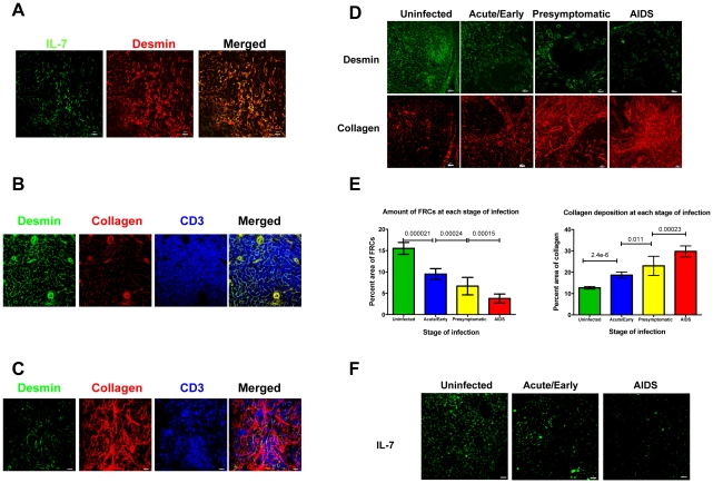
To evaluate this hypothesis, we first show that the FRC network is the major source of IL-7 for T cells in human LTs, as has been demonstrated in mice and monkeys [22], [24]. In LT sections from uninfected individuals stained for IL-7 and desmin, a marker for FRCs, IL-7 largely co-localizes with the FRC network on which lymphocytes, antigen presenting cells and other cells within LTs migrate (Figure 1A). This architecture allows T cells to efficiently access survival factors such as IL-7 and self-antigen-major histocompatibility complex as well as chemokines “posted” on their path. Thus, in the lymph node (LN) sections from HIV-1-uninfected individuals stained for type I collagen, desmin and T cells, the collagen within the FRC network co-localizes with desmin, and the T cells visibly contact the FRC network (Figure 1B). In contrast, HIV-1 infection is associated with stage-specific progressive decreases in the FRC network (Figure 1D-E) and thus the available source of IL-7 (Figure 1F). The depletion of FRCs correlates with a parallel increase in collagen deposited outside the FRC network, so that as infection progresses, fewer and fewer T cells are in contact with and have access to IL-7 on the FRC network (Figure 1C) compared with uninfected populations (Figure 1B).
Figure 1. Collagen deposition and loss of the FRC network impede access to and source of IL-7 in HIV-1 infection.
A. FRCs are the major producers of IL-7. LN sections (representative image for one HIV negative subject of 5) stained for desmin (red) and IL-7 (green). Merged confocal image shows co-localization of IL-7 and FRCs in T cell area. Scale bar, 10 µm. B–C. Collagen deposition and loss of the FRC network disrupts interaction between T cells and FRCs. Confocal images of LN sections from an uninfected subject (representative image for one subject of 5) immunofluorescently stained for desmin (green), collagen (red) and CD3 (blue). The merged image shows that FRCs colocalize with collagen and T cells are in contact with the FRC network (B). Confocal images of LN sections from a subject at AIDS stage (representative image for one subject of 6). The merged image shows that the loss of FRCs and associated collagen deposition leads to loss of the contact between FRCs and T cells, which instead contact mainly extra-FRC collagen. Scale bar, 20 µm (C). D. Confocal images of LN sections from HIV negative subjects and from subjects at different stage of HIV infection immunofluorescently stained for desmin (green) and collagen (red), showing the gradual loss of FRCs in the T cell zone within LTs, which is associated with extensive collagen deposition during HIV infection. Scale bar, 20 µm. E. Quantification of average amount of FRCs and collagen deposition at each stage of infection, showing the gradual loss of FRCs and collagen deposition. Error bars represent the s.d. F. Confocal images of LN sections from HIV negative subjects and from subjects at different stage of HIV infection immunofluorescently stained for IL-7 (green), showing that the gradual loss of IL-7 in the T cell zone is associated with gradual depletion of FRCs (E). Scale bar, 20 µm.
T cell apoptosis increases with decreased availability of IL-7
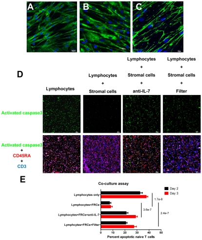
Because survival of naïve T cells is dependent on access to IL-7 [21]–[23], the collagen deposition-restricted access to and loss of the FRC network itself should result in an increase in apoptosis proportional to the extent of collagen deposition and decreased IL-7 source as infection progresses. We first demonstrate that contact with FRCs as a source of IL-7 is critical for T cell survival in an ex vivo culture system. We established monolayers of desmin+ IL-7+ FRC-like cells from stromal cells isolated from human tonsils (Figure 2A–B), and show that IL-7 localizes to the surfaces of live cells stained without permeabilization (Figure 2C). Only about 10% of naïve T cells underwent apoptosis if co-cultured with autologous IL-7+ FRC-like cells compared to about 30–40% of naïve T cells cultured for 2–3 days without stromal cells. We show that the enhanced survival is contact dependent and is mediated mainly via IL-7, as antibody blocking of IL-7 or separation of T cells and the FRCs by transwells leads to increased apoptosis in naïve T cells (Figure 2D–E). However, the blockade of IL-7 does not fully recapitulate the apoptosis level in the naïve T alone culture, suggesting that other survival factors such as CCL19 produced by FRCs may independently support the survival of naïve T cells [22].
Figure 2. Naïve T cells need to contact FRCs to get access to IL-7 for survival.
(A–C) IL-7 is produced and presented on the surface of stromal cells. A-B. Confocal images of monolayer of fixed and permeablized stromal cells isolated from human tonsil immunofluorescently stained for (A) IL-7 (green) or (B) desmin (green) and DAPI (blue) at one-day post passage. Scale bar, 30 µm. C. Confocal image of live stromal cells (DAPI: blue) staining showing the IL-7 (green) on the surface of stromal cells. Scale bar, 10 µm. D-E. FRC-like stromal cells enhance the survival of naïve T cells via IL-7. D. Triple fluorescently stained activated caspase 3+ (green), CD45RA+ (red) and CD3+ (blue) cells in an ex vivo culture system showing that stromal cells enhance the survival of naïve T cell by mechanisms dependent on IL-7 and cell contact. 2x10⁵ lymphocytes from human tonsil were cultured with or without stromal cells for 2–3 days. Naïve T cell apoptosis is reduced in co-cultures with stromal cells (+ stromal cells) compared to cultures without stromal cells. Apoptosis in the naïve T cell population increases with IL-7 blocking antibody (anti-IL-7) or when lymphocytes are separated from stromal cells by a transwell filter (Filter) compared to co-cultures with stromal cells. Scale bar, 60 µm. E. Quantification of the percentages of activated caspase 3+CD45RA+CD3+ naïve T cells in total T cell population at day 2 and day 3 cultures. Values are the mean of the percent apoptotic naïve T cells ± s.d. ANOVA comparison was done on the average percentages of day 2 and day 3.
These ex vivo co-culture results support the concept that naïve T cells need to contact FRCs in order to gain access to IL-7 to maintain their survival. Therefore, the loss of FRCs as well as the loss of the contact between naïve T cells and FRCs together in vivo would be expected to increase apoptosis and thereby deplete naïve T cells. We tested this hypothesis in LTs from HIV-1 infected patients and found that the stage-dependent increases in naïve T cell apoptosis (Figure 3A) were associated with depletion of both naïve CD4+ and CD8+ T cells (Figure 3B–C), and that stage-dependent decreases in the FRC network (Figure 1D) were associated both with apoptosis and naïve T cell depletion (Figure 3D–E).
Figure 3. Loss of FRCs is associated with loss of naïve T cells within LTs.
A. Confocal images of LN sections from subjects at different stage of HIV infection triple immunofluorescently stained for TUNEL (green), CD45RA (red) and CD3 (Blue), showing the gradual loss of CD45RA+CD3+ naïve T cells is associated increased apoptosis in the naïve T cell population within LTs during HIV infection. Scale bar, 10 µm. B. Confocal images of LN sections from subjects at different time points post HIV infection double immunofluorescently stained for CD45RA (green) and CD4 or CD8 (red), showing both naïve CD4 and CD8 T cells are depleted within LTs. Scale bar, 20 µm. C. Quantitative image analysis of the number of apoptotic naïve T cells and the number of naïve T cells (CD45RA+CD3+), showing that increased apoptosis in the naïve T cell population is associated with depletion of naïve T cells (total n = 37, p<0.0001, R2 = 0.5373). D. Quantification of FRCs (the percent area staining positive for desmin in T cell zone) and the number of apoptotic naïve T cells (TUNEL+CD45RA+CD3+), showing that the depletion of FRCs is associated with increased apoptosis in naïve T cell populations (total n = 37, p<0.0001, R2 = 0.5843). E. Quantitative image analysis of FRCs and the number of naïve T cells within LTs, showing that the loss of naïve T cells is associated with loss of FRCs (total n = 37, p<0.0001, R2 = 0.5166). Values are the mean of measurement ± s.d.
Restoration of LT structure and increases in naïve T cells depend on the timing of initiation of HAART
Because the LT damage-mediated naïve T cell depletion mechanism now documented in both HIV-1 infection and SIV infection [24] is cumulative and progressive, the later stage of infection, the greater the damage to LT structure. Thus, if treatment does not restore the LT structure that supports survival of naïve CD4+ T cells, the LT damage mediated naïve T cell depletion could adversely affect immune reconstitution, even with suppression of viral replication and immune activation by HAART. Conversely, the lesser extent of LT damage in early infection could improve immune reconstitution with HAART, if initiating treatment were to restore LT structure and improve naïve T cell survival.
To test these predictions, we examined the effects of HAART initiated in the acute/early, pre-symptomatic and AIDS stages of infection on LT structure, naïve T cell apoptosis and restoration of naïve CD4+ T cell populations. Because loss of the FRC network and fibrosis are less in the acute/early stage than at later stages of HIV-1 infection (Figure 1), we would expect that the preservation of LT structure in acute/early HIV-1 infection would result in decreased apoptosis and greater increases in naïve T cells if HAART is initiated at this stage. We indeed found that the loss of the FRC network and collagen deposition prior to initiating HAART are associated with significantly increased naïve T cell apoptosis after 6 months of HAART (p = 0.0016 and p = 0.0292 respectively) (Figure 4A–B). Furthermore, the level of naïve T cell apoptosis both before and after treatment is significantly associated with fewer naïve T cells in LTs (p = 0.0012). Taken together, these data suggest the extent of LT damage is associated with the extent of inhibition of naïve T cell apoptosis after HAART. We note that this now documents in LTs the previously reported predictive relationship between collagen in LTs and naïve CD4+ T cell increases in peripheral blood [27].
Figure 4. The extent of LT destruction before HAART predicts the extent of restoration of naïve T cells after HAART.
A. The area that FRCs occupy before HAART is negatively associated with the number of apoptotic naïve T cells after 6 months of HAART. B. The collagen area before HAART is positively associated with the number of apoptotic naïve T cells after 6 months of HAART.
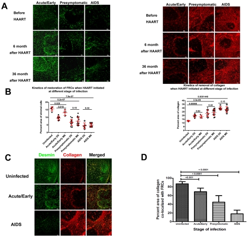
We also find that HAART initiated in the acute/early stage infection results in the greatest restoration of FRCs after 6 months of HAART, albeit not to the level in HIV-1 uninfected population (Figure 5A–B). The restoration of LT structure is a slow process as even after 36 months of HAART the area occupied by the FRC network increases but still remains significantly less than in HIV-1 uninfected individuals. Minimal recovery of the FRC network is seen in HIV-1 infected patients starting HAART at chronic stages (Figure 5A). At 6 months, there is no significant effect on removal of collagen deposition in LTs (Figure 5A–B), but there is increased collagen co-localizing with the FRC network, as opposed to collagen deposits outside the network, albeit not to the same levels seen in HIV-1 uninfected individuals where 90 percent of collagen is within the FRC network (Figure 5C-D). We further found that the new WHO guidelines for initiating HAART at CD4 counts of 350 cells/µl have a sound rationale in preservation of the FRC network, since only when therapy had been initiated at or above 350 cells/µl could we detect significant improvement of FRCs after 6-month HAART (Figure S1).
Figure 5. Restoration of LT structure is slow and incomplete after HAART and is associated with the timing of initiation of HAART.
A. Representative confocal images of immunofluorescent staining for desmin (green) and collagen (red), showing the different extent of restoration of stromal cell network and collagen normalization after 6 months of HAART. Scale bar, 20 µm. B. Quantification of the average area of FRCs and collagen before and after 6 months of HAART in patients receiving the HAART at different stages of infection, showing the different extent of restoration of LTs is associated with the timing of initiation of HAART. Error bars represent the s.d. C. Representative confocal images of immunofluorescent staining for desmin (green) and collagen (red), showing that the different extent of restoration of the FRC network and collagen normalization after 6 months of HAART as represented by the percent area not covered by FRCs. Scale bar, 20 µm. D. Quantification of the percent area covered by FRCs.
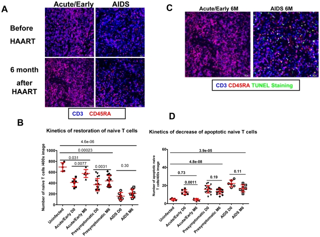
HAART initiated in acute/early stage of infection is also associated with greatest decreases in apoptosis and optimal restoration of naïve T cell populations in LTs (Figure 6). In contrast, naïve T cell numbers in patients who initiated HAART in the AIDS stage of infection did not increase significantly, and apoptosis in naïve T cell populations remained high (Figure 6). These stage-related correlations apply as well to peripheral CD4+ T cell counts in patients receiving HAART for 12 months. The increase in peripheral CD4+ T cell counts to the level of counts in HIV-1 uninfected individuals depended on initiating HAART during the acute/early stage of infection (Figure S2A). We also found that this stage-dependent restoration of peripheral CD4+ T cells can be predicted by the extent of fibrosis before initiation of HAART, again suggesting that fibrosis is one key factor that limits immune reconstitution after long-term HAART (Figure S2B).
Figure 6. Incomplete LT restoration is associated with high level of apoptosis in naïve T cell populations and incomplete restoration of naïve T cells.
A. Confocal images of LN sections from uninfected subjects and patients receiving 6 months of HAART at either acute or AIDS phase of infection, immunofluorescently stained for CD45RA (red) and CD3 (blue), showing the different extent of restoration of naïve T cells. Scale bar, 10 µm. B. Quantification of number of naïve T cells before and after HAART at each stage of infection, showing the different kinetics of restoration of naïve T cells. Error bars represent the s.d. C. Confocal images of LN sections from patients receiving 6 months of HAART at either acute or AIDS phase of infection immunofluorescently stained for CD45RA (red), CD3 (blue) and TUNEL staining of apoptotic cells (green), showing the higher level of apoptosis in naïve T cell populations after 6 months of HAART when HAART was started during chronic phase of infection. Scale bar, 10 µm. D. Quantification of average number of apoptotic naïve T cells before and after HAART at each stage of infection, showing the different kinetics of decrease of apoptotic naïve T cells.
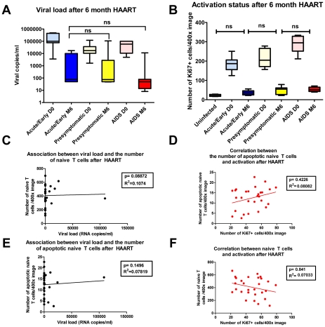
We also assessed the relationships between viral load and the residual Ki67 level to the extent of restoration of naïve T cells when HAART was initiated at different stages. We found that increases in naïve T cells and decreases in apoptosis do not correlate with HAART-mediated suppression of viral replication and immune activation. HAART initiated at all stages of infection can potently and comparably inhibit viral replication and associated chronic immune activation (Figure 7A and B). There is therefore no evidence that these processes are playing important roles in the stage-specific effects of time of initiation on apoptosis and recovery of naïve T cells within LTs. We in fact found no significant association between viral load and the number of naïve T cells or the number of apoptotic naïve T cells, nor any association between activation represented by the number of Ki67+ cells and the number of naïve T cells or the number of apoptotic naïve T cells after 6 month HAART (Figure 7C–F).
Figure 7. The extent of restoration of naïve T cells after HAART is not associated with viral load or immune activation.
A. Viral load before and after HAART in patients receiving HAART at different stages of infection, showing the inhibition of viral replication is similar for patients at different stage of infection. B. Quantification of the number of Ki67+ cells before and after HAART in patients receiving HAART at different stages of infection, showing the inhibition of immune activation by HAART is similar for patients at different stage of infection (ns, not significant). C, E. Association between the viral load and number of naïve T cells and association between viral load and the number of apoptotic naïve T cells after HAART are not significant. D, F. Association between the number of Ki67+ cells and naïve T cells and association between the number of Ki67+ cells and the number of apoptotic naïve T cells after HAART are not significant.
Discussion
It has generally been thought that viral and immune-cell mediated killing of CD4+ T cells and AICD accompanying chronic immune activation are respectively the major direct and indirect mechanisms of CD4+ depletion in HIV-1 infection, and that the slow and incomplete restoration of naïve CD4+ T cells is a consequence of the restricted capacity of the adult thymus to re-supply naive T cells [1]. Here we describe a novel mechanism that depletes CD4+ T cells, particularly naïve CD4+ T cells before HAART and determines the pace and extent of naïve CD4+ T cell restoration after HAART.
Naïve T cells depend on IL-7 produced and presented by the FRC network for survival. Our IL-7 staining is consistent with a large literature on the FRC network as the site and source of most of the IL-7 [22], [24], [28]–[29], although different from one report on human IL-7 staining in LTs [30], probably due to different reagents and methodologies used. As HIV-1 infection advances, collagen deposition increases and the FRC network is destroyed, which decreases the amount of IL-7 available to support T cell survival. As a consequence, increased apoptosis depletes naïve T cells prior to HAART in proportion to the progressive destruction of LT structure from early to later stages of HIV-1 infection. Because of the progressive and cumulative nature of this pathological process, apoptosis of naïve T cells continues at elevated levels after HAART has been initiated, even though HAART can potently suppress viral replication and at least partially normalize immune activation. The elevated apoptosis level in naïve T cell populations is in proportion to pre-existing damage to LT structure, which is greater in the chronic stages of infection. Thus, predictably, early treatment results in better preservation and restoration of LT structure, which leads to improved survival of naïve T cells and greater increases in naïve T cell numbers. While the limited capacity of the thymus in the adult to supply naïve T cells will certainly limit the pace and extent of reconstitution, the LT structure to which thymic emigrants home thus also determines their subsequent survival. By analogy to earlier tap-and-drain models [31], restoration of naïve T cells will be dependent not only on thymic output and from homeostatic proliferation of naïve T cell populations in LT, but also on the drain of overall apoptosis in this population. In such a model, the elevated level of apoptosis in naïve T cells in secondary LTs limits both the extent and pace of immune reconstitution.
As the diversity of the naïve T cell repertoire is critical to defend against new infections and malignancies, the loss and slow restoration of the naïve T cell population creates “holes” in the T cell repertoire and therefore impairs host defenses even after HIV-1 replication has largely been suppressed [5], [8]–[9]. Thus, therapeutic approaches to prevent or moderate damage to the LT niche and restore a functional FRC network could be particularly beneficial in increasing and preserving naïve T cell populations after HAART. The most straightforward way to do this is through earlier treatment. Our findings also suggest the potential clinical benefit of complementing IL-7 treatment during HIV-1 infection in the restoration of naïve T cell population. Indeed, studies have shown that complementing HAART with IL-7 during both SIV and HIV infection significantly increases the circulating naïve CD4+ T cell number [32]–[34]. Furthermore, it has been shown that IL-7 treatment could normalize the extent of apoptosis in CD4+ and CD8+ T cells from HIV-1-infected individuals via up-regulation of Bcl-2 levels [35]–[36]. These data consistently suggest that insufficient IL-7 is a key contributor in the impaired T cell homeostasis in SIV/HIV infection and limits the reconstitution of T cells.
However, the immediate decline of the absolute numbers of both naïve CD4+ and CD8+ T cells after termination of IL-7 therapy [32]–[34] suggests that complementing IL-7 only provides transient survival benefit for naïve CD4 and CD8 T cells and strongly argues for the development of therapeutic interventions to provide long-term survival benefit for naïve T cells through preservation or restoration of the FRC network where naïve cells can access IL-7. Our findings here clearly suggest that collagen deposition and the consequential loss of FRCs as the major source of IL-7 play critical roles in compromising homeostasis of naïve T cells. Therefore, the restoration of the lymphoid tissue niche could potentially provide long-term survival benefits for naïve T cells. Moreover, the development of adjunctive anti-fibrosis treatment such as pirfenidone and losartan [24], [37]–[42] might additionally avert or revert the consequences of damage to the LT niche and improve immune reconstitution.
Materials and Methods
Ethics statement
This human study was conducted according to the principles expressed in the Declaration of Helsinki. The study was approved by the Institutional Review Board of the University of Minnesota. All patients provided written informed consent for the collection of samples and subsequent analysis.
LN biopsy specimens
Inguinal LN (LN) biopsies from HIV negative individuals and HIV-1-infected individuals at different clinical stages (7 at acute/early stage, 18 at presymptomatic stage and 8 at AIDS stage. Table 1) were obtained for this University of Minnesota Institutional Review Board-approved study. Viral load measurements were obtained the same day as biopsies. Each LN biopsy was immediately placed in fixative (4% neutral buffered paraformaldehyde or Streck's tissue fixative) and paraffin embedded.
Table 1. Demographic characteristics and clinical information of subjects.
| Patient | DiseaseStage | Time after HAART | Race | Gender | Age | Peripheral BloodCD4+ T Cell Count (Cells/µl) | Plasma HIV-1RNA Levels(Copies/ml) | Opportunistic Infections (n = none reported) |
| 1292 | Uninfected | N/A | Caucasian | Male | 28 | 925 | Undetectable | N/A |
| 1476 | Uninfected | N/A | Caucasian | Female | 28 | 704 | Undetectable | N/A |
| 1472 | Uninfected | N/A | Caucasian | Female | 52 | 837 | Undetectable | N/A |
| 1425 | Uninfected | N/A | Caucasian | Male | 43 | 1351 | Undetectable | N/A |
| 1442 | Uninfected | N/A | Caucasian | Female | 45 | 1124 | Undetectable | N/A |
| 1430 | Acute | D0 | Caucasian | Male | 26 | 683 | 3610 | n |
| 1430 | Acute | M6 | 702 | 17400 | ||||
| 1458 | Acute | D0 | Caucasian | Male | 51 | 400 | 439000 | n |
| 1458 | Acute | M6 | 671 | <50 | ||||
| 1329 | Acute | D0 | Caucasian | Male | 59 | 370 | 484694 | n |
| 1329 | Acute | M6 | 871 | <50 | ||||
| 1329 | Acute | M36 | 789 | <50 | ||||
| 1469 | Acute | D0 | Caucasian | Male | 44 | 180 | >100,000 | n |
| 1469 | Acute | M6 | 321 | 7547 | ||||
| 1449 | Acute | D0 | Caucasian | Male | 30 | 333 | >100000 | n |
| 1435 | Acute | D0 | Caucasian | Male | 42 | 410 | >100,000 | Unknown |
| 1435 | Acute | M6 | 663 | <50 | ||||
| 1391 | Acute | D0 | Black or African American | Male | 37 | 414 | 24718 | n |
| 1391 | Acute | M6 | 765 | <50 | ||||
| 1437 | AIDS | D0 | Caucasian | Male | 47 | 214 | 656 | n |
| 1437 | AIDS | M6 | 235 | <50 | ||||
| 1438 | AIDS | D0 | Caucasian | 49 | 147 | 4960 | n | |
| 1438 | AIDS | M6 | 151 | <50 | ||||
| 1438 | AIDS | M36 | 216 | <50 | ||||
| 1406 | AIDS | D0 | Black or African American | Male | 45 | 188 | 10684 | n |
| 1406 | AIDS | M6 | 209 | 11438 | ||||
| 1413 | AIDS | D0 | Black or African American | Male | 50 | 42 | 59401 | Unknown |
| 1413 | AIDS | M6 | 121 | 8 | ||||
| 1462 | AIDS | M6 | 143 | <50 | n | |||
| 1327 | AIDS | D0 | Black or African American | Female | 40 | 112 | 12046 | n |
| 1327 | AIDS | M6 | 180 | 14 | ||||
| 1419 | AIDS | D0 | Caucasian | Male | 37 | 157 | 61432 | n |
| 1419 | AIDS | M6 | 320 | 79 | ||||
| 1463 | Pre | D0 | Black or African American | Male | 23 | 259 | 27200 | n |
| 1463 | Pre | M6 | 599 | <50 | ||||
| 1447 | Pre | D0 | Caucasian | Male | 37 | 640 | 12100 | Unknown |
| 1447 | Pre | M6 | 776 | 24300 | ||||
| 1468 | Pre | D0 | Caucasian | Male | 30 | 875 | 2150 | n |
| 1335 | Pre | D0 | Caucasian | Male | 32 | 400 | 15284 | n |
| 1335 | Pre | M36 | 458 | <50 | ||||
| 1428 | Pre | D0 | Caucasian | Male | 30 | 363 | 38600 | Unknown |
| 1428 | Pre | M6 | 379 | <50 | ||||
| 1479 | Pre | D0 | Caucasian | Male | 42 | 273 | 1650 | n |
| 1479 | Pre | M6 | 479 | <75 | ||||
| 1464 | Pre | D0 | Caucasian | Male | 34 | 202 | 122000 | n |
| 1464 | Pre | M6 | 450 | 72 | ||||
| 1669 | Pre | D0 | Caucasian | Male | 23 | 434 | 6506 | n |
| 1669 | Pre | M6 | 524 | 85 | ||||
| 1293 | Pre | D0 | Caucasian | Male | 36 | 905 | 14225 | Unknown |
| 1293 | Pre | M6 | 1842 | <50 | ||||
| 1293 | Pre | M36 | 2251 | <50 | ||||
| 1408 | Pre | D0 | Caucasian | Male | 39 | 685 | 20014 | n |
| 1408 | Pre | M6 | 592 | 9815 | ||||
| 1680 | Pre | M6 | Caucasian | Male | 38 | 539 | <48 | n |
| 1448 | Pre | D0 | Black or African American | Male | 51 | 543 | 10000 | Unknown |
| 1448 | Pre | M6 | 335 | 2790 | ||||
| 1727 | Pre | D0 | Caucasian | Male | 39 | 336 | 17600 | n |
| 1436 | Pre | D0 | Caucasian | Male | 63 | 248 | 46400 | n |
| 1436 | Pre | M6 | 297 | 893 | ||||
| 1407 | Pre | D0 | Caucasian | Male | 35 | 372 | 31922 | n |
| 1407 | Pre | M6 | 353 | 108996 | ||||
| 1679 | Pre | D0 | Caucasian | Male | 36 | 620 | 17388 | n |
| 1679 | Pre | M6 | 721 | <48 | ||||
| 1317 | Pre | D0 | Caucasian | Male | 31 | 399 | 120469 | n |
| 1317 | Pre | M6 | 779 | 303 | ||||
| 1766 | Pre | D0 | Black or African American | Male | 36 | 389 | 6810 | n |
| 1455 | Pre | D0 | Black or African American | Male | 23 | 209 | 19400 | n |
| 1455 | Pre | M6 | 324 | <50 | ||||
| 1459 | Pre | D0 | Caucasian | Male | 36 | 286 | >100000 | n |
Note: Definition of classification of HIV infection stage: Acute/Early stage: patients are RNA+ and antibody negative or have serologic proof of infection within the previous 4 months. AIDS stage: patients whose CD4 count is <200cells/µL. Presymptomatic stage: patients between acute/early stage and AIDS stage.
Immunofluorescence staining and Quantitative Image Analysis (QIA)
All staining procedures were performed as previously described [24], [43] using 5–30 µm tissue sections mounted on glass slides. Tissues were deparaffinized and rehydrated in deionized water. Heat-induced epitope retrieval was performed using a high-pressure cooker (125°C) in either DIVA Decloaker or EDTA Decloaker (Biocare Medical), followed by cooling to room temperature. Tissues for collagen type I staining required pre-treatment with 20 µg/ml proteinase K (Roche Diagnostics) in proteinase K buffer (0.2 M Tris, pH 7.4, 20 mM CaCl2) for 15–20 min at room temperature. Tissue sections were then blocked with SNIPER Blocking Reagent (Biocare Medical) for 30 min at room temperature. Primary antibodies were diluted in TNB (0.1M Tris-HCl, pH 7.5; 0.15 M NaCl; 0.05% Tween 20 with Dupont blocking buffer) and incubated overnight at 4°C. After the primary antibody incubation, sections were washed with phosphate buffered saline (PBS) and then incubated with fluorochrome-conjugated secondary antibodies in TNB for 2 hr at room temperature. Finally, sections were washed with PBS, nuclei were counterstained blue with DAPI, and mounted using Aqua Poly/Mount (Polysciences Inc.). Immunofluorescent micrographs were taken using an Olympus BX61 Fluoview confocal microscope with the following objectives: x20 (0.75 NA), x40 (0.75 NA), and x60 (1.42 NA); images were acquired and mean fluorescence intensities were analyzed using Olympus Fluoview software (version 1.7a).
Isotype-matched negative control antibodies in all instances yielded negative staining results (see Table 2, which lists the primary antibodies and antigen retrieval methodologies).
Table 2. List of primary antibodies and antigen retrieval methodologies.
| Antibody | Clone/Manufacturer & Catalog # | Antigen-retrieval Pretreatment | Antibody Dilution | Species |
| Desmin | D33/Lab Vision & # MS-376-S1 | Diva Decloaker; High pressurecooker for 30 seconds at 125°C. | 1/200 | Mouse |
| Desmin | Polyclonal/Lab Vision & # RB-9014-P1 | Diva Decloaker; High pressurecooker for 30 seconds at 125°C. | 1/200 | Rabbit |
| CD3 | MCA147/AbD Serotec & # MCA1477 | Diva Decloaker; High pressurecooker for 30 seconds at 125°C. | 1/200 | Rat |
| CD3 | SP7/Thermo Scientific & # RM-9107-S1 | Diva Decloaker; High pressurecooker for 30 seconds at 125°C. | 1/100 | Rabbit |
| IL-7 | 7417/R & D Systems & # MAB207 | Diva Decloaker; High pressurecooker for 30 seconds at 125°C.Proteinase K treatment for 15 min | 1/100 | Mouse |
| CD45RA | 4KB5/Dako & # M0754 | Diva Decloaker; High pressurecooker for 30 seconds at 125°C. | 1/100 | Mouse |
| ActivatedCaspase-3 | 8G10/Cell Signaling Tech. & # 9665 | 1mm EDTA (ph 8); High pressurecooker for 30 seconds at 125°C. | 1/100 | Rabbit |
| Collagen 1 | COL-1/Sigma & # C2456 | Diva Decloaker; High pressure cookerfor 30 seconds at 125°C. Protease K (10 µg/ml). | 1/100 | Mouse |
| Collagen I | Polyclonal/Abcam & # ab292 | Diva Decloaker; High pressure cooker for30 seconds at 125°C. Protease K (10 µg/ml). | 1/200 | Rabbit |
| CD4 | Polyclonal/R & D Systems & # AF-379-NA | Diva Decloaker; High pressure cooker for30 seconds at 125°C. | 1/100 | Goat |
| CD4 | 1F6/Novacastra & # NCL-CD4–1F6 | Diva Decloaker; High pressure cooker for30 seconds at 125°C. | 1/100 | Mouse |
| CD8 | SP16/Neomarkers & # RM-9116-s | Diva Decloaker; High pressure cooker for30 seconds at 125°C. | 1/100 | Rabbit |
| Ki-67 | SP6/Neomarkers & # RM-9106-S1 | Diva Decloaker; High pressure cooker for30 seconds at 125°C. | 1/200 | Rabbit |
| IgG IsotypeControls | Dako, Jackson ImmunoResearch | Diva Decloaker; High pressure cooker for30 seconds at 125°C. Protease K (10 µg/ml). | 1/50-1/200 | Mouse, Rabbit, Rat, Goat |
A list of ID numbers for genes and proteins used in the paper: Desmin: 1674, CD3: 916, Interleukin-7: 3574, CD45RA: 151460, Caspase3: 600636, Collagen Type I: 120150, CD4: 186940, CD8: 925, Ki-67: 176741.
Quantitative image analysis (QIA) was performed using 10–20 randomly acquired, high-powered images (X200 or X400 magnification) by either manually counting the cells in each image or by determining the percentage of LT area occupied by positive fluorescence signal using an automated action program in Adobe Photoshop CS with tools from Reindeer Graphics.
Ex vivo culture system
The experimental protocols used here for human tissue samples had full IRB approval (Institutional Review Board: Human Subjects Committee, Research Subjects' Protection Program, University of Minnesota) and informed written consent was obtained from individual patients, or the legal guardians of minors, for the use of tissue in research applications prior to the initiation of surgery. Fresh human palatine tonsil tissues were obtained from routine tonsillectomies and processed within 1–2 h of completion of surgery. Viable tonsil lymphocyte suspensions were prepared by forcing cut tissue pieces through a metal sieve and collecting the released single cell suspension in complete RPMI medium (10% heat inactivated fetal calf serum, 1x l-glutamine, penicillin, and streptomycin solution; Invitrogen). The cells were washed and immediately cryopreserved. By culturing the stroma left on the metal sieve in complete RPMI medium, adherent proliferating fibroblasts were first visible after 2–5 days in culture, and confluent monolayers developed after 10–25 days. These primary stromal populations were readily released with trypsin, and the cells were further expanded and passaged using routine procedures for adherent cells. Some stromal cells were fixed in Streck's tissue fixative at one day prior to co-culture for analysis of intracellular desmin and IL-7 expression. For live stromal cells staining and imaging, stromal cells were directly incubated with antibody against IL-7 at 4°C without heat antigen retrieval and subject to secondary fluorochrome-conjugated-antibody staining. For co-culture of lymphocytes and stromal cells, 2×10⁵ lymphocytes isolated from human tonsil were cultured in chamber slides without stromal cells, with autologous stromal cells (2×10⁴cells/well), with stromal cells and IL-7 blocking antibody or with stromal cells but separated by transwells for 2 to 3 days. After co-culture, the slides were fixed in Streck's tissue fixative and stained for activated caspase3, CD45RA and CD3 to quantify the number of apoptotic naïve T cells by QIA as described above.
Statistical analysis
To test for differences in FRCs and collagen across all stages a 1-way ANOVA was used and post-hoc comparisons were made with Welch's modified 2 sample t-tests with a Bonferroni correction (hence p-values are reported for differences between stages). A similar analysis was used to test for differences from the data that arose from the ex vivo culture system.
To test for associations between FRCs and apoptotic naïve cell counts mixed models were used with FRCs as the explanatory variable in addition to clinical stage of infection (since it is associated with both FRCs and apoptotic naïve cell counts). Random effects were included in these models since all of the data (i.e. all time points) were used to fit these models and random effects provide a simple way to incorporate correlation among measurements from the same subject into the model. Continuous variables were log transformed prior to fitting the model and restricted maximum likelihood was used to obtain parameter estimates. The same approach was used to test for an association between apoptotic naïve cell counts and naïve counts. A similar approach was used to test for an association between both Ki67 levels and viral load and apoptotic naïve cell counts and naïve counts except that FRCs and collagen were included in the model in addition to clinical stage of infection.
To test if baseline FRCs or collagen are predictive of apoptotic naïve cell counts and naïve counts at 6 months post initiation of HAART, linear regression models were used that included the baseline value of the variable we were trying to predict at 6 months since such baseline values are potentially related to the value of the variable at 6 months and the baseline levels of FRCs or collagen. All continuous variables were first log transformed and standard model diagnostics were conducted.
One sample t-tests were used to test for changes over the first 6 months of therapy for each stage. Spearman's rank correlation was used to test for associations that were potentially nonlinear (but monotone).
Supporting Information
Significant restoration of FRCs is associated with early initiation of HAART. Bar plot shows that the increase of FRCs is significant when HAART is initiated when peripheral CD4+ T cells is above 350 cells/µl. In contrast to that, the increase of FRCs is insignificant when HAART is started when peripheral CD4+ T cells is below 350 cells/µl (*, p<0.05; ns, not significant).
(TIF)
Incomplete restoration of peripheral CD4 count after 12 month HAART is associated with initiation of HAART during chronic stage of infection. A. Plot shows that the level of peripheral CD4 count is not significantly different from that in uninfected subjects when HAART is initiated during acute/early stage of infection after 12 month HAART. In contrast to that, when HAART is started during chronic stage of infection, the level of peripheral CD4 count is still significantly lower than that in uninfected subjects after 12 month HAART (*, p<0.05; **, p<0.01; ***, p<0.0001; ns, not significant). B. The percent area of collagen before HAART is negatively associated with the number of peripheral CD4 count after 12 months of HAART.
(TIF)
Acknowledgments
We thank C. O'Neill and T. Leonard for help in preparing the manuscript and figures; Ann Sequin and Katelyn Hanneman for help in organizing patients' information. We also thank all of the donor participants in this study.
Footnotes
The authors have declared that no competing interests exist.
This work was supported in part by NIH research grants AI028246, AI048484 and AI056997 to A.T.H. and AI074340 to T.W.S. The funders had no role in study design, data collection and analysis, decision to publish, or preparation of the manuscript.
References
- 1.Haase AT. Population biology of HIV-1 infection: viral and CD4+ T cell demographics and dynamics in lymphatic tissues. Annu Rev Immunol. 1999;17:625–656. doi: 10.1146/annurev.immunol.17.1.625. [DOI] [PubMed] [Google Scholar]
- 2.Andersson J, Fehniger TE, Patterson BK, Pottage J, Agnoli M, et al. Early reduction of immune activation in lymphoid tissue following highly active HIV therapy. AIDS. 1998;12:F123–129. doi: 10.1097/00002030-199811000-00004. [DOI] [PubMed] [Google Scholar]
- 3.Zhang ZQ, Notermans DW, Sedgewick G, Cavert W, Wietgrefe S, et al. Kinetics of CD4+ T cell repopulation of lymphoid tissues after treatment of HIV-1 infection. Proc Natl Acad Sci U S A. 1998;95:1154–1159. doi: 10.1073/pnas.95.3.1154. [DOI] [PMC free article] [PubMed] [Google Scholar]
- 4.Lewden C, Salmon D, Morlat P, Bevilacqua S, Jougla E, et al. Causes of death among human immunodeficiency virus (HIV)-infected adults in the era of potent antiretroviral therapy: emerging role of hepatitis and cancers, persistent role of AIDS. Int J Epidemiol. 2005;34:121–130. doi: 10.1093/ije/dyh307. [DOI] [PubMed] [Google Scholar]
- 5.Barbaro G, Barbarini G. HIV infection and cancer in the era of highly active antiretroviral therapy (Review). Oncol Rep. 2007;17:1121–1126. [PubMed] [Google Scholar]
- 6.Martin M, Echevarria S, Leyva-Cobian F, Pereda I, Lopez-Hoyos M. Limited immune reconstitution at intermediate stages of HIV-1 infection during one year of highly active antiretroviral therapy in antiretroviral-naive versus non-naive adults. Eur J Clin Microbiol Infect Dis. 2001;20:871–879. doi: 10.1007/s100960100631. [DOI] [PubMed] [Google Scholar]
- 7.Gea-Banacloche JC, Clifford Lane H. Immune reconstitution in HIV infection. AIDS. 1999;13(Suppl A):S25–38. [PubMed] [Google Scholar]
- 8.Engels EA, Brock MV, Chen J, Hooker CM, Gillison M, et al. Elevated incidence of lung cancer among HIV-infected individuals. J Clin Oncol. 2006;24:1383–1388. doi: 10.1200/JCO.2005.03.4413. [DOI] [PubMed] [Google Scholar]
- 9.Grulich AE, van Leeuwen MT, Falster MO, Vajdic CM. Incidence of cancers in people with HIV/AIDS compared with immunosuppressed transplant recipients: a meta-analysis. Lancet. 2007;370:59–67. doi: 10.1016/S0140-6736(07)61050-2. [DOI] [PubMed] [Google Scholar]
- 10.Palefsky JM, Holly EA, Efirdc JT, Da Costa M, Jay N, et al. Anal intraepithelial neoplasia in the highly active antiretroviral therapy era among HIV-positive men who have sex with men. AIDS. 2005;19:1407–1414. doi: 10.1097/01.aids.0000181012.62385.4a. [DOI] [PubMed] [Google Scholar]
- 11.Kaufmann GR, Perrin L, Pantaleo G, Opravil M, Furrer H, et al. CD4 T-lymphocyte recovery in individuals with advanced HIV-1 infection receiving potent antiretroviral therapy for 4 years: the Swiss HIV Cohort Study. Arch Intern Med. 2003;163:2187–2195. doi: 10.1001/archinte.163.18.2187. [DOI] [PubMed] [Google Scholar]
- 12.Schnittman SM, Lane HC, Greenhouse J, Justement JS, Baseler M, et al. Preferential infection of CD4+ memory T cells by human immunodeficiency virus type 1: evidence for a role in the selective T-cell functional defects observed in infected individuals. Proc Natl Acad Sci U S A. 1990;87:6058–6062. doi: 10.1073/pnas.87.16.6058. [DOI] [PMC free article] [PubMed] [Google Scholar]
- 13.Woods TC, Roberts BD, Butera ST, Folks TM. Loss of inducible virus in CD45RA naive cells after human immunodeficiency virus-1 entry accounts for preferential viral replication in CD45RO memory cells. Blood. 1997;89:1635–1641. [PubMed] [Google Scholar]
- 14.Roederer M, Raju PA, Mitra DK, Herzenberg LA. HIV does not replicate in naive CD4 T cells stimulated with CD3/CD28. J Clin Invest. 1997;99:1555–1564. doi: 10.1172/JCI119318. [DOI] [PMC free article] [PubMed] [Google Scholar]
- 15.Rabin RL, Roederer M, Maldonado Y, Petru A, Herzenberg LA. Altered representation of naive and memory CD8 T cell subsets in HIV-infected children. J Clin Invest. 1995;95:2054–2060. doi: 10.1172/JCI117891. [DOI] [PMC free article] [PubMed] [Google Scholar]
- 16.Roederer M, Dubs JG, Anderson MT, Raju PA, Herzenberg LA. CD8 naive T cell counts decrease progressively in HIV-infected adults. J Clin Invest. 1995;95:2061–2066. doi: 10.1172/JCI117892. [DOI] [PMC free article] [PubMed] [Google Scholar]
- 17.Sempowski GD, Haynes BF. Immune reconstitution in patients with HIV infection. Annu Rev Med. 2002;53:269–284. doi: 10.1146/annurev.med.53.082901.104032. [DOI] [PubMed] [Google Scholar]
- 18.Douek DC, McFarland RD, Keiser PH, Gage EA, Massey JM, et al. Changes in thymic function with age and during the treatment of HIV infection. Nature. 1998;396:690–695. doi: 10.1038/25374. [DOI] [PubMed] [Google Scholar]
- 19.Haynes BF, Hale LP, Weinhold KJ, Patel DD, Liao HX, et al. Analysis of the adult thymus in reconstitution of T lymphocytes in HIV-1 infection. J Clin Invest. 1999;103:921. doi: 10.1172/jci5201e1. [DOI] [PMC free article] [PubMed] [Google Scholar]
- 20.Walker RE, Carter CS, Muul L, Natarajan V, Herpin BR, et al. Peripheral expansion of pre-existing mature T cells is an important means of CD4+ T-cell regeneration HIV-infected adults. Nat Med. 1998;4:852–856. doi: 10.1038/nm0798-852. [DOI] [PubMed] [Google Scholar]
- 21.Bajenoff M, Egen JG, Koo LY, Laugier JP, Brau F, et al. Stromal cell networks regulate lymphocyte entry, migration, and territoriality in lymph nodes. Immunity. 2006;25:989–1001. doi: 10.1016/j.immuni.2006.10.011. [DOI] [PMC free article] [PubMed] [Google Scholar]
- 22.Link A, Vogt TK, Favre S, Britschgi MR, Acha-Orbea H, et al. Fibroblastic reticular cells in lymph nodes regulate the homeostasis of naive T cells. Nat Immunol. 2007;8:1255–1265. doi: 10.1038/ni1513. [DOI] [PubMed] [Google Scholar]
- 23.Schluns KS, Kieper WC, Jameson SC, Lefrancois L. Interleukin-7 mediates the homeostasis of naive and memory CD8 T cells in vivo. Nat Immunol. 2000;1:426–432. doi: 10.1038/80868. [DOI] [PubMed] [Google Scholar]
- 24.Zeng M, Smith AJ, Wietgrefe SW, Southern PJ, Schacker TW, et al. Cumulative mechanisms of lymphoid tissue fibrosis and T cell depletion in HIV-1 and SIV infections. J Clin Invest. 2011;121:998–1008. doi: 10.1172/JCI45157. [DOI] [PMC free article] [PubMed] [Google Scholar]
- 25.Kaufmann GR, Bloch M, Finlayson R, Zaunders J, Smith D, et al. The extent of HIV-1-related immunodeficiency and age predict the long-term CD4 T lymphocyte response to potent antiretroviral therapy. AIDS. 2002;16:359–367. doi: 10.1097/00002030-200202150-00007. [DOI] [PubMed] [Google Scholar]
- 26.Moore RD, Keruly JC. CD4+ cell count 6 years after commencement of highly active antiretroviral therapy in persons with sustained virologic suppression. Clin Infect Dis. 2007;44:441–446. doi: 10.1086/510746. [DOI] [PubMed] [Google Scholar]
- 27.Schacker TW, Reilly C, Beilman GJ, Taylor J, Skarda D, et al. Amount of lymphatic tissue fibrosis in HIV infection predicts magnitude of HAART-associated change in peripheral CD4 cell count. AIDS. 2005;19:2169–2171. doi: 10.1097/01.aids.0000194801.51422.03. [DOI] [PubMed] [Google Scholar]
- 28.Kroncke R, Loppnow H, Flad HD, Gerdes J. Human follicular dendritic cells and vascular cells produce interleukin-7: a potential role for interleukin-7 in the germinal center reaction. Eur J Immunol. 1996;26:2541–2544. doi: 10.1002/eji.1830261040. [DOI] [PubMed] [Google Scholar]
- 29.Zhou YW, Aritake S, Tri Endharti A, Wu J, Hayakawa A, et al. Murine lymph node-derived stromal cells effectively support survival but induce no activation/proliferation of peripheral resting T cells in vitro. Immunology. 2003;109:496–503. doi: 10.1046/j.1365-2567.2003.01693.x. [DOI] [PMC free article] [PubMed] [Google Scholar]
- 30.Napolitano LA, Grant RM, Deeks SG, Schmidt D, De Rosa SC, et al. Increased production of IL-7 accompanies HIV-1-mediated T-cell depletion: implications for T-cell homeostasis. Nat Med. 2001;7:73–79. doi: 10.1038/83381. [DOI] [PubMed] [Google Scholar]
- 31.Ho DD, Neumann AU, Perelson AS, Chen W, Leonard JM, et al. Rapid turnover of plasma virions and CD4 lymphocytes in HIV-1 infection. Nature. 1995;373:123–126. doi: 10.1038/373123a0. [DOI] [PubMed] [Google Scholar]
- 32.Fry TJ, Moniuszko M, Creekmore S, Donohue SJ, Douek DC, et al. IL-7 therapy dramatically alters peripheral T-cell homeostasis in normal and SIV-infected nonhuman primates. Blood. 2003;101:2294–2299. doi: 10.1182/blood-2002-07-2297. [DOI] [PubMed] [Google Scholar]
- 33.Leone A, Rohankhedkar M, Okoye A, Legasse A, Axthelm MK, et al. Increased CD4+ T cell levels during IL-7 administration of antiretroviral therapy-treated simian immunodeficiency virus-positive macaques are not dependent on strong proliferative responses. J Immunol. 2010;185:1650–1659. doi: 10.4049/jimmunol.0902626. [DOI] [PMC free article] [PubMed] [Google Scholar]
- 34.Levy Y, Lacabaratz C, Weiss L, Viard JP, Goujard C, et al. Enhanced T cell recovery in HIV-1-infected adults through IL-7 treatment. J Clin Invest. 2009;119:997–1007. doi: 10.1172/JCI38052. [DOI] [PMC free article] [PubMed] [Google Scholar]
- 35.Sportes C, Hakim FT, Memon SA, Zhang H, Chua KS, et al. Administration of rhIL-7 in humans increases in vivo TCR repertoire diversity by preferential expansion of naive T cell subsets. J Exp Med. 2008;205:1701–1714. doi: 10.1084/jem.20071681. [DOI] [PMC free article] [PubMed] [Google Scholar]
- 36.Vassena L, Proschan M, Fauci AS, Lusso P. Interleukin 7 reduces the levels of spontaneous apoptosis in CD4+ and CD8+ T cells from HIV-1-infected individuals. Proc Natl Acad Sci U S A. 2007;104:2355–2360. doi: 10.1073/pnas.0610775104. [DOI] [PMC free article] [PubMed] [Google Scholar]
- 37.Gao X, He X, Luo B, Peng L, Lin J, et al. Angiotensin II increases collagen I expression via transforming growth factor-beta1 and extracellular signal-regulated kinase in cardiac fibroblasts. Eur J Pharmacol. 2009;606:115–120. doi: 10.1016/j.ejphar.2008.12.049. [DOI] [PubMed] [Google Scholar]
- 38.Moreno M, Gonzalo T, Kok RJ, Sancho-Bru P, van Beuge M, et al. Reduction of advanced liver fibrosis by short-term targeted delivery of an angiotensin receptor blocker to hepatic stellate cells in rats. Hepatology. 2010;51:942–952. doi: 10.1002/hep.23419. [DOI] [PubMed] [Google Scholar]
- 39.Yao HW, Zhu JP, Zhao MH, Lu Y. Losartan attenuates bleomycin-induced pulmonary fibrosis in rats. Respiration. 2006;73:236–242. doi: 10.1159/000090140. [DOI] [PubMed] [Google Scholar]
- 40.Taniguchi H, Ebina M, Kondoh Y, Ogura T, Azuma A, et al. Pirfenidone in idiopathic pulmonary fibrosis. Eur Respir J. 2010;35:821–829. doi: 10.1183/09031936.00005209. [DOI] [PubMed] [Google Scholar]
- 41.Azuma A. Pirfenidone: antifibrotic agent for idiopathic pulmonary fibrosis. Expert Rev Respir Med. 2010;4:301–310. doi: 10.1586/ers.10.32. [DOI] [PubMed] [Google Scholar]
- 42.Diop-Frimpong B, Chauhan VP, Krane S, Boucher Y, Jain RK. Losartan inhibits collagen I synthesis and improves the distribution and efficacy of nanotherapeutics in tumors. Proc Natl Acad Sci U S A. 2011;108:2909–2914. doi: 10.1073/pnas.1018892108. [DOI] [PMC free article] [PubMed] [Google Scholar]
- 43.Estes JD, Gordon SN, Zeng M, Chahroudi AM, Dunham RM, et al. Early resolution of acute immune activation and induction of PD-1 in SIV-infected sooty mangabeys distinguishes nonpathogenic from pathogenic infection in rhesus macaques. J Immunol. 2008;180:6798–6807. doi: 10.4049/jimmunol.180.10.6798. [DOI] [PMC free article] [PubMed] [Google Scholar]
Associated Data
This section collects any data citations, data availability statements, or supplementary materials included in this article.
Supplementary Materials
Significant restoration of FRCs is associated with early initiation of HAART. Bar plot shows that the increase of FRCs is significant when HAART is initiated when peripheral CD4+ T cells is above 350 cells/µl. In contrast to that, the increase of FRCs is insignificant when HAART is started when peripheral CD4+ T cells is below 350 cells/µl (*, p<0.05; ns, not significant).
(TIF)
Incomplete restoration of peripheral CD4 count after 12 month HAART is associated with initiation of HAART during chronic stage of infection. A. Plot shows that the level of peripheral CD4 count is not significantly different from that in uninfected subjects when HAART is initiated during acute/early stage of infection after 12 month HAART. In contrast to that, when HAART is started during chronic stage of infection, the level of peripheral CD4 count is still significantly lower than that in uninfected subjects after 12 month HAART (*, p<0.05; **, p<0.01; ***, p<0.0001; ns, not significant). B. The percent area of collagen before HAART is negatively associated with the number of peripheral CD4 count after 12 months of HAART.
(TIF)


