Abstract
OBJECTIVES/HYPOTHESIS
The CD44 family of receptors includes multiple variant isoforms, several of which have been linked to malignant properties including migration, invasion, and metastasis. The objective of this study was to investigate the role of the CD44 v3, v6, and v10 variant isoforms in head and neck squamous cell carcinoma (HNSCC) tumor progression behaviors.
STUDY DESIGN
Laboratory study involving cell cultures and clinical tissue specimens.
METHODS
Analysis of the expression of standard CD44s and the CD44 variant isoforms v3, v6, and v10 was carried out in the HNSCC cell line, HSC-3. The role of CD44 isoforms in migration, proliferation, and cisplatin resistance was determined. Immunohistochemical analysis was performed on clinical tissue specimens obtained from a series of 82 HNSCC patients. The expression of standard CD44s and the CD44 v3, v6, and v10 variants in primary tumor specimens (n=82) and metastatic cervical lymph nodes (n=24) were analyzed with respect to various clinicopathologic variables.
RESULTS
HSC-3 cells express at least 4 CD44 isoforms, and these CD44 isoforms mediate migration, proliferation, and cisplatin sensitivity. Compared to primary tumors, a significantly greater proportion of metastatic lymph nodes demonstrated strong expression of CD44 v3 (lymph node: 14/24 vs. primary tumor: 38/82), CD44 v6 (lymph node: 18/24 vs. primary tumor: 26/82), and CD44 v10 (lymph node: 14/24 vs. primary tumor: 16/82), while expression of standard CD44 was not significantly different in metastatic lymph nodes and primary tumors (lymph node: 10/24 vs. primary tumor: 60/82). Expression of CD44 variant isoforms were associated with advanced T stage (v3 and v6), regional (v3) and distant (v10) metastasis, perineural invasion (v6), and radiation failure (v10). CD44 v6 and CD44 v10 were also significantly associated with shorter disease-free survival.
CONCLUSION
CD44 isoforms mediate migration, proliferation, and cisplatin sensitivity in HNSCC. Furthermore, expression of certain CD44 variants may be important molecular markers for HNSCC progression, and should be investigated as potential therapeutic targets for therapy.
Keywords: head and neck squamous cell carcinoma, CD44, hyaluronan
INTRODUCTION
Head and neck squamous cell carcinoma (HNSCC) is the 6th most common cancer worldwide.1 Despite greater emphasis on multi-modality therapy including surgery, radiation and chemotherapy, advanced stage HNSCC continues to have poor 5-year survival rates (0-40%) that have not significantly improved in the last 30 years. To improve outcomes for this deadly disease requires a better understanding of the mechanisms underlying HNSCC tumor growth, metastasis, and treatment resistance.
Tumor cell progression is characterized by increased proliferation, invasion of adjacent tissues, migration, and metastasis. Tumor progression behaviors are mediated by both intracellular signaling pathways and interactions between cancer cells and their microenvironment. Much research has been carried out to identify the molecules that mediate tumor progression. CD44, a family of alternatively spliced transmembrane cell surface adhesion molecules expressed on many types of cells, has been studied regarding its role in mediating tumor progression in a variety of solid tumors including HNSCC.2,3
The human CD44 gene contains 19 exons.2-5 Up to ten exons (primarily exons 6-14) may be alternatively spliced to give rise to multiple variant CD44 (eg. CD44 v3, CD44 v6, CD44 v10, etc.) isoforms, which along with standard CD44s (∼85 kD isoform with no variable exons) make up the CD44 class of receptors (FIGURE 1). Alternative splicing and post-translational modifications are tightly regulated and permit expression of multiple different CD44 isoforms. The many splicing possibilities of the variable exons of CD44 could theoretically give rise to a vast number of CD44 variants, although relatively few have been described. Various CD44 variant isoforms are differentially expressed in normal and malignant cells, and confirmation of CD44 isoform expression in HNSCC, both in tissue specimens and established cell lines, is well documented.5-10
FIGURE 1. Schematic exon map of human CD44 gene.

Up to 10 possible variable exons may be inserted between exon 5 and exon 15 of CD44. Exon 7 contains the CD44 v3 exon; exon 10 contains the v6 exon; exon 14 contains the v10 exon.
All isoforms of the CD44 membrane receptor share a common ligand-binding region for hyaluronan (HA), a glycosaminoglycan component of the extracellular matrix.2,3 Recently, HA has been studied with regard to its ability to promote various CD44-mediated signaling pathways.2-5,8,9 HA-CD44 signaling has been linked to tumor progression, including invasion and metastasis, in many solid tumors.8,9,11,12 In HNSCC cell lines, HA-CD44 promotes phospholipase C, topoisomerase II, and epidermal growth factor- (EGFR)-mediated signaling and chemotherapy resistance.13-17 Since all CD44 isoforms are capable of HA interaction, HA-mediated signaling may involve standard CD44s or other CD44 variant isoforms.
Overexpression of several CD44 variant isoforms has been associated with tumor progression, suggesting that these CD44 isoforms may have unique signaling properties. In colon cancer, CD44 v3 has been shown to promote invasion and resistance to apoptosis, while CD44 v6 was associated with metastasis and decreased disease-free survival.6,9,20 In lung cancer, there is preferential CD44 variant expression in squamous cell carcinoma and bronchioalveolar carcinoma, where the v5 and v6 variants appear to promote metastasis.21,22 Numerous reports have shown that CD44 variants promote breast cancer progression, including the association of CD44 v3-containing isoforms and breast cancer metastasis.6,23,25
The purpose of this study was to elucidate the role of CD44 variant isoforms in HNSCC. In the current investigation, our hypothesis was that CD44 variant isoforms promote in vitro tumor progression behaviors in HNSCC. Furthermore, we hypothesized that CD44 variant isoforms would be associated with negative clinicopathologic variables in HNSCC patients. We analyzed the role of CD44 variant isoforms in an oral tongue squamous cell carcinoma cell line, HSC-3, as well as in a panel of 82 HNSCC primary tumor and 24 metastatic lymph node clinical specimens.
MATERIALS AND METHODS
Cell Culture, Antibodies, and Reagents
The cell line, HSC-3 (JCRB, Japan), was established in 1985 from a primary oral tongue squamous cell carcinoma removed from a 64 year old male patient. The HSC-3 cells were maintained in Dulbecco’s Modified Eagle’s medium supplemented with 10% fetal bovine serum. Monoclonal rat anti-human CD44 antibody (clone: 020; isotype: IgG2b), which recognizes an epitope common to all CD44 isoforms, was obtained from CMB-TECH, Inc., (San Francisco, CA). Polyclonal rabbit anti-human CD44v3 antibody (Cat No. 217599), which was produced using a synthetic peptide of the human CD44 variant isoform containing the v3 exon (exon 7), was obtained from CalBiochem (EMD Biosciences, San Diego, CA). Polyclonal rabbit anti-human CD44v6 antibody (Cat. No. 2176604), which was produced using a synthetic peptide of the human CD44 variant isoform containing the v6 exon (exon 10), was obtained from CalBiochem (EMD Biosciences, San Diego, CA). Polyclonal rabbit anti-human CD44v10 antibody (Cat. No. 217685), which was produced using a synthetic peptide corresponding to amino acids from human CD44v10 (exon 14), was obtained from CalBiochem (EMD Biosciences, San Diego, CA). High molecular mass Healon HA polymers (∼500,000-Da polymers) were prepared by gel filtration chromatography using a Sephacryl S1000 column. The purity of the high molecular mass HA polymers used in our experiments was further verified by anion exchange high performance liquid chromatography.
Preparations of CD44siRNA and Transfection of Cell Cultures
Preparation of CD44siRNA and scrambled siRNA were described previously.26 CD44 target sequence (5′-AACTCCATCTGTGCAGCAAAC-3′) and scrambled sequences (5′-AAAAACGGTAGATGCATCAGC-3′) were used. HSC-3 cells were transfected with siRNA using siPORT Lipid as transfection reagent (Silencer™ siRNA transfection kit; Ambion, TX) according to the protocol provided by Ambion. Cells were incubated with 50 pmol of siRNA for at least 48 hours before biochemical experiments and/or functional assays were conducted as described below.
Quantitative Real-Time RT-PCR
Quantitative real-time RT-PCR (qPCR) was performed using an Applied Biosystems Prism 7300 Sequence Detection System with SYBR Green PCR Master Mix (Applied Biosystems, Inc., Foster City, CA, USA). Primers were designed to be specific for CD44 variants:
CD44s: forward primer 5′-TCCAACACCTCCCAGTATGACA-3′ and reverse primer 5′-GGCAGGTCTGTGACTGATGTACA-3′;
CD44v3: forward primer 5′-GCAGGCTGGGAGCCAAAT-3′ and reverse primer 5′-GAGGTGTCTGTCTCTTTCATCTTCATT-3′;
CD44v6: forward primer 5′-GGAACAGTGGTTTGGCAACAG-3′ and reverse primer 5′-TTGGGTGTTTGGCGATATCC-3′;
CD44v10: forward primer 5′-CAGGTGGAAGAAGAGACCCAAA-3′ and reverse primer 5′-GGATGAAGGTCCTGCTTTCCTT-3′
Total RNA was isolated from HSC-3 cells using Tripure reagent (Roche Applied Science) and first-stranded cDNAs were synthesized from 2 μg total RNA using Superscript™ III first-strand synthesis system (Invitrogen) in a 20 μl-reaction. One μl of reverse transcription product was used for qPCR. The thermal cycler conditions were 10 min at 95 °C, followed by two-step (15 seconds at 95 °C and 1 minute at 60 °C) PCR for 40 cycles, and a dissociation analysis step at the end to ensure the purity of the PCR product. Amplification data were analyzed with an Applied Biosystems Prism Sequence Detection Software (Version 2.1). The cycle threshold (CT) value corresponding to the PCR cycle number at which fluorescence emission in real time reached a threshold above the baseline emission was determined for each gene of interest and normalized to a cycle threshold for a housekeeping gene (36B4) determined in parallel. The 36B4 is a human acidic ribosomal phosphoprotein P0 whose expression was not changed in the tumor cells. The difference of CT between the target gene and 36B4 for each sample was calculated (ΔCT ) and the expression level of the target gene relative to 36B4 was given by 2-ΔCT. All reactions were prepared in triplicate and three independent sets of samples were used in each experiment.
Immunoblotting
After growing in serum-free media for 24 hours, HSC-3 cells that had been transfected with 50 pmol of CD44 siRNA, or non-transfected cells, were solubilized in 50 mM HEPES (pH 7.5), 150 mM NaCl, 20 mM MgCl2, 1.0% NP-40, 0.2 mM Na3VO4, 0.2 mM phenylmethylsulfonyl fluoride, 10 μg/ml leupeptin, and 5 μg/ml aprotinin. After brief centrifugation, the samples were electrophoresed on 4-12% Tris-Glycine gels (Novex), and blotted onto nitrocellulose. After blocking non-specific sites with 5% milk, the nitrocellulose filters were incubated with anti-CD44 antibody, followed by incubation with horseradish peroxidase (HRP)-labeled anti-rabbit IgG. The blots were then developed by the enhanced chemiluminescence system (GE Healthcare Biosciences, Piscataway, NJ).
Tumor Cell Migration Assays
Twenty-four Transwell units (8-μm porosity polycarbonate filters, CoStar Corp., Cambridge, MA) were used for monitoring in vitro cell migration. HSC-3 cells, which were transfected with 50 pmol of CD44siRNA or 50 pmol CD44-related scrambled sequences, or no transfection, were plated in the upper chamber of the Transwell unit (1 × 10⁴ cells/unit in triplicates). The medium containing 50 μg/ml HA was placed in the lower chamber of the Transwell unit. After 18-hour incubation at 37 °C in a humidified 95% air/5% CO2 atmosphere, cells were fixed in 4% paraformaldehyde (in PBS), stained in 0.1% Trypan blue and those on the upper side of the filter were removed by wiping with a cotton swap. The number of cells that migrated to the lower side of the polycarbonate filters were determined by standard cell number counting methods. Easy assay was repeated at least 3 times. The number of migrated cells for untreated cells is designated as 100%.
Tumor Cell Growth Assays
HSC-3 cells (untransfected or transfected with CD44siRNA or with non-targeting scrambled siRNA, 3 × 10³ cells/well) were plated in 96-well culture plates in 0.1 ml of Dulbecco’s modified Eagle’s medium (high glucose) for 24 hours at 37 °C in 5% CO2, 95% air, followed by treatment with increasing concentration of cisplatin (6.25 ×10⁻⁸ to 6.4 ×10⁻⁵ M) for 48 hours. The in vitro growth of these cells was analyzed by measuring increases in cell number using the 3-(4,5-dimethylthiazol-2-yl)-2,5-diphenyltetrazolium bromide (MTT) assays (CellTiter 96® nonradioactive cell proliferation assay according to the procedures provided by Promega, Madison, WI). Subsequently, viable cell-mediated reaction products were recorded by a Molecular Devices (Spectra Max 250) Spectrometer at a wavelength of 570 nm. The percentage of absorbance relative to untreated controls (i.e. cells treated without cisplatin) was plotted as a linear function of drug concentration. The 50% inhibitory concentration (IC50) was identified as a concentration of drug required to achieve a 50% growth inhibition relative to untreated controls. In some experiments, HSC-3 cells were treated with 5-20 μg/ml antibodies (rat anti-CD44 antibody, rabbit anti-CD44v3 antibody, rabbit anti-CD44v6 antibody, and rabbit anti-CD44v10 antibody), with or without treatment with cisplatin 2.5 μM for 48 hours. The in vitro growth of these cells was similarly analyzed by MTT assay.
Clinical Tumor Samples
IRB approval was obtained from our institution’s Committee on Human Research. The clinical tissue specimens were portions of cervical lymph nodes, primary tumors, and normal mucosa (distant from primary tumor) of patients undergoing surgical treatment of squamous cell carcinomas from multiple primary sites of the upper aerodigestive tract at a university-affiliated Veterans Affairs Medical Center from September 1, 2001 to June 30, 2006. Eighty-two primary tumors and 24 metastatic lymph nodes from 82 patients were analyzed in this study. The presence of ≥80% cancer cells in the procured samples was confirmed by a clinical pathologist. The clinical and pathological characteristics of patients are summarized in TABLE 1. The median follow-up period for living patients was 28 months (range: 3- 78 months).
TABLE 1. Characteristics of study population.
| No. of patients (%) | |
|---|---|
| Total | 82 (100) |
| Median age (years) | 59 (range: 31-90) |
| Gender Male Female |
80 (97.6) 2 (2.4) |
| Primary Tumor Site Oral Cavity Oropharynx Larynx |
42 (51.2) 22 (26.8) 18 (22) |
| Histologic grading Well-differentiated Moderate- Poorly- |
15 (18.2) 54 (65.9) 13 (15.9) |
| T classification T1 T2 T3 T4 |
15 (18.3) 29 (35.4) 26 (31.7) 12 (14.6) |
| Pathological staging I II III IV |
14 (17.1) 18 (22) 23 (28) 27 (32.9) |
| Lymph node involvement Negative Positive |
58 (70.7) 24 (29.3) |
| Distant metastasis Negative Positive |
73 (89) 9 (11) |
| Prior radiation No Yes |
68 (82.9) 14 (17.1) |
Immunohistochemistry
The tissue specimens were fixed in 4% paraformaldehyde in 0.1 M phosphate buffer for 24 hours at 4°C and embedded in paraffin. Five μm-thick tissue sections were placed on positively charged glass slides (Fisher Scientific, Pittsburgh, PA, USA). Immunohistochemical stains were performed using the Vectastain ABC kit (Vector Laboratories, Burlingame, CA, USA), according to the manufacturer’s protocol. Anti-CD44 antibody, anti-CD44v3 antibody, anti-CD44v6 antibody, or anti-CD44v10 antibody (dilution factor from 1:100 up to 1:1000 determined by titration) were applied to tissue sections and incubated overnight at 4°C. Secondary biotinylated antibody and streptavidin-HRP conjugate complex were applied for 60 and 30 minutes, respectively. After washing in buffer, the chromogen diaminobenzidine was applied for 5 minutes followed by a counterstain with Mayer’s hematoxylin. Negative controls included substituting the primary antisera with preimmune sera from the same species and omitting the primary antibody, or by using an antibody of irrelevant specificity.
Semiquantitative Analysis of CD44 Variant Expression
Semi-quantitative analysis of CD44 variant isoform expression was performed on 82 HNSCC primary tumors and 24 metastatic lymph nodes. Regions of tissue analysis for immunohistochemistry were confirmed to represent squamous cell carcinoma on hematoxylin and eosin staining performed and examined by a clinical pathologist. Specimens were stained with a panel of anti-CD44 antibodies as described above. Tumor specimens were reviewed by two blinded observers and grouped according to the following system: None: rare or no cells stained; Weak: few cells (<50%) stained; Strong: majority of cells stained.
Statistical Analysis
The statistical analysis was performed by an independent biostatistician. CD44s, CD44v3, CD44v6, and CD44v10 expression in both primary tumors and lymph nodes were analyzed statistically with respect to various demographic and clinical outcome parameters using Fisher’s exact testing. Lymph node CD44 expression and primary tumor CD44 expression in the study patients were analyzed using McNemar’s test to determine whether the degree of CD44 expression was significantly different in the two groups. The patient parameters examined included gender, age, presence of tobacco abuse, primary tumor T status, grade of differentiation, presence of lymph nodes, distant metastasis, recurrence after radiotherapy, disease free survival, and overall survival. Disease-free survival, sub-classified by various groupings, was plotted on Kaplan-Meier graphs with log-rank testing.
RESULTS
CD44 variant isoforms are expressed in the HNSCC cell line, HSC-3
Variable protein and mRNA expression of CD44 isoforms in a panel of HNSCC cell lines was previously reported.15 In the current investigation, we performed an analysis of CD44 isoform expression in the HNSCC cell line, HSC-3. Total RNA was extracted from the HSC-3 cells. Quantitative real-time RT-PCR reactions were performed using primers which amplify standard CD44 (CD44s), v3-containing CD44 isoforms, v6-containing CD44 isoforms, and v10-containing CD44 isoforms (FIGURE 2). The CD44 mRNA expression in the HSC-3 cell line was expressed as a relative ratio to the mRNA expression of the housekeeping gene, 36B4. 36B4 is a human acidic ribosomal phosphoprotein P0 whose expression was not changed in the tumor cells. Variable expression of mRNAs of the four CD44 isoforms examined was seen in HSC-3 cells. The CD44v10 variant exon mRNA was the most highly expressed (FIGURE 2, (4)). CD44s, CD44 v3, and CD44 v6 had similar, approximately 3-fold less mRNA expression in HSC-3 cells (FIGURE 2, (1), (2), and (3), respectively).
FIGURE 2. CD44 isoform RNA expression in HSC-3 cells.
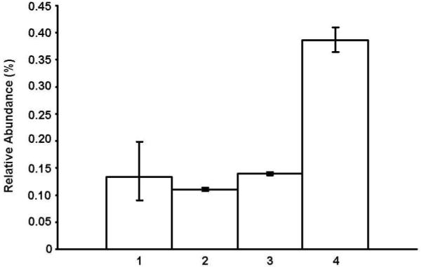
Quantitative real-time RT-PCR reactions were performed on total RNA extracts from HSC-3 cells using primers specific for CD44s (1), CD44 v3 (2), CD44 v6 (3), and CD44 v10 (4). RNA levels are expressed relative to the expression of 36B4, a human acidic ribosomal phosphoprotein P0 whose expression was unchanged in the tumor cells. Error bars were determined from standard deviations.
Next, lysates from HSC-3 cells that were transfected with CD44 siRNA, or non-transfected cells, were obtained and immunoblotted with anti-CD44 antibody, or anti-actin antibody as a loading control (FIGURE 3). The non-transfected HSC-3 cells demonstrated protein expression of multiple CD44 isoforms including CD44s and multiple CD44v variant isoforms (FIGURE 3, lane 1). However, HSC-3 cells transfected with CD44 siRNA showed loss of both CD44s and CD44v bands on anti-CD44 immunoblotting (FIGURE 3, lane 2), confirming CD44 protein expression in HSC-3 cells as well as demonstrating our ability to “knock-down” CD44 isoform function in subsequent tumor cell assays. Taken together, the quantitative real-time RT-PCR and immunoblotting results demonstrate that the HSC-3 cell line expresses multiple CD44 isoforms.
FIGURE 3. CD44 isoform protein expression in HSC-3 cells.
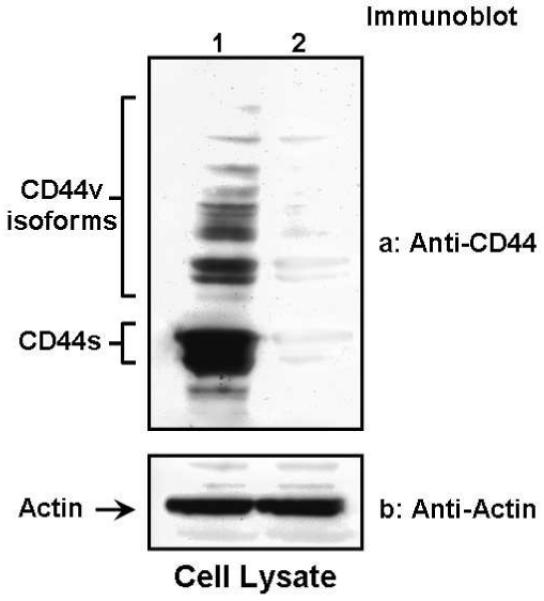
Protein lysates from HSC-3 cells transfected with CD44 siRNA (lane 2) or non-transfected cells (lane 1) were immunoblotted with anti-CD44 antibody (panel a) or anti-actin antibody (panel b) as a loading control.
CD44 isoforms mediate HSC-3 cell migration
The introduction of HA to HNSCC cell cultures results in increased tumor cell migration.14,15 Since all CD44 isoforms share a common ligand-binding domain for HA, we examined the role of the CD44 isoform receptor family on HSC-3 cell migration. Suppression of CD44 with siRNA was used to examine the role of CD44 receptor isoforms in HSC-3 cells on migration activity using in vitro Transwell migration assays (FIGURE 4). With HA as chemoattractant, we observed that untransfected (control) HSC-3 cells actively migrated (FIGURE 4, (1)). Similar migration activity was seen after HSC-3 cells were transfected with the control scrambled siRNA sequence (FIGURE 4, (2)). However, significantly reduced migration activity was observed when HSC-3 cells had been transfected with CD44 siRNA sequence (FIGURE 4, (3)), indicating that CD44 isoforms play a role in HNSCC tumor cell migration.
FIGURE 4. In vitro tumor cell migration assays.
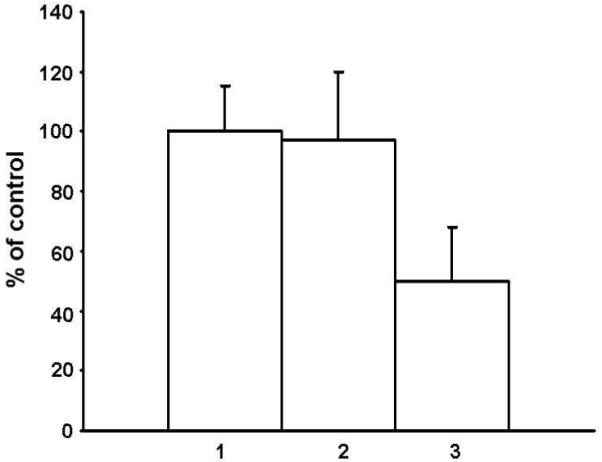
Using hyaluronan as a chemoattractant, Transwell migration assays of HSC-3 cells transfected with CD44 siRNA (3) or scrambled sequence siRNA (2) were performed as described in Methods. The migration of non-transfected HSC-3 cells (1) served as a reference control (designated 100%). Error bars were determined from standard deviations.
CD44 isoforms mediate HSC-3 cell proliferation and cisplatin sensitivity
The introduction of HA to HNSCC cell cultures results in increased tumor cell proliferation and cisplatin resistance.13-15 Suppression of CD44 with siRNA was used to examine the role of CD44 receptor isoforms in HSC-3 cell proliferation and cisplatin sensitivity in the absence of HA treatment. Tumor cell growth with cisplatin was measured via MTT assay in cells that had been transfected with CD44 siRNA, or scrambled sequence siRNA, or untransfected cells (FIGURE 5A). In the presence of increasing concentrations of cisplatin alone, there was progressive HSC-3 cell death (IC50 of 5.5 μM) in the untransfected control group. Transfection of HSC-3 cells with scrambled siRNA did not significantly alter the cisplatin sensitivity of the cell cultures (IC50 of 5.0 μM). However, transfection of HSC-3 cells with CD44 siRNA significantly increased the cisplatin sensitivity of the HNSCC cells at least 10-fold (IC50 of 0.4 μM), suggesting that CD44 receptors help mediate tumor cell cisplatin response.
FIGURE 5. In vitro tumor cell growth assays.
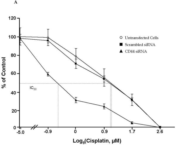
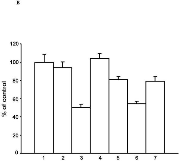
A) HSC-3 cells (untransfected or transfected with CD44siRNA or with non-targeting scrambled siRNA) were cultured for 24 hours followed by treatment with increasing concentration of cisplatin for 48 hours. The in vitro growth of these cells was analyzed by MTT assays. The 50% inhibitory concentration (IC50) was identified as a concentration of drug required to achieve a 50% growth inhibition relative to untreated controls. Error bars were determined from standard deviations. B) Untreated HSC-3 cells (1) and HSC-3 cells that were treated with normal rat immunoglobulin (2) (control for CD44 antibody), anti-CD44 antibody (3), normal rabbit immunoglobulin (4) (control for CD44 v3, v6, and v10 antibodies), anti-CD44v3 antibody (5), anti-CD44v6 antibody (6), or anti-CD44v10 antibody (7) underwent MTT assays to determine in vitro tumor cell growth. Error bars were determined from standard deviations.
Next, we examined the role of specific variant-containing isoforms of CD44 on tumor cell proliferation and cisplatin response. Tumor cell growth at 48 hours was measured via MTT assay after cells had been treated with anti-CD44 antibody, anti-CD44 v3 antibody, anti-CD44 v6 antibody, or anti-CD44 v10 antibody (FIGURE 5B). In the control group (FIGURE 5B, (1)) of HSC-3 cells (media alone, no antibody), there was robust cell growth that was not significantly reduced when cells were treated with non-specific normal rat (FIGURE 5B, (2)) or normal rabbit (FIGURE 5B, (4)) immunoglobulin. However, when tumor cells were treated with anti-CD44 antibody (binds to and blocks common HA-binding domain found in all isoforms of CD44), cell growth was reduced by about 50% (FIGURE 5B, (3)). Interestingly, when HSC-3 cells were treated with the CD44 variant antibodies (v3, v6, and v10), which bind to the respective variant exon domains but do not interfere with HA-binding to the CD44 receptor, a reduction in tumor growth was seen, although to a lesser degree than seen with anti-CD44 antibody. Among the 3 variant CD44 antibodies, the greatest reduction in tumor cell growth was seen with CD44 v6 (FIGURE 5B, (6)), with a lesser reduction seen with CD44 v3 (FIGURE 5B, (5)) and CD44 v10 (FIGURE 5B, (7)). These results suggest that CD44 variant isoforms may have unique signaling properties that mediate tumor growth in an HA-independent manner.
To assess whether specific variant-containing isoforms of CD44 may regulate cisplatin response in HSC-3 cells, the preceding experiment was repeated in the presence of increasing concentrations of cisplatin (TABLE 2). In the presence of cisplatin alone, HSC-3 cells had an IC50 of 5.0 μM. When cells were also treated with anti-CD44 antibody, HSC-3 cells exhibited increased cisplatin sensitivity with an IC50 of 1.3 μM. HSC-3 cells that were treated with CD44 v3, v6, and v10 also exhibited an increase in cisplatin sensitivity although to a lesser degree than seen with anti-CD44 antibody. Among the 3 variant CD44 antibodies, the greatest increase in cisplatin sensitivity was seen with CD44 v6 (IC50 of 1.5 μM), followed by CD44 v10 (IC50 of 2.5 μM) and CD44 v3 (IC50 of 4.0 μM). These results suggest that CD44 variant isoforms may have unique signaling properties that mediate tumor cisplatin response in an HA-independent manner. Taken together, these findings support the notion that CD44 variant isoforms play an important role in the regulation of HNSCC cell proliferation and cisplatin sensitivity.
TABLE 2. CD44 variant isoform inhibition and cisplatin sensitivity.
HSC-3 cells were treated with normal immunoglobulin (rat or rabbit), anti-CD44 antibody, anti-CD44v3 antibody, anti-CD44v6 antibody, or anti-CD44v10 antibody, followed by treatment with increasing concentrations of cisplatin. The in vitro growth of these cells was analyzed by MTT assay. The 50% inhibitory concentration (IC50) was identified as a concentration of drug required to achieve a 50% growth inhibition relative to untreated controls. IC50 are given +/- standard deviations
| IC50 (μM), Cisplatin | |
|---|---|
| Ctrl | 5.0 ± 0.6 |
| Normal IgG (rat or rabbit) | 4.8 ± 0.5 |
| CD44s | 1.3 ± 0.2 |
| CD44v3 | 4.0 ± 0.4 |
| CD44v6 | 1.5 ± 0.3 |
| CD44v10 | 2.5 ± 0.4 |
CD44s -v3, -v6, and -v10 isoforms are expressed in HNSCC primary tumors and lymph nodes
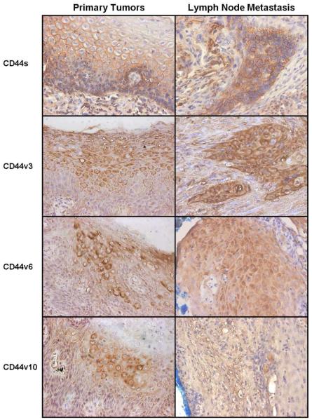
In vitro studies of HSC-3 cells indicate that multiple CD44 variant isoforms are likely expressed in HNSCC. We further observed that CD44 isoforms play a role in tumor progression behaviors. Next, we examined whether similar CD44 variant isoforms were expressed in clinical HNSCC tissue specimens. A series of 82 primary HNSCC tumors and 24 metastatic cervical lymph nodes were immunostained with antibodies for CD44s, CD44 v3, CD44 v6, and CD44 v10, as described in METHODS. FIGURE 6 shows representative examples of the immunohistochemical staining of HNSCC primary tumors and lymph nodes with the panel of antibodies. Note that there is mostly membrane staining with the CD44s antibody, while more cytoplasmic staining is seen with the CD44 variant antibodies.
FIGURE 6. CD44 variant isoform expression in head and neck squamous cell carcinoma (HNSCC) primary tumors and lymph nodes.
A panel of anti-CD44 variant antibodies (CD44s, CD44v3, CD44v6, and CD44v10) were used to examine CD44 isoform protein expression in HNSCC primary tumors and metastatic lymph nodes. Representative examples of primary tumors and lymph nodes stained for each of the CD44 antibodies are shown (original magnification x200). CD44 expression was scored as “strong,” “weak,” or “none,” as described in Methods. The example shown for CD44v10 expression in a lymph node was classified as weak, while the other examples shown were classified as strong.
Association of CD44 variant-containing isoforms with clinicopathologic variables
Semi-quantitative analysis of CD44 variant isoform expression was performed on the primary tumors and lymph nodes. Confirmation that specimens analyzed contained >80% tumor cells was performed by a clinical pathologist. All tumor specimens were reviewed by two observers blinded to the study variables and grouped according to the following system: None: rare or no cells stained; Weak: few cells (<50%) stained; Strong: majority of cells stained. CD44s, CD44v3, CD44v6, and CD44v10 expression in both primary tumors and lymph nodes were analyzed statistically with respect to various demographic and clinical outcome parameters (TABLE 1). The patient parameters examined included gender, age, presence of tobacco abuse, primary tumor T status, grade of differentiation, presence of lymph nodes, distant metastasis, recurrence after radiotherapy, disease free survival, and overall survival.
Standard CD44s expression was not associated with any of the clinical patient parameters. No association of any of the CD44 variant isoforms was found with respect to grade of tumor differentiation, age, gender, or history of tobacco abuse. TABLE 3 shows that CD44 v3, v6, and v10 were strongly expressed in a higher proportion of lymph nodes than primary tumors, and this difference was statistically significant (p=.03, p=.004, and p=.03, respectively). TABLE 4 demonstrates that strong CD44 v3 and v6 expression were associated with advanced T stage (p=.01 and p<.0001, respectively). Strong CD44 v3 expression in primary tumors was associated with positive lymph node metastasis (p=.03, data not shown). TABLE 5 and TABLE 6 indicate that strong CD44 v10 expression in primary tumors was associated with radiation failure (p<.0001) and with distant metastasis (p=.004). Primary tumor CD44 v6 expression was significantly associated with perineural invasion (p=.02, data not shown).
TABLE 3. CD44 variant expression in primary tumors and lymph nodes.
| Primary tumor, n =82 | Metastatic lymph node, n =24 | |||||
|---|---|---|---|---|---|---|
| None | Weak | Strong | None | Weak | Strong | |
| CD44s | 8 | 14 | 60 | 8 | 6 | 10 |
| CD44 v3 | 24 | 20 | 38 | 4 | 6 | 14 |
| CD44 v6 | 10 | 46 | 26 | 4 | 2 | 18 |
| CD44 v10 | 50 | 16 | 16 | 8 | 2 | 14 |
TABLE 4. CD44 variant expression in early vs. advanced primary tumors.
| Patients with early (T1-T2) vs. advanced (T3-T4) primary tumors | |||||||
|---|---|---|---|---|---|---|---|
| T1-T2, n=44 | T3-T4, n=38 | ||||||
| None | Weak | Strong | None | Weak | Strong | ||
| CD44s | 3 | 9 | 32 | CD44s | 5 | 5 | 28 |
| CD44 v3 | 15 | 15 | 14 | CD44 v3 | 9 | 5 | 24 |
| CD44 v6 | 4 | 36 | 4 | CD44 v6 | 6 | 10 | 22 |
| CD44 v10 | 27 | 9 | 8 | CD44 v10 | 23 | 7 | 8 |
TABLE 5. Primary tumor CD44 variant expression in previously irradiated patients.
| Patients with prior radiation or chemotherapy, n=14 | |||
|---|---|---|---|
| None | Weak | Strong | |
| CD44s | 1 | 4 | 9 |
| CD44 v3 | 7 | 3 | 4 |
| CD44 v6 | 4 | 5 | 4 |
| CD44 v10 | 2 | 4 | 8 |
TABLE 6. Primary tumor CD44 variant expression in patients with distant metastasis.
| Patients with distant metastasis, n=9 | |||
|---|---|---|---|
| None | Weak | Strong | |
| CD44s | 0 | 2 | 7 |
| CD44 v3 | 3 | 4 | 2 |
| CD44 v6 | 2 | 3 | 4 |
| CD44 v10 | 1 | 4 | 4 |
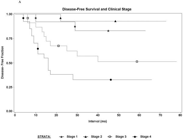
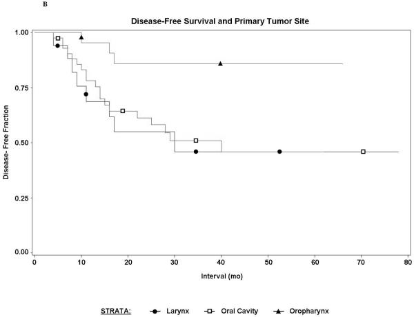
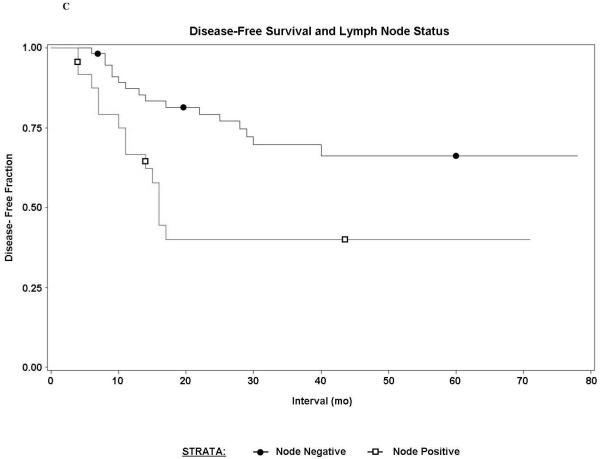
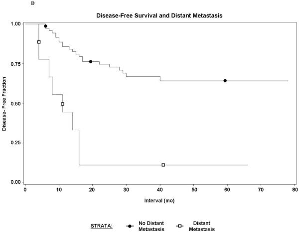
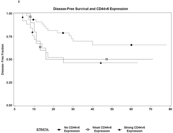
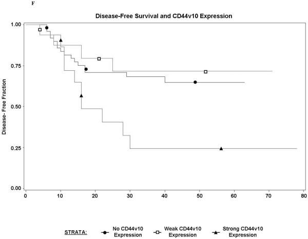
We analyzed our study population with respect to disease free survival and overall survival. As expected, overall tumor stage correlated with shorter disease free interval (p=.0001). Non-oropharyngeal primary site (p=.02), positive cervical lymph nodes (p=.002), distant metastasis (p<.0001), and CD44 v6 (p=.03) and CD44 v10 expression (p=.04) in primary tumors were all significantly associated with worse disease free survival (FIGURE 7). Prior radiation therapy (p=.9), CD44s expression (p=.9), and CD44 (standard and variant) expression in metastatic lymph nodes were not significantly associated with disease free interval or overall survival (data not shown). Our analysis of CD44s, v3, v6, and v10 expression support the notion that various CD44 variant isoforms are associated with advanced primary tumor stage, metastasis, treatment failure, and reduced disease-free interval.
FIGURE 7. Disease-free survival analysis of study population.
A) Disease free survival rates in patients, grouped by clinical stage (p=.0001); B) Disease free survival rates in patients, grouped by primary tumor site (p=.02); C) Disease free survival rates in patients with or without positive cervical lymph nodes (p=.002); D) Disease free survival rates in patients with or without distant metastasis (p<.0001); E) Disease free survival rates in patients with or without primary tumor expression of CD44 v6 (p=.03); F) Disease free survival rates in patients with or without primary tumor expression of CD44 v10 (p=.04).
DISCUSSION
Our results demonstrate that CD44 isoforms play a role in mediating HNSCC progression behaviors including tumor cell migration, proliferation, and cisplatin response. Supporting our in vitro data is the finding that the CD44 v3, -v6, and -v10 variant-containing isoforms are significantly correlated with various clinicopathologic variables including advanced T stage, regional and distant metastasis, radiation failure, perineural invasion, and shorter disease-free survival.
Proliferation, migration, and drug resistance characterize tumor progression in HNSCC cells. Oncogenic signaling and cytoskeleton functions are directly involved in these in vitro tumor progression behaviors, which result in the observed clinical features of advanced HNSCC. Clinical features of advanced HNSCC include adjacent tissue invasion, metastasis, and treatment resistance, which lead to increased disease recurrence and decreased survival. A number of studies have aimed to identify those molecules which are expressed in epithelial tumor cells and mediate these tumor progression behaviors. Among such molecules is CD44, a family of glycoprotein receptors expressed in many tumor cells.2-10,27 As transmembrane proteins, CD44 isoforms are capable of interactions with both the cell cytoplasm and the extracellular milieu. Among the major components of the extracellular matrix are hyaluronan, and all CD44 isoforms share a common extracellular binding domain for HA. All CD44 isoforms also share common cytoplasmic domains, most notably ankyrin-binding regions, which are capable of interacting with cytoskeleton proteins to activate cell signaling pathways.27,28 CD44 isoforms also interact with other receptor and non-receptor kinases, including EGFR, ErbB2, src, and transforming growth factor (TGF)-β.3 The ability of CD44 receptors to provide a direct link between the extracellular matrix and the cytoskeleton, coupled with their ability to interact with a multitude of signaling kinases, explains how one family of molecules is able to mediate a diversity of tumor progression functions.
The mechanism of CD44 receptors to promote tumor migration, proliferation, and survival through HA-mediated signaling has been described.4,5,8,9,13-17 HA binding to the extracellular domain of CD44 promotes interaction of CD44 with the cytoskeleton, particularly ankyrin through the conserved ankyrin-binding domain of the CD44 cytoplasmic region. This CD44-ankyrin interaction causes activation of various intracellular signaling pathways leading to the onset of cytoskeleton function.29-32 One of the important CD44-ankyrin mediated intracellular signaling pathways is the release of intracellular Ca2+ stores, which in turn, results in calmodulin-dependent activation, promoting tumor cell migration.13-16 Other CD44-ankryin mediated functions include cell adhesion and degradation of the extracellular matrix to allow tumor cell invasion.27 CD44 has also been shown to be physically linked to other receptor kinases in tumor cells including EGFR, ErbB2, and TGF-β, implicating a role for HA and CD44 in these well-described oncogenic signaling pathways.14,17,31 CD44 and ErbB2 are physically linked via disulfide bonds in ovarian tumor cells.31 HA binding to the CD44-ErbB2 complex activates ErbB2 tyrosine kinase activity and promotes ovarian tumor cell growth and migration. HA and CD44 interact with EGFR in HNSCC to promote various EGFR-mediated downstream signaling pathways.14,17 CD44-EGFR mediates extracellular signal-regulated kinase (ERK) phosphorylation, which has been shown to lead to increased tumor cell growth, migration, and chemotherapy resistance in HNSCC.
CD44 isoforms are expressed to varying degrees in both normal and tumor cells, suggesting a normal cellular function for many of the different CD44 receptor isoforms. How similar CD44-mediated signaling pathways can promote both normal and malignant cellular behaviors can be explained by several mechanisms. The mechanisms to regulate CD44 signaling include variable N-/O-linked glycosylation in the CD44 extracellular domain and modulation of the CD44 cytoplasmic domain by cytoskeletal proteins such as ankyrin. It is thought, however, that the most important mechanism that results in differing CD44-mediated signaling is by alteration of the CD44 receptor through additional exon-coded structures (via an alternative splicing process), leading to production of CD44 variant isoforms. In cancer cells, this latter mechanism can result in overexpression or novel expression of certain CD44 variant isoforms.27 The insertion of various variant exons to the extracellular component of the CD44 receptor has been shown to alter the signaling properties of the CD44 receptor by providing additional binding domains for molecules other than HA.3,27 The insertion of variant exons to the standard CD44 receptor may also result in changes to the HA-binding affinity of the CD44 isoform, further altering the receptor’s signaling behavior. We describe below some of the unique signaling properties of some of the more well-described CD44 variant isoforms.
Several CD44 variant exons are known to contain binding domains for various growth factor ligands. For example, the v3 exon (exon 7) is known to contain important glycosaminoglycan (GAG) attachment sites, and it has been postulated that tumor cells with CD44 receptors exhibiting these GAG sequences are involved with heparin binding growth factors such as basic fibroblast growth factor (bFGF), vascular endothelial growth factor (VEGF), and heparin binding-epidermal growth factor.8,23,24 The CD44 v3 isoform is expressed in the metastatic breast tumor cell line Met-1, where it was shown to bind VEGF, suggesting that this isoform may promote breast tumor-associated angiogenesis.8,23 CD44 v3 also appears to be linked to several other tumorigenic molecules to promote tumor cell migration and the invasive tumor cell phenotype. In particular, CD44 v3 co-localizes with the active form of matrix metalloproteinase-9 (MMP-9) in Met-1 cells, promoting degradation of the extracellular matrix to facilitate tumor cell invasion.8 This isoform also upregulates cytoskeleton function, through ankyrin, to activate the membrane-associated actomyosin contractile system and mediate tumor cell migration. The role of CD44 v3 isoforms in HNSCC progression is highlighted by studies showing an association of v3-containing isoforms with HNSCC growth, migration, and matrix metalloproteinase expression.15,33 Transfection of a CD44 v3 isoform into a non-expressing HNSCC cell line also resulted in significantly increased tumor cell migration, but not proliferation.34 In the current investigation, CD44 v3 isoforms were expressed in HSC-3 cells. While treatment of anti-CD44 v3 antibody had less influence on in vitro proliferation or cisplatin sensitivity, tissue analysis revealed CD44 v3 isoforms were preferentially expressed in metastatic lymph nodes. Additionally, strong CD44 v3 isoform expression in primary tumors was significantly associated with advanced T stage and positive lymph nodes, but did not correlate with disease-free interval.
CD44 v6-containing isoforms also appear to promote tumor progression. Transfection of CD44 v6 converted non-metastatic rat carcinoma cells into metastatic cells, while co-injection of anti-CD44 v6 antibody into these same cells suppressed their metastatic behavior.35-36 The CD44 v6 splice variant was also found to stimulate sustained increased mitogen activated protein (MAP) kinase levels and subsequent downstream Ras signaling, resulting in increased tumor cell proliferation.35 In the current investigation, CD44 v6 isoforms were expressed in HSC-3 cells, and treatment of anti-CD44 v6 antibody resulted in decreased proliferation and increased cisplatin sensitivity. CD44 v6 isoforms were also preferentially expressed in metastatic lymph nodes, and strong expression in primary tumors was significantly associated with advanced T stage, perineural invasion, and decreased disease-free survival.
CD44 v10-containing isoforms have been studied regarding tumor progression in breast and renal cell carcinoma.23,37 Although all CD44 isoforms share HA binding domains, certain CD44 variant isoforms, such as CD44 v10, exhibit significantly reduced affinity for HA binding.27 It is thought that the reduction in HA-mediated cell adhesion in tumor cells expressing CD44 v10 may be the earliest event in the onset of tumor migration and invasion. The unique structure of CD44 v10 may also cause constitutive activation of CD44-cytoskeleton interactions which induce tumor cell migration and invasion. Studies have revealed that CD44 v10-transfected breast tumor cells display higher migration/invasion potential, produce higher levels of bFGF and interleukin-8, and exhibit more potent tumor growth potential than parental control cells.27 In the current investigation, CD44 v10 isoforms were strongly expressed in HSC-3 cells, and treatment of anti-CD44 v10 antibody resulted in moderately decreased proliferation and increased cisplatin sensitivity. CD44 v10 isoforms were also preferentially expressed in metastatic lymph nodes, and strong expression in primary tumors was significantly associated with distant metastasis, radiation failure, and decreased disease-free survival.
Considering the numerous studies regarding the unique signaling properties of CD44 variant isoforms, it is not surprising that the presence of high levels of CD44 variant isoforms is emerging as an important tumor marker in a number of solid malignancies. Overexpression of CD44 v3-containing isoforms was found to correlate with increased histologic grade and metastasis in breast cancer, increased metastasis in melanoma, and increased invasion and metastasis in colon cancer.6,9,20,23-35,38 CD44 v6 overexpression has been correlated with advanced tumor stage and poor survival in non-Hodgkin’s lymphoma and colon cancer.20 CD44 v10 correlation with renal cell carcinoma histologic grade, staging, and poor prognosis has been reported.37
The expression of CD44 variant isoforms in HNSCC has also been studied, but their role remains controversial. Whereas some studies have found a correlation between increased CD44 variant expression and HNSCC progression, other studies have reported no correlations or negative correlations. Reategui et al described a novel CD44 v3 isoform in both tissue and soluble form that correlated with HNSCC status.34 Wang et al reported that CD44 v3-containing isoforms were associated with HNSCC lymph node metastasis.15 Kawano and colleagues found correlations between CD44 v6 expression in HNSCC and tumor volume, lymph node metastasis, and shorter survival.39 However, others have reported that down-regulation of various CD44 variant isoforms correlated with a worse prognosis. Kanke et al reported that down-regulation of CD44 v2 correlated with poorer differentiation and shorter overall survival while down-regulation of CD44 v6 correlated with a higher rate of cervical metastasis.40,41 Fonseca et al found down-regulation of CD44 v3 and v6 correlated with tumor grade and pattern of invasion.7 Other groups, including Van Hal et al and Herold-Mende et al, found no correlation with CD44 splice variants and any clinicopathologic variables, and concluded that CD44 variant isoforms do not play a role in HNSCC progression.42,43 There are some possible explanations for these discrepant results. First, different studies have used different antibodies, making comparisons between research groups difficult. Certain CD44 variant domain epitopes may become hidden and not recognized by some antibodies due to post-translational changes (eg. glycosylation) which alter the 3-dimensional conformation of the protein. In addition, assessment of immunostaining positivity is dependent on what region of the tumor that is examined, and it has been reported that there are often large areas within the tumor that do not stain for many CD44 isoforms. Unlike most previous studies of HNSCC variant expression in the literature, our study included in vitro assays and clinical tissue analysis using the same CD44 variant antibodies, and both the in vitro and clinical specimen results in our investigation were consistent with each other. Thus, we believe our data is supportive of the notion that CD44 variant isoforms play an important role in HNSCC progression.
Recent work has shown that CD44 is preferentially expressed in tumor stem cells which have the unique ability to initiate stem cell-specific properties. Prince et al reported that CD44 is one of the important surface markers unique to cancer stem cells in HNSCC.44 The finding that CD44 is a marker for cancer stem cells provides further support to the notion that CD44 receptors are important in HNSCC progression, and suggests CD44 as a possible therapeutic target. What role the various CD44 variant isoforms might play in cancer stem cells remains largely unexplored. Studies of CD44 and cancer stem cells suggest that only a few CD44-positive cells are the critical determinants of tumor survival, which may explain the results of some studies showing minimal expression of CD44 in advanced (large volume) tumors, since these tumors may have proportionately fewer CD44-positive stem cells, but nonetheless, contain the essential tumor cells which mediate a more aggressive clinical phenotype.
Our investigation suggests an important role for CD44 variant isoforms in HNSCC progression. However, further study is warranted to better elucidate the mechanism by which CD44 v3, v6, and v10-containing isoforms mediate HNSCC progression. In addition, a search to identify other possible CD44 variant isoforms that may be important in HNSCC progression is also necessary. Our study identified isoforms in HNSCC cell cultures and tissue specimens that contained the CD44 v3, v6, or v10 exons, but we have not yet determined the exact composition of these CD44 variant isoforms. For example, it is not known whether HSC-3 cells express CD44 v3 (standard + v3 exon only) or CD44v3,8-10 (standard + v3 and v8-10 exons)--both variants have been previously reported in HNSCC--or other yet to be described v3-containing isoforms. Additional work with HSC-3 cells, including sequencing the v3-, v6-, and v10-containing isoforms that are expressed by this cell line, is planned. Similarly, for future HNSCC tissue analysis, obtaining samples that will allow use of RT-PCR techniques to clone and sequence the CD44 variant isoforms that are expressed will help elucidate the role of the unique CD44 variant isoforms that correlate with tumor progression. CD44 variant isoforms identified in clinical specimens may be transfected into HNSCC cell cultures, followed by proliferation, migration, and other assays to provide further understanding of the cellular mechanisms by which these isoforms promote tumor progression.
Our work and others suggest that CD44 variant isoforms are important tumor markers. As we learn more about the oncogenic signaling mechanisms unique to these variant isoforms, the implication that CD44 receptors may be a therapeutic target becomes apparent. Emerging techniques utilizing therapeutic siRNA and anti-sense RNAs can be applied to CD44 receptors. Future work planned includes utilizing an orthotopic nude mouse model of human HNSCC to grow CD44 variant isoform positive cancers that could then undergo anti-CD44 treatments to reduce the size and metastatic potential of the xenograft tumors.
CONCLUSION
In summary, our investigation examined the role of CD44 variant isoforms in HNSCC. In vitro analysis of a HNSCC cell line demonstrated expression of CD44 variant isoforms, where they were found to mediate migration, proliferation, and cisplatin sensitivity. Analysis of a series of HNSCC clinical tissue specimens revealed several CD44 variant isoforms whose expression was associated with various clinicopathologic variables. These data suggest CD44 variant isoforms may be important molecular markers and possible therapeutic targets in HNSCC treatment.
ACKNOWLEDGMENTS
We would like to acknowledge Dr. Darren Cox for reviewing pathology specimens and Dr. Alan Bostrom for his biostatistical analysis. We would also like to thank Christina Camacho for help in preparing graphs and tables.
This work was supported by a Veterans Affairs Career Development Award (SJW), American Academy of Otolaryngology-Head and Neck Surgery/American Head and Neck Society Young Investigator Award (SJW), National Institutes of Health Grants RO1 CA66163, RO1 CA78633 and PO1 AR39448 from the USPHS (LYWB), and a Veterans Affairs Merit Review grant (LYWB).
REFERENCES
- 1.Parkin DM, Pisani P, Ferlay J. Global cancer statistics, 2002. Ca Cancer J Clin. 2005;55:74–108. doi: 10.3322/canjclin.55.2.74. [DOI] [PubMed] [Google Scholar]
- 2.Toole BP, Hascall VC. Hyaluronan and tumor growth. Am J Pathol. 2002;161:745–747. doi: 10.1016/S0002-9440(10)64232-0. [DOI] [PMC free article] [PubMed] [Google Scholar]
- 3.Turley E, Noble P, Bourguignon L. Signaling properties of hyaluronan receptors. J Biol Chem. 2002;277:4589–92. doi: 10.1074/jbc.R100038200. [DOI] [PubMed] [Google Scholar]
- 4.Bourguignon LY, Singleton PA, Diedrich F, et al. CD44 interaction with Na+-H+ exchanger (NHE1) creates acidic microenvironments leading to hyaluronidase-2 and cathepsin B activation and breast tumor cell invasion. J Biol Chem. 2004;279:26991–7007. doi: 10.1074/jbc.M311838200. [DOI] [PubMed] [Google Scholar]
- 5.Bourguignon LY, Zhu H, Zhou B, et al. Hyaluronan promotes CD44v3-Vav2 interaction with Grb2-p185HER2 and induces Rac1 and Ras signaling during ovarian tumor cell migration and growth. J Biol Chem. 2001;276:48679–92. doi: 10.1074/jbc.M106759200. [DOI] [PubMed] [Google Scholar]
- 6.Iida N, Bourguignon LY. New CD44 splice variants associated with human breast cancers. J Cell Physiol. 1995;162:127–33. doi: 10.1002/jcp.1041620115. [DOI] [PubMed] [Google Scholar]
- 7.Fonseca I, Pereira T, Rosa-Santos J, Soares J. Expression of CD44 isoforms in squamous cell carcinoma of the border of the tongue: a correlation with histological grade, pattern of stromal invasion, and cell differentiation. J Surg Onc. 2001;76:115–20. doi: 10.1002/1096-9098(200102)76:2<115::aid-jso1021>3.0.co;2-9. [DOI] [PubMed] [Google Scholar]
- 8.Bourguignon LY, Gunja-Smith Z, Iida N, et al. CD44v3,8-10 is involved in cytoskeleton-mediated tumor cell migration and matrix metalloproteinase (MMP-9) association in metastatic breast cancer cells. J Cell Physiol. 1998;176:206–215. doi: 10.1002/(SICI)1097-4652(199807)176:1<206::AID-JCP22>3.0.CO;2-3. [DOI] [PubMed] [Google Scholar]
- 9.Kuniyasu H, Oue N, Tsutsumi M, Tahara E, Yasui W. Heparan sulfate enhances invasion by human colon carcinoma cell lines through expression of CD44 variant exon 3. Clin Cancer Res. 2001;7:4067–72. [PubMed] [Google Scholar]
- 10.Wibulswas A, Croft D, Pitsillides A, et al. Influence of epitopes CD44v3 and CD44v6 in the invasive behavior of fibroblast-like synoviocytes derived from rheumatoid arthritic joints. Arthritis Rheum. 2002;46:2059–64. doi: 10.1002/art.10421. [DOI] [PubMed] [Google Scholar]
- 11.Misra S, Ghatak S, Zoltan-Jones A, Toole B. Regulation of multi-drug resistance in cancer cells by hyaluronan. J Biol Chem. 2003;278:25285–88. doi: 10.1074/jbc.C300173200. [DOI] [PubMed] [Google Scholar]
- 12.Bates RC, Edwards NS, Burns GF, Fisher DE. A CD44 survival pathway triggers chemoresistance via lyn kinase and phosphoinositide 3-kinase/Akt in colon carcinoma cells. Cancer Res. 2001;61:5275–83. [PubMed] [Google Scholar]
- 13.Wang SJ, Bourguignon LY. Hyaluronan-CD44 promotes phospholipase C-mediated Ca2+ signaling and cisplatin resistance in head and neck cancer. Arch Otolaryngol Head Neck Surg. 2006;132:19–24. doi: 10.1001/archotol.132.1.19. [DOI] [PubMed] [Google Scholar]
- 14.Wang SJ, Bourguignon LY. Hyaluronan and the interaction between CD44 and epidermal growth factor receptor in oncogenic signaling and chemotherapy resistance in head and neck cancer. Arch Otolaryngol Head Neck Surg. 2006;132:771–778. doi: 10.1001/archotol.132.7.771. [DOI] [PubMed] [Google Scholar]
- 15.Wang SJ, Wreesmann VB, Bourguignon LY. Association of CD44 v3 containing isoforms in tumor cell growth, migration, matrix metalloproteinase expression, and lymph node metastasis in head and neck cancer. Head Neck. 2007;29:550–558. doi: 10.1002/hed.20544. [DOI] [PubMed] [Google Scholar]
- 16.Wang SJ, Peyrollier K, Bourguignon LY. The influence of hyaluronan-CD44 interaction on topoisomerase II activity and etoposide cytotoxicity in head and neck cancer. Arch Otolaryngol Head Neck Surg. 2007;133:281–288. doi: 10.1001/archotol.133.3.281. [DOI] [PubMed] [Google Scholar]
- 17.Bourguignon LY, Gilad E, Brightman A, Diedrich F, Singleton P. Hyaluronan-CD44 interaction with leukemia-associated RhoGEF and epidermal growth factor receptor promotes Rho/Ras co-activation, phospholipase C epsilon-Ca2+ signaling, and cytoskeleton modification in head and neck squamous cell carcinoma cells. J Biol Chem. 2006;281:14026–14040. doi: 10.1074/jbc.M507734200. [DOI] [PubMed] [Google Scholar]
- 18.Ni H, Leong A, Cheon D, Hooi S. Expression of CD44 variants in colorectal carcinoma quantified by real-time reverse transcriptase-polymerase chain reaction. J Lab Clin Med. 2002;139:59–65. doi: 10.1067/mlc.2002.120425. [DOI] [PubMed] [Google Scholar]
- 19.Lakshman M, Subramaniam V, Rubenthiran U, Jothy S. CD44 promotes resistance to apoptosis in human colon cancer cells. Exp Mol Pathol. 2004;77:18–25. doi: 10.1016/j.yexmp.2004.03.002. [DOI] [PubMed] [Google Scholar]
- 20.Kuhn S, Koch M, Nübel T, et al. A complex of EpCAM, claudin-7, CD44 variant isoforms, and tetraspanins promotes colorectal cancer progression. Mol Cancer Res. 2007;5:553–567. doi: 10.1158/1541-7786.MCR-06-0384. [DOI] [PubMed] [Google Scholar]
- 21.Pirinen R, Hirvikoski P, Bohn J, et al. Reduced expression of CD44v3 variant isoform is associated with unfavorable outcome in non-small cell lung carcinoma. Hum Pathol. 2000;31:1088–95. doi: 10.1053/hupa.2000.16277. [DOI] [PubMed] [Google Scholar]
- 22.Mizera-Nyczak E, Dyszkiewicz W, Heider KH, Zeromski J. Isoform expression of CD44 adhesion molecules, Bcl-2, p53 and Ki-67 proteins in lung cancer. Tumour Biol. 2001;22:45–53. doi: 10.1159/000030154. [DOI] [PubMed] [Google Scholar]
- 23.Kalish ED, Iida N, Moffat FL, Bourguignon LY. A new CD44v3-containing isoform is involved in tumor cell growth and migration during human breast carcinoma progression. Front Biosci. 1999;4:1–8. doi: 10.2741/kalish. [DOI] [PubMed] [Google Scholar]
- 24.Bourguignon LY, Zhu H, Shao L, Zhu D, Chen YW. Rho-kinase (ROK) promotes CD44v(3,8-10)-ankyrin interaction and tumor cell migration in metastatic breast cancer cells. Cell Motil Cytoskeleton. 1999;43:269–287. doi: 10.1002/(SICI)1097-0169(1999)43:4<269::AID-CM1>3.0.CO;2-5. [DOI] [PubMed] [Google Scholar]
- 25.Iida N, Bourguignon LY. Coexpression of CD44 variant (v10/ex14) and CD44S in human mammary epithelial cells promotes tumorigenesis. J Cell Physiol. 1997;171:152–160. doi: 10.1002/(SICI)1097-4652(199705)171:2<152::AID-JCP5>3.0.CO;2-N. [DOI] [PubMed] [Google Scholar]
- 26.Bourguignon LY, Ramez M, Gilad E, et al. Hyaluronan-CD44 interaction stimulates keratinocyte differentiation, lamellar body formation/secretion, and permeability barrier homeostasis. J Invest Dermatol. 2006;126:1356–1365. doi: 10.1038/sj.jid.5700260. [DOI] [PubMed] [Google Scholar]
- 27.Bourguignon LY, Zhu D, Zhu H. CD44 isoform-cytoskeleton interaction in oncogenic signaling and tumor progression. Front Biosci. 1998;3:d637–49. doi: 10.2741/a308. [DOI] [PubMed] [Google Scholar]
- 28.Singleton PA, Bourguignon LY. CD44 interaction with ankyrin and IP3 receptor in lipid rafts promotes hyaluronan-mediated Ca2+ signaling leading to nitric oxide production and endothelial cell adhesion and proliferation. Exp Cell Res. 2004;295:102–118. doi: 10.1016/j.yexcr.2003.12.025. [DOI] [PubMed] [Google Scholar]
- 29.Bourguignon LY. Hyaluronan-mediated CD44 activation of RhoGTPase signaling and cytoskeleton function promotes tumor progression. Semin Cancer Biol. 2008;18:251–259. doi: 10.1016/j.semcancer.2008.03.007. [DOI] [PMC free article] [PubMed] [Google Scholar]
- 30.Bourguignon LY, Peyrollier K, Xia W, Gilad E. Hyaluronan-CD44 interaction activates stem cell marker Nanog, Stat-3-mediated MDR1 gene expression, and ankyrin-regulated multidrug efflux in breast and ovarian tumor cells. J Biol Chem. 2008;283:17635–17651. doi: 10.1074/jbc.M800109200. [DOI] [PMC free article] [PubMed] [Google Scholar]
- 31.Bourguignon LY, Gilad E, Peyrollier K. Heregulin-mediated ErbB2-ERK signaling activates hyaluronan synthases leading to CD44-dependent ovarian tumor cell growth and migration. J Biol Chem. 2007;282:19426–19441. doi: 10.1074/jbc.M610054200. [DOI] [PubMed] [Google Scholar]
- 32.Zhu D, Bourguignon LY. Interaction between CD44 and the repeat domain of ankyrin promotes hyaluronic acid-mediated ovarian tumor cell migration. J Cell Physiol. 2000;183:182–195. doi: 10.1002/(SICI)1097-4652(200005)183:2<182::AID-JCP5>3.0.CO;2-O. [DOI] [PubMed] [Google Scholar]
- 33.Franzmann E, Weed DT, Civantos F, et al. A novel CD44 v3 isoform is involved in head and neck squamous cell carcinoma progression. Otolarngol Head Neck Surg. 2001;124:426–32. doi: 10.1067/mhn.2001.114674. [DOI] [PubMed] [Google Scholar]
- 34.Reategui EP, de Mayolo AA, Das PM, et al. Characterization of CD44v3-containing isoforms in head and neck cancer. Cancer Biol Ther. 2006;5:1163–1168. doi: 10.4161/cbt.5.9.3065. [DOI] [PubMed] [Google Scholar]
- 35.Günthert U, Hofmann M, Rudy W, et al. A new variant of glycoprotein CD44 confers metastatic potential to rat carcinoma cells. Cell. 1991;65:13–24. doi: 10.1016/0092-8674(91)90403-l. [DOI] [PubMed] [Google Scholar]
- 36.Seiter S, Arch R, Reber S, et al. Prevention of tumor metastasis formation by anti-variant CD44. J Exp Med. 1993;177:443–455. doi: 10.1084/jem.177.2.443. [DOI] [PMC free article] [PubMed] [Google Scholar]
- 37.Li N, Tsuji M, Kanda K, Murakami Y, Kanayama H, Kagawa S. Analysis of CD44 isoform v10 expression and its prognostic value in renal cell carcinoma. BJU Int. 2000;85:514–518. doi: 10.1046/j.1464-410x.2000.00483.x. [DOI] [PubMed] [Google Scholar]
- 38.Dome B, Somlai B, Ladanyi A, Fazekas K, Zoller M, Timar J. Expression of CD44v3 splice variant is associated with the visceral metastatic phenotype of human melanoma. Virchows Arch. 2001;439:628–35. doi: 10.1007/s004280100451. [DOI] [PubMed] [Google Scholar]
- 39.Kawano T, Nakamura Y, Yanoma S. Expression of E-cadherin, and CD44s and CD44v6 and its association with prognosis in head and neck cancer. Auris Nasus Larynx. 2004;31:35–41. doi: 10.1016/j.anl.2003.09.005. et a; [DOI] [PubMed] [Google Scholar]
- 40.Kanke M, Fujii M, Kameyama K, et al. Role of CD44 variant exon 6 in invasion of head and neck squamous cell carcinoma. Arch Otolaryngol Head Neck Surg. 2000;126:1217–1223. doi: 10.1001/archotol.126.10.1217. [DOI] [PubMed] [Google Scholar]
- 41.Kanke M, Fujii M, Kameyama K, et al. Clinicopathological significance of expression of CD44 variants in head and neck squamous cell carcinoma. Jpn J Cancer Res. 2000;91:410–5. doi: 10.1111/j.1349-7006.2000.tb00960.x. [DOI] [PMC free article] [PubMed] [Google Scholar]
- 42.Herold-Mende C, Seiter S, Born AI, et al. Expression of CD44 splice variants in squamous epithelia and squamous cell carcinomas of the head and neck. J Pathol. 1996;179:66–73. doi: 10.1002/(SICI)1096-9896(199605)179:1<66::AID-PATH544>3.0.CO;2-5. [DOI] [PubMed] [Google Scholar]
- 43.Van Hal NL, van Dongen GA, Stigter-Van Walsum M, Snow GB, Brakenhoff RH. Characterization of CD44v6 isoforms in head-and-neck squamous-cell carcinoma. Int J Cancer. 1999;82:837–845. doi: 10.1002/(sici)1097-0215(19990909)82:6<837::aid-ijc12>3.0.co;2-h. [DOI] [PubMed] [Google Scholar]
- 44.Prince ME, Sivanandan R, Kaczorowski A, et al. Identification of a subpopulation of cells with cancer stem cell properties in head and neck squamous cell carcinoma. Proc Natl Acad Sci U S A. 2007;104:973–978. doi: 10.1073/pnas.0610117104. [DOI] [PMC free article] [PubMed] [Google Scholar]


