Abstract
Background
We have shown previously that acute infection with the respiratory pathogen, pneumonia virus of mice (PVM), results in local production of the proinflammatory chemokine, CCL3, and that neutrophil recruitment in response to PVM infection is reduced dramatically in CCL3 -/- mice.
Results
In this work, we demonstrate that CCL3-mediated neutrophil recruitment is coordinated by interferon-gamma (IFNγ). Neutrophil recruitment in response to PVM infection was diminished five-fold in IFNγ receptor gene-deleted mice, although neutrophils from IFNγR -/- mice expressed transcripts for the CCL3 receptor, CCR1 and responded functionally to CCL3 ex vivo. Similarly, in the absence of PVM infection, CCL3 overexpression alone could not elicit neutrophil recruitment in the absence of IFNγ. Interestingly, although supplemental IFNγ restored neutrophil recruitment and resulted in a sustained weight loss among CCL3-overexpressing IFNγ -/- mice, CCL3-mediated neutrophil recruitment alone did not result in the pulmonary edema or respiratory failure characteristic of severe viral infection, suggesting that CCL3 and IFN-γ together are sufficient to promote neutrophil recruitment but not pathologic activation.
Conclusion
Our findings reveal a heretofore unrecognized hierarchical interaction between the IFNγ and CCL3, which demonstrate that IFNγ is crucial for CCL3-mediated neutrophil recruitment in vivo.
Background
Most respiratory virus infections are relatively benign and self-limited events. However, infection with highly pathogenic viruses can result in more severe sequelae, in which disease progresses to respiratory failure due to uncontrolled inflammation, pulmonary edema, and damage to lung tissue [1-5].
As part of an ongoing effort to understand inflammatory responses during severe respiratory virus infection, we have developed an inhalation model using the natural rodent pathogen, pneumonia virus of mice (PVM). Originally identified by Horsfall and colleagues [6,7], PVM is a pneumovirus (family Paramyxoviridae) that is closely related to respiratory syncytial virus (RSV), and is among the few characterized mouse models of virus-induced acute respiratory distress syndrome (ARDS) [7-9]. Among the prominent features of this infection, a minimal intranasal inoculum (30 – 100 pfu) results in robust virus replication within bronchial epithelial cells that is accompanied by profound granulocyte recruitment. In the absence of pharmacologic intervention, PVM infection progresses to pulmonary edema and respiratory compromise, similar to the more severe forms of RSV infection experienced by human infants [10,11]. In our earlier studies, we identified the chemokine CCL3 (MIP-1α) as a crucial component of this inflammatory response. PVM not only elicits production of CCL3 by infected bronchial epithelial cells [12], mice devoid of CCL3 or its receptor, CCR1, recruit dramatically fewer neutrophils to airways [13]. Blockade of the CCL3/CCR1 proinflammatory signaling pathway in conjunction with antiviral therapy resulted in improved survival in response to an otherwise lethal virus inoculum [14,15]. As CCL3 is only one of several major pro-inflammatory signaling pathways activated by PVM infection [12], there is certainly the possibility of additive, synergistic, or hierarchical means to promote and to amplify the ongoing inflammatory response.
Although first identified as a component of the antiviral response to Sindbis virus [16], the role of the Th1 cytokine, interferon-γ (IFNγ) in pneumovirus infection remains uncertain. IFNγ is readily detected in bronchoalveolar lavage fluid and nasal washings from RSV-infected infants [17,18], and minimal or absent response has been correlated with poor clinical outcome [19-24]. IFNγ is also detected in BAL fluid of BALB/c mice in response to challenge with RSV virions [25,26] and plays a role in limiting the inflammatory response to secondary challenge and in generating the allergic histopathology in response to formalin-fixed RSV vaccine antigens and virion components [27,28]. Likewise, local production of IFNγ is a prominent response to PVM infection [12,29,30], although its role in modulating the primary inflammatory response has not yet been fully explored.
In this manuscript, we explore the role of IFNγ in modulating the inflammatory response to PVM infection, and utilize overexpression analysis to begin a dissection of the independent and interdependent contributions of both IFN-γ and CCL3 to the process of neutrophil recruitment in vivo.
Results
Microarray profiling of IFNγ expression in response to PVM infection
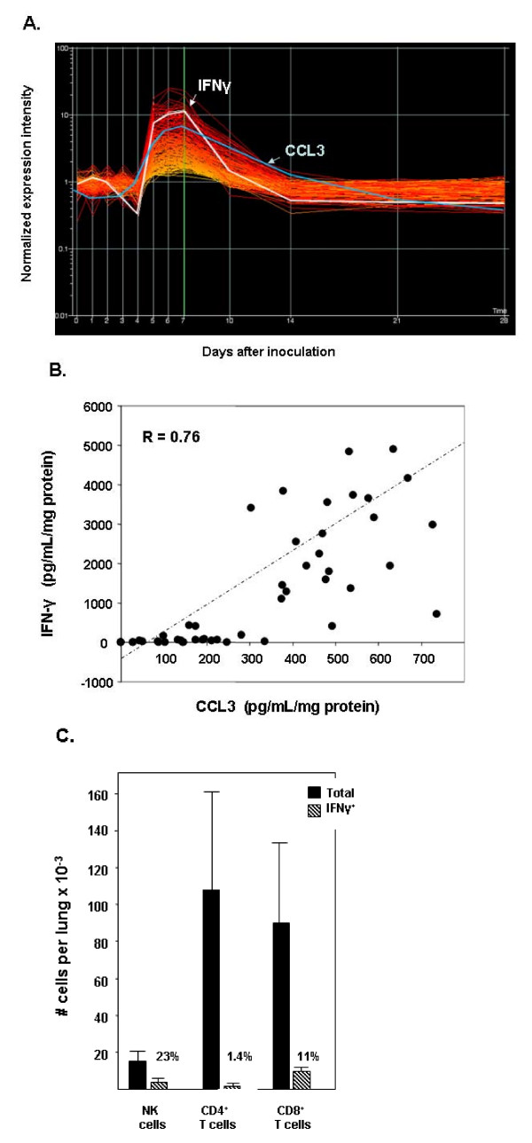
Transcript encoding the cytokine IFNγ was detected in mouse lung tissue at various time points in response to PVM infection [12]. In response to a non-lethal inoculum of PVM strain J3666, IFNγ mRNA was detected above baseline levels beginning on day 5. IFN-γ mRNA levels peak at day 7 after inoculation, and fall rapidly to baseline between days 7 – 14. Shown in Figure 1A are profiles of the 203 transcripts (of total 45,101 transcripts on the 430_2 mouse chip) that display kinetic expression correlations of 0.900 or greater with the IFN-γ profile, as per the 'find similar' algorithm of Genespring GX 7.3. Selected transcripts, categorized by function, are listed in Table 1. Among the transcripts that correlate with the IFNγ profile are 17 characterized interferon-response genes. Most intriguing is the close correlation (0.965) between the expression patterns of IFNγ and CCL3 (MIP-1α). CCL3 is essential for granulocyte recruitment in response to PVM infection [13]. As shown in Figure 1B, there is a significant correlation between levels of immunoreactive IFNγ and CCL3 in lung tissue from individual PVM-infected mice.
Figure 1.
(A) Expression of transcripts in mouse lung tissue in response to PVM infection: IFN-γ and IFN-γ correlating profiles. Baseline expression in uninfected mice (day 0) is set at 1.0 and normalized expression (per gene, per chip) is shown for days 1 – 7, 10, 14, 21 and 28 after inoculation. Profiles of 203 transcripts with patterns that correlate with that the profile of IFN-γ (0.900 to 0.969) are shown in yellow to red, respectively, and identified by name in Table 1. The expression profile of CCL3 (MIP-1α), a chemokine crucial for neutrophil recruitment in response to PVM infection, is overdrawn with a blue line (correlation 0.965). (B) Correlation of IFN-γ and CCL3 protein levels in individual PVM-infected mice. IFN-γ and CCL3 detected by ELISA in lung tissue homogenates from individual mice days 0 – 28 after inoculation with 30 pfu PVM (n = 43) are as shown. (C) IFNγ+ NK and T cells detected in lungs of PVM-infected mice. Total and IFNγ+ NK cells, CD4+ T cells, and CD8+ T cells (± sd) detected per lung on day 6 after inoculation with 10 pfu PVM.
Table 1.
Expression profiles that correlate with IFN-γ in PVM-infected mouse lung tissue.
| Transcript | Symbol | Acc. No. | Correl. |
| Interferon-γ and related transcripts | |||
| Interferon-γ | Ifng | K00083 | 1.000 |
| Interferon inducible protein 1 | Ifi1 | NM_008326 | 0.961 |
| Interferon-stimulated protein | Isg20 | BC022751 | 0.953 |
| Interferon-γ induced GTPase | Igtp | NM_018738 | 0.953 |
| Interferon-induced transmembrane protein 6 | Ifitm6 | BB193024 | 0.950 |
| CXC chemokine ligand 11 (IP-9) | Cxcl11 | NM_019494 | 0.949 |
| Interferon inducible protein 47 | Ifi47 | NM_008330 | 0.940 |
| Interferon activatible protein 203 | Ifi203 | AI607873 | 0.932 |
| Interferon activated gene 205 | Ifi205 | AI481797 | 0.929 |
| Interferon induced protein with tetratricopeptide repeats 1 | Ifit1 | NM_008331 | 0.929 |
| Interferon consensus sequence binding protein 1 | Icsbp1 | BG069095 | 0.926 |
| Interferon regulatory factor 7 | Irf7 | NM_016850 | 0.922 |
| Interferon activated gene 205 | Ifi205 | AI481797 | 0.916 |
| Interferon regulatory factor 5 | Irf5 | NM_012057 | 0.914 |
| Interferon activated gene 203 | Ifi203 | NM_008328 | 0.910 |
| Interferon-induced protein with tetratricopeptide repeats, 3 | Ifit3 | NM_010501 | 0.909 |
| Interferon induced protein with tetratricopeptide repeats 2 | Ifit2 | NM_008332 | 0.901 |
| Other inflammation-associated transcripts | |||
| CC Chemokine ligand 3 (MIP-1α) | Ccl3 | NM_011337 | 0.965 |
| Toll-like receptor 2 | Tlr2 | NM_011905 | 0.959 |
| Interleukin-13 receptor alpha 1 | Il13ra1 | S80963 | 0.959 |
| Suppressor of cytokine signaling 3 | Socs3 | NM_007707 | 0.951 |
| Galectin-9 | Lgals9 | NM_010708 | 0.948 |
| Interleukin-1 receptor antagonist | Il1rn | M57525 | 0.947 |
| Regulator of G-protein signaling 19 interacting protein 1 | Rgs19ip1 | NM_018771 | 0.943 |
| Interleukin-6 | Il6 | NM_031168 | 0.937 |
| CD244 natural killer cell receptor 2B4 | Cd244 | NM_018729 | 0.928 |
| CSF2 receptor | Csf2rb2 | NM_007781 | 0.928 |
| Fc receptor, IgG, high affinity, I | Fcgr1 | AF143181 | 0.926 |
| CC chemokine receptor 1 | Ccr1 | AV231648 | 0.926 |
| Pentaxin-related gene | Ptx3 | NM_008987 | 0.926 |
| CXC chemokine ligand 13 (BLC) | Cxcl13 | AF030636 | 0.921 |
| CXC chemokine ligand 2 (MIP-2α) | Cxcl2 | NM_009140 | 0.919 |
| CXC chemokine ligand 5 (ENA-78) | Cxcl1 | BB554288 | 0.914 |
| Arginase II | Arg2 | NM_009705 | 0.904 |
| Signal transducer and activator of transcription 1 | Stat1 | AW214029 | 0.904 |
| Regulator of G-protein signaling 1 | Rgs1 | NM_015811 | 0.903 |
| CC chemokine receptor-like 2 | Ccrl2 | AJ318863 | 0.902 |
| Various | |||
| Membrane-spanning 4-domains, subfamily A, member 8A | Ms4a8a | NM_022430 | 0.969 |
| Chondroitin sulfate proteoglycan 2 | Cspg2 | BM251152 | 0.963 |
| Fas death domain-associated protein | Daxx | NM_007829 | 0.960 |
| O-acyltransferase domain containing 1 | Oact1 | AV366860 | 0.960 |
| Mitogen activated protein kinase kinase kinase kinase 1 | Map4k1 | BB546619 | 0.960 |
| Lymphocyte cytosolic protein 2 | Lcp2 | BC006948 | 0.959 |
| Solute carrier family 15, member 3 | Slc15a3 | NM_023044 | 0.956 |
| Indoleamine-pyrrole 2,3 dioxygenase | Indo | NM_008324 | 0.954 |
| Proteosome subunit beta type 9 | Tap1 | AW048052 | 0.952 |
| Phospholipase A1 member A | Pla1a | NM_134102 | 0.949 |
| Methylene tetrahydrofolate dehydrogenase | Mthfd2 | BG076333 | 0.949 |
| Pre-B colony enhancing factor 1 | Pbef1 | AW989410 | 0.948 |
| Thioredoxin reductase 1 | Txnrd1 | BB284199 | 0.948 |
| CGG triplet repeat binding protein 1 | Cggbp1 | BI080272 | 0.945 |
| Sphingosine kinase 1 | Sphk1 | AF068749 | 0.944 |
| Pyrophosphatase | Pyp | NM_026438 | 0.944 |
| 2'-5' oligoadenylate synthetase 1G | Oas1g | BC018470 | 0.943 |
| Ubiquitin D | Ubd | NM_023137 | 0.943 |
| Apolipoprotein D | Apod | NM_007470 | 0.940 |
| Membrane-spanning 4-domains, subfamily A, member 4C | Ms4a4b | NM_029499 | 0.936 |
| AT rich interacting domain 5A | Arid5a | BC027152 | 0.935 |
| Hemopoietic cell kinase | Hck | NM_010407 | 0.933 |
| Histocompatibility 2, complement component factor B | H2-Bf | NM_008198 | 0.933 |
| ATP binding cassette | Abcc5 | BB436535 | 0.933 |
| Cholesterol 25-hydroxylase | Ch25h | NM_009890 | 0.932 |
| BING 4 protein | Bing4 | C78559 | 0.932 |
| Thymidylate kinase, LPS inducible | Tyki | AK004595 | 0.930 |
| Tripartite motif protein 30 | Trim30 | BM240719 | 0.929 |
| Tissue specific transplantation antigen 30 | Tsta3 | NM_031201 | 0.929 |
| Syndecan binding protein | Sdcbp | AV227603 | 0.928 |
| Prostaglandin-endoperoxide synthase 2 | Ptgs2 | M94967 | 0.926 |
| Traf binding protein | T2bp | BB277065 | 0.925 |
| Two pore segment channel 2 | Tpcn2 | BC025890 | 0.925 |
| Early growth response 2 | Egr2 | X06746 | 0.925 |
| GLI pathogenesis-related 2 | Glipr2 | BM208214 | 0.925 |
| Cytochrome p450, family 7, subfamily b | Cyp7b1 | NM_007825 | 0.924 |
| Rab20, Ras oncogene | Rab20 | BG066967 | 0.923 |
| Solute carrier 39 | Slc39a14 | BB399837 | 0.922 |
| Dual specificity phosphatase 3 | Dusp3 | BQ266434 | 0.922 |
| Ribosome binding protein 1 | Rrbp1 | AF273691 | 0.922 |
| Spermidine synthase | Srm | NM_009272 | 0.921 |
| Ubiquitin-specific protease 18 | Usp18 | NM_011909 | 0.920 |
| Lipocalin | Lcn2 | X14607 | 0.920 |
| Jun-B oncogene | Junb | NM_008416 | 0.919 |
| Guanylate nucleotide binding protein 3 | Gbp3 | NM_018734 | 0.919 |
| Pre-B cell colony-enhancing factor 1 | Pbef1 | AW989410 | 0.917 |
| Membrane-spanning 4-domains subfamily A, member 6B | Ms4a6b | NM_027209 | 0.917 |
| SLAM family member 7 | Slamf7 | AK016183 | 0.915 |
| Ras and Rab interactor 1 | Rin1 | BC011277 | 0.915 |
| Class II transactivator | C2ta | AF042158 | 0.913 |
| Myxovirus resistance I | Mx1 | M21039 | 0.910 |
| Chloride channel calcium activated 2 | Clca1 | AF108501 | 0.910 |
| Rap2C, member of RAS oncogene family | Rap2c | AK008416 | 0.910 |
| Tumor necrosis factor, alpha induced protein 2 | Tnfaip2 | NM_009396 | 0.908 |
| SLAM family member 8 | Slamf8 | BC024587 | 0.908 |
The microarray analysis software package, Genespring GX 7.3 'find similar' function was used to inspect all transcript profiles for patterns related to that displayed by IFN-γ. The minimum correlation considered to be similar was set at 0.900 (see Figure 1A).
Detection IFNγ+ NK and T cells in PVM infected lung tissue
Both total and IFNγ+ subsets of NK cells, CD4+ and CD8+ T cells were enumerated in single cell suspensions of lung tissue from PVM-infected BALB/c mice evaluated at day 6 after inoculation with 10 pfu PVM strain J3666 [Figure 1C]. Only a small fraction (<2%) of the CD4+ T cells detected at this time point stained positively for IFNγ, in contrast to the larger fraction of IFNγ+CD8+ T cells detected (9.9 ± 0.6 × 10³ cells, 11% of total CD8+ T cells). Interestingly, 23% of the total NK cells (3.4 ± 0.9 × 10³ cells) stained positively for IFNγ, an increase from 0.3 ± 0.08 × 10³ cells, or 4% of the total NK cells detected in a single lung from uninfected mice (data not shown).
IFNγ-dependent responses to PVM infection
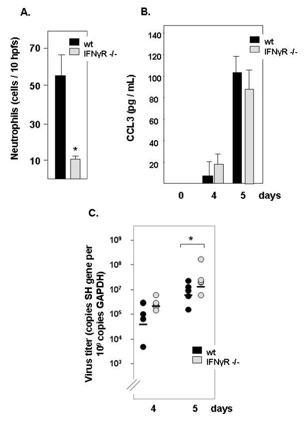
Wild type and IFNγ receptor gene deleted (IFNγR -/-) mice were infected with PVM and various parameters relating to the inflammatory response were assessed. Neutrophil recruitment to the airways was markedly diminished in IFNγR -/- mice [Figure 2A], reduced from 54 ± 11 per 10 hpfs among wild type to 10 ± 1.3 hpfs among IFNγR -/- mice, as determined on cytospin preparations of cells in BAL fluid (p < 0.001). These findings are consistent with those of Frey and colleagues [30], who described reduced inflammation in association with reduced IFNγ production in the lungs of PVM infected, T-cell deficient mice. Given our earlier studies on the essential role of CCL3 in eliciting neutrophil recruitment, it is interesting to note that the absence of IFNγ signaling had no impact on local production of this chemokine in response to PVM infection [Figure 2B]. IFNγ was also detected in response to PVM infection in both wild type and in IFNγR-/- mice, albeit at higher levels among the latter group, most likely due to the absence of feedback inhibition (data not shown). The diminished neutrophil recruitment, while significant, was not as profound as that observed in mice subjected to complete blockade of CCL3-mediated signaling, in which we observed 104-105 fold-diminished neutrophil recruitment [14,15]. As might be anticipated from the diminished inflammatory response, we observe a statistically significant increase in virus titer among the IFNγR-/- mice [Figure 2C], although this difference is likewise not as dramatic as that observed in response to complete blockade of CCL3 signaling.
Figure 2.
Neutrophil recruitment in response to PVM infection is diminished in IFN-γR gene-deleted mice. (A) Neutrophils detected in BAL fluid 5 days after inoculation; hpf, high power field; *p < 0.001;(B) Detection of CCL3 in BAL fluid; *p < 0.001 (C) Virus copy number detected in lung tissue determined by quantitative RT-PCR.
Receptor expression and responses of neutrophils from IFNγR gene-deleted mice
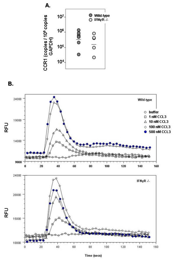
As part of an initial attempt to determine whether neutrophils from IFNγR -/- mice were capable of responding to CCL3, we explored receptor expression and ligand-mediated calcium flux in neutrophils isolated from both gene-deleted and wild type mice. As shown in Figure 3A, both wild type and IFNγR-/- neutrophils express transcripts encoding CCR1, the major receptor for CCL3; no significant difference in absolute copy number was determined. Likewise, CCL3 induced dose-dependent intracellular calcium flux in both gene-deleted and wild type neutrophils [Figure 3B], demonstrating that neutrophils from IFNγR-/- mice have the innate capacity to respond to this chemoattractant ligand; the EC50s and maximum calcium fluxes detected were indistinguishable between the wild type and gene-deleted strains.
Figure 3.
Comparison of wild type and IFNγR gene-deleted neutrophils. (A) Expression of CCR1 transcript in wild type and IFNγR gene-deleted neutrophils (n = 9 and 6 independent samples, respectively) determined by quantitative RT-PCR; horizontal line denotes mean copy number. (B) Calcium flux (RFU) measured in response to increasing concentrations (0 – 500 nM) of CCL3.
Overexpression of CCL3
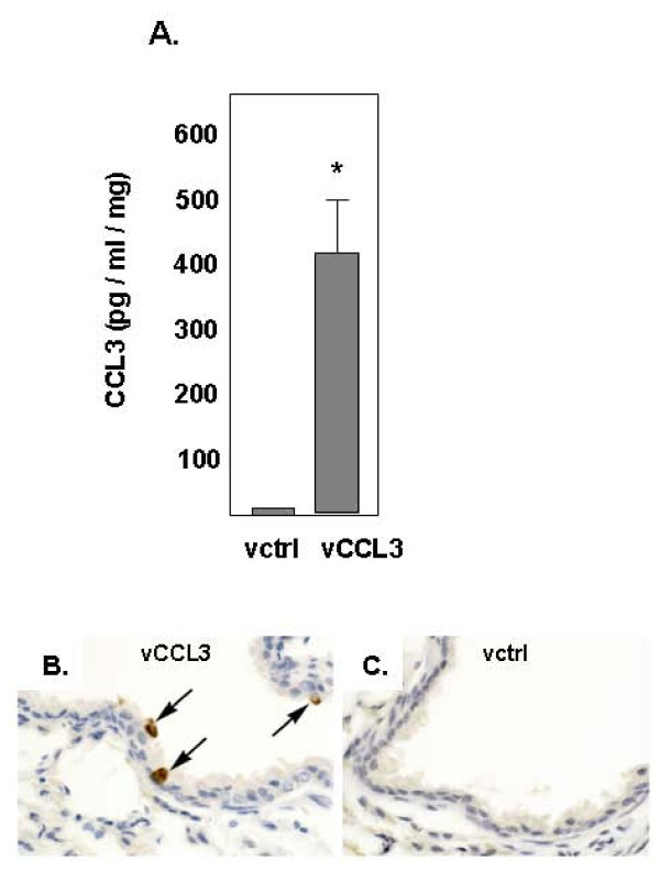
In order to examine the independent and interdependent contributions of CCL3 and IFNγ to the process of neutrophil recruitment in vivo, we generated a method for overexpression of CCL3 in vivo. CCL3 was detected in lung tissue homogenates [Figure 4A], reaching levels similar to those detected in lung tissue of mice in response to PVM infection [12]. Immunoreactive CCL3 was detected in bronchial epithelial cells [Figure 4B]. No CCL3-positive cells were detected in lung tissue from mice challenged with control vector (vctrl) [Figure 4C].
Figure 4.
Heterologous expression of CCL3 in mouse lungs. (A) Detection of immunoreactive CCL3 in lungs of mice on day 9 after challenge via intranasal inoculation with the CCL3 overexpression vector (vCCL3) or control vector (vctrl), *p < 0.01. (B) Lung tissue from mice challenged with vCCL3, immunohistochemical localization of CCL3 within bronchiolar epithelial cells (at arrows), (C) Lung tissue from mice challenged with vctrl.
Inflammatory responses to IFNγ and CCL3
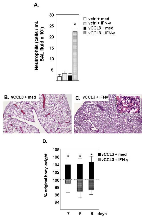
We examined neutrophil recruitment in response to CCL3 overexpression in IFNγ gene-deleted mice (IFNγ -/-) with and without IFNγ supplementation. As shown in Figure 5A, few neutrophils are detected in BAL fluid at baseline (vctrl) and no recruitment over baseline is observed in response to IFNγ alone. Likewise, overexpression of CCL3 in the absence of IFNγ does not elicit neutrophil recruitment. Neutrophil recruitment (~10 – fold over baseline) was observed in response to CCL3 expression only in the presence of IFNγ. At the microscopic level, no inflammation was observed in lung tissue of IFNγ -/- mice in response to CCL3 overexpression alone [Figure 5B]. In contrast, significant pathology was observed in lung tissue of IFNγ -/- mice expressing CCL3 and supplemented with exogenous IFNγ. Findings include moderate peribronchiolar granulocytic infiltration and substantial parenchymal involvement but minimal edema fluid within the bronchioles and in the parenchymal tissue [Figure 5C]. Interestingly, weight loss is sustained among the mice overexpressing CCL3 while receiving supplemental IFNγ over the 9 day examination period [Figure 5D], but, despite the substantial inflammatory response, we observe no progression to respiratory failure up to and including t = 14 days.
Figure 5.
Neutrophil recruitment in response to CCL3 is ablated in IFN-γ gene-deleted mice. (A) Neutrophils detected in BAL fluid of IFNγ gene-deleted (IFNγ -/-) mice (+vctrl +med (medium; RPMI + 10% FCS vehicle control); open bar), + IFNγ (+vctrl + IFNγ, grey-shaded bar), +vCCL3 +med (black bar), or +vCCL3 + IFNγ (black-shaded bar); *p < 0.01 vs. other conditions, day 9 after challenge with vCCL3 or vctrl. (B, C) Microscopic images of lung tissue from IFN-γ -/- mice challenged with (B) vCCL3 + med or (C) vCCL3 + IFNγ; original magnification, 20×. Inset, original magnification 63×, documenting neutrophil recruitment. (D) Change in body weight in response to CCL3 overexpression ± IFN-γ; *p < 0.01 at time points shown.
Discussion
In previous work, we demonstrated that the actions of the chemokine, CCL3, signaling via its receptor CCR1, were crucial for granulocyte recruitment to the lungs in response to PVM infection [13-15]; CCL3 has also been shown to be a crucial mediator of granulocyte recruitment in mouse models of influenza [31]. Paradoxically, CCL3 gene-deletion results in augmented neutrophil and eosinophil recruitment in response to Cryptococcus neoformans infection [32]. Here we show that CCL3-mediated neutrophil recruitment depends directly on IFNγ signaling, both in the setting of acute virus infection and in response to heterologous CCL3 expression in the respiratory epithelium.
Granulocyte recruitment is a primary finding in severe respiratory virus infection; activation of granulocytes can result in the release of proinflammatory cytokines and proteolytic enzymes that can contribute to the ongoing lung damage [33-37]. Interestingly, although neutrophils are recruited to the lung parenchyma in response to CCL3 via coordination by IFNγ, these cytokines alone clearly are not sufficient to induce the inflammatory state that ultimately promotes lung damage and respiratory failure. Thus, despite our findings demonstrating improved survival from PVM infection with CCR1 blockade [15], and those of He and colleagues [38], who likewise demonstrated that CCR1 antagonism provided protection against neutrophil-mediated lung injury in a mouse model of acute pancreatitis, the results presented here, in which we observe neutrophil recruitment but minimal clinical disease, suggest that neutrophil recruitment and neutrophil activation are to some extent distinct and discrete signaling events. It will be crucial to identify the proinflammatory mediators that activate and well as those that recruit neutrophils in order to have a complete picture of the proinflammatory state characteristic of PVM infection.
The experimental studies performed in this manuscript utilize both IFNγ and IFNγR gene-deleted mice, which are in BALB/c and C57BL/6 background strains, respectively. PVM infection has been explored systematically in several inbred strains of mice by Anh and colleagues [39] who determined that the C57BL/6 strain is somewhat more resistant to infection than BALB/c, but that both of these inbred strains can ultimately succumb to the sequelae of severe disease. We have used both of these strains extensively for our studies (reviewed in [7-9]) and both respond to PVM infection with robust virus replication in lung tissue, granulocyte recruitment and local production of proinflammatory cytokines, including CCL3 and IFNγ; no systematic differences, other than the aforementioned susceptibility to infection, have been detected.
Both CCL3 and IFNγ have been detected in human studies and in mouse models of other severe respiratory virus infections, including avian influenza, SARS coronavirus, and human respiratory syncytial virus [17,18,40-47], although the potential for interplay between these specific signaling pathways has not been considered previously. Our data suggest that that IFNγ and CCL3 signaling pathways, both crucial features of the response to pneumovirus infection, interact in a hierarchical fashion, as IFNγ does not elicit neutrophil recruitment on its own [Figure 5A], but is crucial for CCL3 to function effectively. Interactions between IFNγ and CCL3 may occur at the level of signal transduction, or via alterations to the neutrophil itself. As has been documented clearly, CCL3 can function alone to induce changes in calcium concentration and chemotactic responses in mouse neutrophils in vitro [48]. The current literature on interactions of IFNγ with granulocytes was recently reviewed [49]. Among the possibilities that may address our findings, Hansen and Finbloom [50] reported that human neutrophils express IFNγ receptors and Bonecchi and colleagues [51] have shown that human neutrophils respond to IFNγ with increased expression of a variety of mediators and receptors, including the primary CCL3 receptor, CCR1. It is unclear whether mouse neutrophils respond in a similar fashion, and whether or not these defined molecular responses take place in vivo, although we have shown here that neutrophils from IFNγR gene-deleted mice express transcripts for CCR1 and mobilize intracellular calcium in response to CCL3 when examined ex vivo. We have not yet explored the possibility that the IFNγ coordinates neutrophil recruitment in response to CCL3 in a more indirect fashion, possibly via one or more intermediary cytokines. An example of this phenomenon was reported by Khader and colleagues [52], who demonstrated that Mycobacterium tuberculosis-infected dendritic cells from IL-12p40 gene-deleted mice that were unresponsive to a CCL19 gradient were also overproducing the cytokine IL-10. Most intriguing, addition of IL-10 to wild-type dendritic cells reproduced the inhibited chemotaxis response.
Conclusion
In summary, we demonstrate here that CCL3, a proinflammatory mediator produced in response to RSV and shown to be a crucial in recruiting neutrophils in response to the mouse pneumovirus, PVM, functions via a hierarchical relationship with IFNγ. Specifically, CCL3 recruits neutrophils to the lung in vivo only in coordination with IFNγ-mediated signaling pathways. The mechanism via which IFNγ modulates neutrophil responses to CCL3 is an intriguing subject for future exploration.
Methods
Microarray analysis
Generation of gene microrarray data was as described previously [12]. Data collected were evaluated using the microarray software analysis package Genespring GX 7.3. The 'find similar' function was used to inspect all 45,101 transcript profiles in order to detect kinetic profiles similar to that of IFNγ. The minimum correlation to be considered a similar profile was set at 0.900. The higher the correlation coefficient (maximum 1.000 for complete overlap), the more similar the gene expression profiles.
Mouse, virus and vector stocks
BALB/c and C57BL/6 mice were purchased from Taconic Laboratories (Germantown, NY and Rockville, MD). Homozygous IFNγ gene-deleted (IFNγ -/-) mice [53] on a BALB/c background and IFNγ receptor gene-deleted (IFNγR -/-) mice [54] on a C57BL/6 background were purchased from Jackson Laboratories, Bar Harbor, ME. All animal studies were performed as per approved protocols CHUA #634 (SUNY Upstate) or LAD 8E (NIAID). PVM strain J3666 was passaged, stored and quantitated as described previously [13]. Mice were anaesthetized and inoculated by intranasal challenge with 30 – 100 plaque forming units (pfu) PVM also as previously described. For challenge with recombinant vectors (described as follows), dilutions of secondary stock aliquots of vCCL3 and vctrl (described in the section to follow) were prepared in RPMI cell culture medium. Under brief anaesthesia, mice were inoculated with 150 μl of stock (50 μl/dose × 3 doses) to achieve challenges of 1.0 – 1.5 × 10¹¹ pfu per mouse. On days indicated, mice in each challenge group were sacrificed by cervical dislocation and bronchoalveolar lavage (BAL) fluid, total lung protein and total lung RNA were harvested. For some experiments, mice received 15 μg recombinant murine IFNγ (R&D Systems, Minneapolis, MN) diluted in tissue culture medium (RPMI + 10% fetal calf serum) or tissue culture medium (vehicle) via intraperitoneal injection one day prior to intranasal challenge with the vCCL3 or vctrl which yielded 323 ± 28 pg IFNγ/mg lung on day 4 post-inoculation.
Flow cytometric determination of IFNγ+ NK and T cells in mouse lung tissue
Whole lungs of BALB/c mice (uninfected or day 6 after inoculation with 10 pfu PVM, n = 5 per datapoint) were cut into ~3 mm3 pieces in HBSS buffer (Invitrogen) and pressed through a 100 micron cell strainer (BD Biosciences, San Jose, CA) to obtain single cell suspensions. Cells were suspended in RPMI-1640 medium supplemented with 10% fetal calf serum, 2 mM glutamine, 100 U/mL penicillin, 100 U/mL streptomycin, 50 μM 2-mercaptoethanol, 1 mM sodium pyruvate, and nonessential amino acids (all from Invitrogen) and incubated for 6 hrs at 37°C at a density of 1 × 10⁶ cells/ml with 1 μM ionomycin, 20 ng/ml phorbol-12-myristate acetate (EMD Biosciences, San Diego, CA) and 10 μg/ml brefeldin A (Sigma-Aldrich Co., St. Louis, MO). DNAse I (Sigma) was added for 5 minutes and then cells were washed once and stained with violet LIVE/DEAD Fixable Dead Cell stain (Invitrogen) for 30 minutes on ice, washed in PBS, fixed in 4% PFA, and stored at -80°C until analysis. Intracellular cytokine staining was performed as described previously [55]. Cells were stained with I-Ad FITC, DX5-PE, CD3-PE-Cy5, CD4 PerCP/Cy5.5, IFNγ PE-Cy7, and CD8 APC-Cy7 (BD Biosciences) in PBS with 0.1% BSA, 0.1% saponin (Sigma) and 5% nonfat dry skim milk. Controls were stained with isotype matched antibodies. Samples were acquired with a 4-laser LSR II flow cytometer (BD Biosciences) and analyzed on FlowJo software (Tree Star, Inc., San Carlos, CA). Viable lymphocytes were identified by typical forward and side scatter and negative staining for LIVE/DEAD violet. T cells were identified as I-A-, CD3+ and either CD4+ or CD8+ as indicated; NK cells were identified as I-A-, CD3-, DX5+. Quadrant statistical markers were based on corresponding isotype matched controls. Samples consisted of a known fraction (typically 1/4) of the cells obtained from a whole lung, and the entire sample was analyzed (typically 2 – 4 × 10⁵ events) yielding the absolute number of cells per lung.
Isolation of neutrophils from wild type and IFNγR-/- mice
Neutrophils were isolated from wild type and IFNγR-/- mice as described [56]. Briefly, 2 mL intraperitoneal injection of thioglycollate was administered and 4 hours later, mice were sacrificed and cells were harvested by peritoneal flush with 10 mL PBS (without calcium or magnesium). Cells were washed, red blood cells lysed with distilled water, and viability determined at >95% by trypan blue exclusion. Further isolation via Ficoll/Hypaque density gradient centrifugation yielded neutrophil purities of 85 – 99% as determined by modified Giemsa staining of cytospin preparations.
Absolute quantification of CCR1 expression
Total RNA was isolated from neutrophils elicited from wild type and IFNγR -/- mice using the RT2 qPCR-Grade RNA Isolation Kit (SuperArray Bioscience Corporation). The cDNA was prepared using the 1st strand cDNA Synthesis Kit for RT-PCR (AMV; Roche Applied Science). QPCR was performed using the TaqMan Universal PCR Maser Mix (Applied BioSystems) with primer-probe pairs for GAPDH (TaqMan Rodent GAPDH Control Reagents VIC probe Applied Biosysystems) or CCR1 (chemokine (C-C motif) receptor 1 Mm00438260_s1 FAM labled, Applied Biosystems). The standard curve for mouse GAPDH included serial dilutions of the DECA template GAPDH-Mouse probe (Ambion); the standard curve for mouse CCR1 included serial dilutions of the coding sequence (GenBank Accession # U28404) in pCEP4. Reactions were run in triplicate in the 7500 RealTime PCR System (Applied Biosystems); data presented as copies of CCR1 per 109 copies GAPDH.
Intracellular calcium measurements
Intracellular calcium measurements were performed in a Benchtop Scanning Fluorometer and Integrated Fluid Transfer Workstation (Flexstation; Molecular Devices) as described [56]. Briefly, 2.5 × 10⁵ thioglycollate-elicited neutrophils were suspended in 100 μl of Hank's buffered saline solution with 20 mM HEPES and 100 μl fluorescent dye (FLIPPER calcium 3 assay kit component A; Molecular Devices) in a 96 well plate. The cells were incubated at 37°C for 30 minutes, centrifuged for 5 minutes, and challenged with various concentrations of CCL3, buffer alone (negative control) or f-MLF (positive control). Changes in intracellular calcium concentration were recorded as relative fluorescence units (RFU).
Construction vCCL3 and control (vctrl) overexpression vectors
Generation of overexpression vectors was accomplished using the commercially available AdEasy XL vector system (Stratgene, La Jolla CA) according to the manufacturer's instructions. Briefly, murine CCL3 (GenBank Accesion No. NM 011337) was ligated into the multiple cloning site of the shuttle vector, pShuttleCMV; the corresponding control plasmid, containing the β-galactosidase gene, was supplied with the kit. The constructs were linearized with Pme I and transformed into an E. coli strain, (BJ5183) which contains the replication-incompetent pAD-1 backbone. Transformants were selected for kanamycin resistance, and recombinants subsequently identified by restriction digestion. Once recombinants were identified, they were produced in bulk using the recombination-deficient bacterial strain, XL-10 Gold. Purified recombinant plasmid DNA was digested with Pac I to expose inverted terminal repeats and used to transfect AD-293 cells in which the deleted viral assembly genes are complemented in vivo. The resulting constructs, vctrl and vCCL3 were harvested from the transfected AD-293 cells when more than 90% of the monolayer exhibited cytopathic effects. Secondary stocks were produced in a similar fashion. Titration was performed by standard plaque assay. The concentration of secondary stocks reached titers of ~1012 pfu/ml for each construct. Viral stocks were stored at -80°C prior to use.
Bronchoalveolar lavage (BAL) and differential cell counts
At time points indicated, BAL fluids were harvested from 5 mice by trans-tracheal instillation and removal of pre-chilled phosphate-buffered saline with 0.25% bovine serum albumin (BSA; 0.80 ml instillation with recovery of 0.5 to 0.6 ml per mouse). Neutrophil counts were determined by visual inspection of methanol-fixed cytospin preparations stained with modified Giemsa (DiffQuik, Fisher Scientific, Pittsburgh PA).
Chemokine and cytokine determinations
Concentrations of CCL3 and IFNγ were determined in BAL fluid (pg/ml) isolated as previously described [13] from five mice per datapoint, using commercially available ELISA kits (R&D Systems, Minneapolis, MN). Total protein was determined the Bradford colorimetric assay using bovine serum albumin standards
Gross and microscopic pathology and immunohistochemical detection of CCL3
Paraffin blocks of formalin-fixed lung tissue from mice challenged with vctrl or vCCL3 were paraffin-embedded and sectioned. Standard hematoxylin and eosin staining of formalin-fixed tissue was performed by American Histolabs (Gaithersburg, MD). To detect CCL3 protein expression in situ, slides were incubated with a 1:50 dilution of goat anti-CCL3 (R&D Systems, Minneapolis, MN) followed by a 1:400 dilution of biotinylated rabbit anti-goat Ig and developing reagents (performed by Histoserv, Inc., Germantown, MD).
Virus titer
Quantitative reverse transcriptase PCR to document PVM titer in mouse lung tissue was as described previously [57]. Datapoints are presented as copies of PVM SH gene per 109 copies GAPDH.
Statistical analysis
Experimental datapoints were from triplicate samples, experiments replicated two to three times. Data were evaluated by Student's t-test or Mann-Whitney U-test as appropriate.
Abbreviations
CCL3: CC chemokine ligand 3; IFNγ: interferon-gamma; IFNγR: interferon-gamma receptor; PVM: pneumonia virus of mice; RSV: respiratory syncytial virus; CCR1: CC chemokine receptor 1; BAL: bronchoalveolar lavage; βgal: beta galactosidase; NK: natural killer; CCL19: CC chemokine ligand 19; PFU: plaque forming units; RFU: relative fluorescence units.
Authors' contributions
CB contributed to the initial design of the CCL3 overexpression studies and carried out the experimental work. CP contributed to the design of the virus infection studies and carried out the experimental work, also purified mouse neutrophils and determined CCR1 expression by quantitative RT-PCR. KD contributed to the design of the virus infection and neutrophil purification studies, reviewed the manuscript and assisted in design of the display items. JG assisted with the neutrophil purification protocol and performed the calcium transient studies. CP and BF designed and executed of the flow cytometry and intracellular cytokine staining studies. JD designed and provided direct overview of the CCL3 overexpression studies, and wrote the first draft of the manuscript. HR designed and provided direct overview of the virus infection studies, compiled the subsequent and final drafts of the manuscript and figures and oversaw the manuscript submission and revision.
All authors read and approved the final manuscript.
Author Information
Joseph B. Domachowske, M. D. is Professor of Pediatrics, Microbiology and Immunology at SUNY Upstate Medical University, Syracuse, New York. Helene F. Rosenberg, M. D. is Senior Investigator and Section Chief in the Laboratory of Allergic Diseases, National Institute of Allergy and Infectious Diseases, National Institutes of Health, Bethesda, Maryland. Drs. Domachowske and Rosenberg have collaborated extensively, and together have developed the pneumonia virus of mice (PVM) model for the study of the sequelae severe respiratory virus infection in a rodent host.
Acknowledgments
Acknowledgements
The authors thank Ms. Leslie Pesnicak for assistance with the intraperitoneal inoculations. Grant support provided by Children's Miracle Network of NY to JBD and NIAID DIR funding to HFR.
Contributor Information
Cynthia A Bonville, Email: bonvillc@upstate.edu.
Caroline M Percopo, Email: percopoc@niaid.nih.gov.
Kimberly D Dyer, Email: kdyer@niaid.nih.gov.
Jiliang Gao, Email: jgao@niaid.nih.gov.
Calman Prussin, Email: cprussin@niaid.nih.gov.
Barbara Foster, Email: bfoster@cablespeed.com.
Helene F Rosenberg, Email: hrosenberg@niaid.nih.gov.
Joseph B Domachowske, Email: domachoj@upstate.edu.
References
- Bauer TT, Ewig S, Rodloff AC, Müller E. Acute respiratory distress syndrome and pneumonia: a comprehensive review of clinical data. Clin Infect Dis. 2006;43:748–756. doi: 10.1086/506430. [DOI] [PMC free article] [PubMed] [Google Scholar]
- Hammer J, Numa A, Newth CJ. Acute respiratory distress syndrome caused by respiratory syncytial virus. Pediatr Pulmonol. 1997;23:176–183. doi: 10.1002/(SICI)1099-0496(199703)23:3<176::AID-PPUL2>3.0.CO;2-M. [DOI] [PubMed] [Google Scholar]
- Xu T, Qiao J, Zhao L, Wang G, He G, Li K, Tian Y, Gao M, Wang J, Wang H, Dong C. Acute respiratory distress syndrome induced by avian influenza A (H5N1) virus in mice. Am J Respir Crit Care Med. 2006;174:1011–1017. doi: 10.1164/rccm.200511-1751OC. [DOI] [PubMed] [Google Scholar]
- Chen CY, Lee CH, Liu CY, Wang JH, Wang LM, Perng RP. Clinical features and outcomes of severe acute respiratory syndrome and predictive factors for acute respiratory distress syndrome. J Chin Med Assoc. 2005;68:4–10. doi: 10.1016/S1726-4901(09)70124-8. [DOI] [PMC free article] [PubMed] [Google Scholar]
- Salto-Tellez M, Tan E, Lim B. ARDS in SARS: cytokine mediators and treatment implications. Cytokine. 2005;29:92–94. doi: 10.1016/j.cyto.2004.09.002. [DOI] [PMC free article] [PubMed] [Google Scholar]
- Ginsburg HS, Horsfall FL., Jr Characteristics of the multiplication cycle of pneumonia virus of mice (PVM) J Exp Med. 1951;93:151–160. doi: 10.1084/jem.93.2.151. [DOI] [PMC free article] [PubMed] [Google Scholar]
- Rosenberg HF, Bonville CA, Easton AJ, Domachowske JB. The pneumonia virus of mice infection model for severe respiratory syncytial virus infection: identifying novel targets for therapeutic intervention. Pharmacol Ther. 2005;105:1–6. doi: 10.1016/j.pharmthera.2004.09.001. [DOI] [PubMed] [Google Scholar]
- Easton AJ, Domachowske JB, Rosenberg HF. Pneumonia virus of mice. In: Cane P, editor. Perspectives in Medical Virology. Vol. 12. 2006. pp. 299–319. [DOI] [Google Scholar]
- Rosenberg HF, Domachowske JB. Pneumonia virus of mice: severe respiratory infection in a natural host. Immunol Lett. 2008;118:6–12. doi: 10.1016/j.imlet.2008.03.013. [DOI] [PMC free article] [PubMed] [Google Scholar]
- Bennett NJ, Ellis JA, Bonville CA, Rosenberg HF, Domachowske JB. Immunization strategies for the prevention of pneumovirus infections. Expert Rev Vaccines. 2007;6:169–182. doi: 10.1586/14760584.6.2.169. [DOI] [PubMed] [Google Scholar]
- Thomas LH, Friedland JS, Sharland M. Chemokines and their receptors in respiratory disease: a therapeutic target for respiratory syncytial virus infection. Expert Rev Anti Infect Ther. 2007;5:415–425. doi: 10.1586/14787210.5.3.415. [DOI] [PubMed] [Google Scholar]
- Bonville CA, Bennett NJ, Koehnlein M, Haines DM, Ellis JA, DelVecchio AM, Rosenberg HF, Domachowske JB. Respiratory dysfunction and proinflammatory chemokines in the pneumonia virus of mice (PVM) model of viral bronchiolitis. Virology. 2006;349:87–95. doi: 10.1016/j.virol.2006.02.017. [DOI] [PubMed] [Google Scholar]
- Domachowske JB, Bonville CA, Gao JL, Murphy PM, Easton AJ, Rosenberg HF. The chemokine macrophage-inflammatory protein-1alpha and its receptor CCR1 control pulmonary inflammation and antiviral host defense in paramyxovirus infection. J Immunol. 2000;165:2677–2682. doi: 10.4049/jimmunol.165.5.2677. [DOI] [PubMed] [Google Scholar]
- Bonville CA, Easton AJ, Rosenberg HF, Domachowske JB. Altered pathogenesis of severe pneumovirus infection in response to combined antiviral and specific immunomodulatory agents. J Virol. 2003;77:1237–1244. doi: 10.1128/JVI.77.2.1237-1244.2003. [DOI] [PMC free article] [PubMed] [Google Scholar]
- Bonville CA, Lau VK, DeLeon JM, Gao JL, Easton AJ, Rosenberg HF, Domachowske JB. Functional antagonism of chemokine receptor CCR1 reduces mortality in acute pneumovirus infection in vivo. J Virol. 2004;78:7984–7989. doi: 10.1128/JVI.78.15.7984-7989.2004. [DOI] [PMC free article] [PubMed] [Google Scholar]
- Wheelock EF. Interferon-Like Virus-Inhibitor Induced in Human Leukocytes by Phytohemagglutinin. Science. 1965;149:310–311. doi: 10.1126/science.149.3681.310. [DOI] [PubMed] [Google Scholar]
- Pitrez PM, Brennan CS, Sly PD. Inflammatory profile in nasal secretions of infants hospitalized with acute lower airway tract infections. Respirology. 2005;10:365–370. doi: 10.1111/j.1440-1843.2005.00721.x. [DOI] [PubMed] [Google Scholar]
- Melendi GA, Laham FR, Monsalvo AC, Casellas JM, Israelí V, Polack NR, Kleeberger SR, Polack FP. Cytokine profiles in the respiratory tract during primary infection with human metapneumovirus, respiratory syncytial virus, or influenza virus in infants. Pediatrics. 2007;120:e410–e415. doi: 10.1542/peds.2006-3283. [DOI] [PubMed] [Google Scholar]
- Semple MG, Dankert HM, Ebrahimi B, Correia JB, Booth JA, Stewart JP, Smyth RL, Hart GA. Severe respiratory syncytial virus bronchiolitis in infants is associated with reduced airway interferon gamma and substance P. PLoS ONE. 2007;2:e1038. doi: 10.1371/journal.pone.0001038. [DOI] [PMC free article] [PubMed] [Google Scholar]
- Renzi PM, Turgeon JP, Marcotte JE, Drblik SP, Berube D, Gagnon MF, Spier S. Reduced interferon-gamma production in infants with bronchiolitis and asthma. Am J Respir Crit Care Med. 1999;159:1417–1422. doi: 10.1164/ajrccm.159.5.9805080. [DOI] [PubMed] [Google Scholar]
- Aberle JH, Aberle SW, Dworzak MN, Mandl SW, Rebhandl W, Vollnhofer G, Kundi M, Popow-Kraupp T. Reduced interferon-gamma expression in peripheral blood mononuclear cells of infants with severe respiratory syncytial virus disease. Am J Respir Crit Care Med. 1999;160:1263–1268. doi: 10.1164/ajrccm.160.4.9812025. [DOI] [PubMed] [Google Scholar]
- Bont L, Heijnen CJ, Kavelaars A, van Aalderen WM, Brus F, Draaisma JM, Pekelharing-Berghuis M, van Diemen-Steenvoorde RA, Kimpen JL. Local interferon-gamma levels during respiratory syncytial virus lower respiratory tract infection are associated with disease severity. J Infect Dis. 2001;184:355–358. doi: 10.1086/322035. [DOI] [PubMed] [Google Scholar]
- Legg JP, Hussain IR, Warner JA, Johnston SL, Warner JO. Type 1 and type 2 cytokine imbalance in acute respiratory syncytial virus bronchiolitis. Am J Respir Crit Care Med. 2003;168:633–639. doi: 10.1164/rccm.200210-1148OC. [DOI] [PubMed] [Google Scholar]
- Lee FE, Walsh EE, Falsey AR, Lumb ME, Okam NV, Liu N, Divekar AA, Hall CB, Mossmann TR. Human infant respiratory syncytial virus (RSV) – specific type 1 and 2 cytokine responses ex vivo during primary RSV infection. J Infect Dis. 2007;195:1779–1788. doi: 10.1086/518249. [DOI] [PubMed] [Google Scholar]
- Lee YM, Miyahara N, Takeda K, Prpich J, Oh A, Balhorn A, Joetham A, Gelfand EW, Dakhama A. IFN-gamma production during initial infection determines the outcome of reinfection with respiratory syncytial virus. Am J Respir Crit Care Med. 2008;177:208–218. doi: 10.1164/rccm.200612-1890OC. [DOI] [PMC free article] [PubMed] [Google Scholar]
- Mejías A, Chávez-Bueno S, Ríos AM, Saavedra-Lozano J, Fonseca Aten M, Hatfield J, Kapur P, Gómez AM, Jafri HS, Ramilo O. Anti-respiratory syncytial virus (RSV) neutralizing antibody decreases lung inflammation, airway obstruction, and airway hyperresponsiveness in a murine RSV model. Antimicrob Agents Chemother. 2004;48:1811–1822. doi: 10.1128/AAC.48.5.1811-1822.2004. [DOI] [PMC free article] [PubMed] [Google Scholar]
- Durbin JE, Durbin RK. Respiratory syncytial virus-induced immunoprotection and immunopathology. Viral Immunol. 2004;17:370–380. doi: 10.1089/vim.2004.17.370. [DOI] [PubMed] [Google Scholar]
- Johnson TR, Mertz SE, Gitiban N, Hammond S, Legallo R, Durbin RK, Durbin JE. Role for innate IFNs in determining respiratory syncytial virus immunopathology. J Immunol. 2005;174:7234–7241. doi: 10.4049/jimmunol.174.11.7234. [DOI] [PubMed] [Google Scholar]
- Claassen EA, Kant PA van der, Rychnavska ZS, van Bleek GM, Easton AJ, Most RG van der. Activation and inactivation of antiviral CD8 T cell responses during murine pneumovirus infection. J Immunol. 2005;175:6597–6604. doi: 10.4049/jimmunol.175.10.6597. [DOI] [PubMed] [Google Scholar]
- Frey S, Krempl CD, Schmitt-Graf A, Ehl S. The role of T cells in virus control and disease after infection with pneumonia virus of mice. J Virol. 2008;82:11619–11627. doi: 10.1128/JVI.00375-08. [DOI] [PMC free article] [PubMed] [Google Scholar]
- Cook DN, Beck MA, Coffman TM, Kirby SL, Sheridan JF, Pragnell IB, Smithies O. Requirement of MIP-1 alpha for an inflammatory response to viral infection. Science. 1995;269:1583–1585. doi: 10.1126/science.7667639. [DOI] [PubMed] [Google Scholar]
- Olszewski MA, Huffnagle GB, McDonald RA, Lindell DM, Moore BB, Cook DN, Toews GB. The role of macrophage inflammatory protein-1 alpha/CCL3 in regulation of T cell-mediated immunity to Cryptococcus neoformans infection. J Immunol. 2000;165:6429–6436. doi: 10.4049/jimmunol.165.11.6429. [DOI] [PubMed] [Google Scholar]
- Nys M, Deby-Dupont G, Habraken Y, Legrand-Poels S, Kohnen S, Ledoux D, Canivet JL, Damas P, Lamy Y. Bronchoalveolar lavage fluids of ventilated patients with acute lung injury activate NF-kappaB in alveolar epithelial cell line: role of reactive oxygen/nitrogen species and cytokines. Nitric Oxide. 2003;9:33–43. doi: 10.1016/j.niox.2003.07.001. [DOI] [PubMed] [Google Scholar]
- Yang KY, Arcaroli JJ, Abraham E. Early alterations in neutrophil activation are associated with outcome in acute lung injury. Am J Respir Crit Care Med. 2003;167:1567–1574. doi: 10.1164/rccm.200207-664OC. [DOI] [PubMed] [Google Scholar]
- Arcaroli J, Yang KY, Yum HK, Kupfner J, Pitts TM, Park JS, Strassheim D, Abraham E. Effects of catecholamines on kinase activation in lung neutrophils after hemorrhage or endotoxemia. J Leukoc Biol. 2002;72:571–579. [PubMed] [Google Scholar]
- Geerts L, Jorens PG, Willems J, De Ley M, Slegers H. Natural inhibitors of neutrophil function in acute respiratory distress syndrome. Crit Care Med. 2001;29:1920–1924. doi: 10.1097/00003246-200110000-00012. [DOI] [PubMed] [Google Scholar]
- Johnson JL, Moore EE, Tamura DY, Zallen G, Biffl WL, Silliman CC. Interleukin-6 augments neutrophil cytotoxic potential via selective enhancement of elastase release. J Surg Res. 1998;76:91–94. doi: 10.1006/jsre.1998.5295. [DOI] [PubMed] [Google Scholar]
- He M, Horuk R, Bhatia M. Treatment with BX471, a nonpeptide CCR1 antagonist, protects mice against acute pancreatitis-associated lung injury by modifying neutrophil recruitment. Pancreas. 2007;34:233–241. doi: 10.1097/mpa.0b013e31802e7598. [DOI] [PubMed] [Google Scholar]
- Anh DB, Faisca P, Desmecht DJ. Differential resistance/susceptibility patterns to pneumovirus infection among inbred mouse strains. Am J Physiol Lung Cell Mol Physiol. 2006;291:L426–L435. doi: 10.1152/ajplung.00483.2005. [DOI] [PubMed] [Google Scholar]
- McNamara PS, Flanagan BF, Hart CA, Smyth RL. Production of chemokines in the lungs of infants with severe respiratory syncytial virus bronchiolitis. J Infect Dis. 2005;191:1225–1232. doi: 10.1086/428855. [DOI] [PubMed] [Google Scholar]
- Harrison AM, Bonville CA, Rosenberg HF, Domachowske JB. Respiratory syncytical virus-induced chemokine expression in the lower airways: eosinophil recruitment and degranulation. Am J Respir Crit Care Med. 1999;159:1918–1924. doi: 10.1164/ajrccm.159.6.9805083. [DOI] [PubMed] [Google Scholar]
- Huang KJ, Su IJ, Theron M, Wu YC, Lai SK, Liu CC, Lei HY. An interferon-gamma related cytokine storm in SARS patients. J Med Virol. 2005;75:185–194. doi: 10.1002/jmv.20255. [DOI] [PMC free article] [PubMed] [Google Scholar]
- Glass WG, Subbarao K, Murphy B, Murphy PM. Mechanisms of host defense following severe acute respiratory syndrome-coronavirus (SARS-CoV) pulmonary infection of mice. J Immunol. 2004;173:4030–4039. doi: 10.4049/jimmunol.173.6.4030. [DOI] [PubMed] [Google Scholar]
- Yen YT, Liao F, Hsiao CH, Kao CL, Chen YC, Wu-Hsieh BA. Modeling the early events of severe acute respiratory syndrome coronavirus infection in vitro. J Virol. 2006;80:2684–2693. doi: 10.1128/JVI.80.6.2684-2693.2006. [DOI] [PMC free article] [PubMed] [Google Scholar]
- To KF, Chan PKS, Chan KF, Lee WK, Lam WY, Wong KF, Tang NLS, Tsang DNC, Sung RYT, Buckley TA, Tam JS, Cheng AF. Pathology of fatal human infection associated with avian influenza A H5N1 virus. J Med Virol. 2001;63:242–246. doi: 10.1002/1096-9071(200103)63:3<242::AID-JMV1007>3.0.CO;2-N. [DOI] [PubMed] [Google Scholar]
- Szretter KJ, Gangappa S, Lu X, Smith C, Wun-Ju S, Zaki SR, Sambhara S, Tumpey TM, Katz JM. Role of host cytokine responses in the pathogenesis of avian H5N1 influenza viruses in mice. J Virol. 2007;81:2736–2744. doi: 10.1128/JVI.02336-06. [DOI] [PMC free article] [PubMed] [Google Scholar]
- Zhou J, Law HKW, Cheung CY, Ng IHY, Peiris JSM, Lau YL. Differential expression of chemokines and their receptors in adult and neonatal macrophages infected with human or avian influenza viruses. J Infect Dis. 2006;194:61–70. doi: 10.1086/504690. [DOI] [PMC free article] [PubMed] [Google Scholar]
- Gao JL, Wynn TA, Chang Y, Lee EJ, Broxmeyer HE, Cooper S, Tiffany HL, Westphal H, Kwon-Chung J, Murphy PM. Impaired host defense, hematopoiesis, granulomatous inflammation and type 1-type 2 cytokine balance in mice lacking CC chemokine receptor 1. J Exp Med. 1997;185:1959–1968. doi: 10.1084/jem.185.11.1959. [DOI] [PMC free article] [PubMed] [Google Scholar]
- Ellis TN, Beaman BL. Interferon-γ activation of polymorphonuclear neutrophil function. Immunology. 2004;112:2–12. doi: 10.1111/j.1365-2567.2004.01849.x. [DOI] [PMC free article] [PubMed] [Google Scholar]
- Hansen BD, Finbloom DS. Characterization of the interaction between recombinant human interferon-γ and its receptor on human polymorphonuclear leukocytes. J Leukoc Biol. 1990;47:64–69. doi: 10.1002/jlb.47.1.64. [DOI] [PubMed] [Google Scholar]
- Bonecchi R, Polentarutti N, Luini W, Borsatti A, Bernasconi S, Locati M, Power C, Proudfoot A, Wells TN, Mackay C, Mantovani A, Sozzani S. Up-regulation of CCR1 and CCR3 and induction of chemotaxis to CC chemokines by IFN-g in human neutrophils. J Immunol. 1999;162:474–479. [PubMed] [Google Scholar]
- Khader SA, Partida-Sanchez S, Bell G, Jelley-Gibbs DM, Swain S, Pearl JE, Ghilardi N, Desauvage FJ, Lund FE, Cooper AM. Interleukin 12p40 is required for dendritic cell migration and T cell priming after Mycobacterium tuberculosis infection. J Exp Med. 2006;203:1805–1815. doi: 10.1084/jem.20052545. [DOI] [PMC free article] [PubMed] [Google Scholar]
- Dalton DK, Pitts-Meek S, Keshav S, Figari IS, Bradley A, Stewart TA. Multiple defects of immune cell function in mice with disrupted interferon-gamma genes. Science. 1993;259:1739–1742. doi: 10.1126/science.8456300. [DOI] [PubMed] [Google Scholar]
- Huang S, Hendriks W, Althage A, Hemmi S, Bluethmann H, Kamijo R, Vilcek J, Zinkernagel RM, Aguet M. Immune response in mice that lack the interferon-gamma receptor. Science. 1993;259:1742–1745. doi: 10.1126/science.8456301. [DOI] [PubMed] [Google Scholar]
- Foster B, Prussin C. Unit 6.24, Detection of intracellular cytokines by flow cytometry. In: Coligan JE, Kruisbeek AM, Marguilies DH, Shevach EM, Strober W, editor. Current Protocols in Immunology. Wiley; 2003. pp. 6.24.1–6.24.16. [Google Scholar]
- Gao JL, Guillabert A, Hu J, Le Y, Urizar E, Seligman E, Fang KJ, Yuan X, Imbault V, Communi D, Wang JM, Parmentier M, Murphy PM, Migeotte I. F2L, a peptide derived from heme-binding protein, chemoattracts mouse neutrophils by specifically activating Fpr2, the low-affinity N-formylpeptide receptor. J Immunol. 2007;178:1450–1456. doi: 10.4049/jimmunol.178.3.1450. [DOI] [PubMed] [Google Scholar]
- Ellis JA, Martin BV, Waldner C, Dyer KD, Domachowske JB, Rosenberg HF. Mucosal inoculation with an attenuated mouse pneumovirus strain protects against virulent challenge in wild type and interferon-gamma receptor deficient mice. Vaccine. 2007;25:1085–1095. doi: 10.1016/j.vaccine.2006.09.081. [DOI] [PMC free article] [PubMed] [Google Scholar]


