Abstract
Background
Promoting survival of transplanted cells or endogenous precursors is an important goal. We hypothesized that a novel approach to promote vascularization would be to create injectable microenvironments within the myocardium that recruit endothelial cells and promote their survival and organization.
Methods and Results
In this study we demonstrate that self-assembling peptides can be injected and that the resulting nanofiber microenvironments are readily detectable within the myocardium. Furthermore, the self-assembling peptide nanofiber microenvironments recruit progenitor cells that express endothelial markers, as determined by staining with isolectin and for the endothelial-specific protein platelet—endothelial cell adhesion molecule-1. Vascular smooth muscle cells are recruited to the microenvironment and appear to form functional vascular structures. After the endothelial cell population, cells that express α-sarcomeric actin and the transcription factor Nkx2.5 infiltrate the peptide microenvironment. When exogenous donor green fluorescent protein—positive neonatal cardiomyocytes were injected with the self-assembling peptides, transplanted cardiomyocytes in the peptide microenvironment survived and also augmented endogenous cell recruitment.
Conclusions
These experiments demonstrate that self-assembling peptides can create nanofiber microenvironments in the myocardium and that these microenvironments promote vascular cell recruitment. Because these peptide nanofibers may be modified in a variety of ways, this approach may enable injectable tissue regeneration strategies.
Keywords: tissue engineering, microenvironment, regeneration
The dominant cause of heart failure is loss of myocardium as a result of infarction and the limited regeneration potential of cardiomyocytes in mammals. Several different approaches in cell transplantation and cardiac tissue engineering have emerged as potential treatments to restore cardiac function.1 Implantation of skeletal muscle cells, bone marrow cells, mesenchymal stem cells, or myoblasts has been reported to stimulate revascularization of ischemic heart tissue and enhance cardiac function.2,3 In addition, cell-seeded grafts have been proposed for in vitro cardiac tissue growth and subsequent in vivo transplantation. These grafts can consist of embryonic or neonatal cardiomyocytes seeded in 3-dimensional scaffolds; the cardiac myocytes cultured in these scaffolds can spatially organize and differentiate into myocardium-like 3-dimensional tissue.4,5 These results suggest that cell therapy and tissue engineering of myocardium have potential for myocardial regeneration or replacement.
However, current approaches to cardiac regeneration face important challenges. Thus far, studies indicate that relatively few transplanted cells survive within the myocardium after injection.6,7 Engineered ex vivo myocardial grafts have properties of normal myocardium, but whether appropriate vascularization and oxygen delivery can occur in surgically implanted cardiac grafts is unclear.4,5 Ultimately, a successful cardiac regeneration strategy will depend on recruitment of both cardiac myocytes and endothelial cells in order to develop the microvasculature for oxygenation and nutrient delivery. Thus far, regeneration strategies have not yet yielded replacement tissue with normal vascular/cardiomyocyte architecture, suggesting that new approaches are needed.
A particularly useful approach to cardiac regeneration would be a method that could employ injection into the injured area in a manner similar to cell injection therapy (rather than surgical implantation of myocardium-like volume) and that would provide a suitable growth environment for endothelial cells. This could possibly be performed with self-assembling peptides, which are short peptides that have unique properties.8 These short peptides, typically 8 to 16 amino acids long, are in solution at low pH and osmolarity but rapidly form fibers on the order of 5 to 10 nm and assemble into a 3-dimensional scaffold at physiological pH and osmolarity.9 They support attachment, growth, and differentiation of many types of mammalian primary cells.10,11 Furthermore, peptide scaffolds can potentially be modified to add growth factors and other cellular signals. In this study we demonstrate that self-assembling peptides can be injected into the myocardium to create 3-dimensional microenvironments. These microenvironments recruit both endogenous endothelial and smooth muscle cells, and exogenously injected cells survive in the microenvironments. These data suggest that the self-assembling peptide approach can create injectable microenvironments that promote vascularization. We present these data as representing an initial step toward injectable cardiac regeneration because further modification of the peptide microenvironment could enable an injectable approach to cardiac repair.
Methods
Peptide Gel
RAD16-II peptide (AcN-RARADADARARADADA-CNH2) was synthesized by Synpep. Biotinylated RAD16-II peptide was synthesized at the Massachusetts Institute of Technology by adding a biotin molecule linked to the peptide by 2 N-e-Fmoc-e-aminocaproic acid groups. Immediately before injection, to initiate self-assembly, peptides were dissolved in sterile sucrose (295 mmol/L) at 1% (wt/vol) and sonicated for 10 minutes.
Myocardial Injection
Adult, male C57BL/6 mice aged 8 to 10 weeks were obtained from Charles River Laboratories (Wilmington, Mass). The animals were anesthetized (50 mg/kg sodium pentobarbital in 20% ethanol), and, after tracheal intubation, the hearts were exposed by separation of the ribs. The peptide gel (10 μL) or Matrigel (10 μL; BD Biosciences) was injected into the free wall of the left ventricle (LV) through a 30-gauge needle while the heart was beating. After injection, the chests were closed, and animals were allowed to recover under a heating lamp. The animal protocols were approved by the Harvard Medical School Standing Committee on Animals.
Neonatal Cardiomyocyte Isolation
One-day-old pups from enhanced green fluorescent protein (eGFP)—expressing mice (Jackson Laboratories, Bar Harbor, Me) were killed; hearts were excised, washed in Hanks’ buffered salt solution, and, after they were minced, placed in trypsin (1 mg/mL) for 6 hours at 4°C. The resulting pellet was then dissolved in collagenase type 2 (0.8 mg/mL) for 1 hour at 37°C. The resulting pellet was resuspended in Medium 199 containing 20% fetal bovine serum and plated for 2 hours. The nonadherent cells were either directly suspended in 1% peptide microenvironment solution (100 000 cells per 10 μL) or collected and analyzed by flow cytometry with the use of an antibody to α-sarcomeric actin to determine myocyte composition.
Embryonic Stem Cell Injection
Embryonic stem cells stably transfected with cardiac-specific α-cardiac myosin heavy chain promoter-driven eGFP were prepared as previously described.12 Undifferentiated embryonic stem cells were detached from culture dishes and suspended in 1% peptide microenvironment solution (100 000 cells per 10 μL) before injection into the LV free wall.
Immunofluorescence/Immunohistoschemistry
At the indicated times, animals were euthanatized by CO2 inhalation, and hearts were excised and fixed with 4% paraformaldehyde. After dehydration, the hearts were embedded in paraffin, and 5-μm sections were made. For staining, paraffin was removed by immersion in xylene, and tissue sections were probed with the use of isolectin-fluorescein (Vector Laboratories) or antibodies to α-sarcomeric actin (Sigma; catalog No. A-2172), α-smooth muscle actin (Sigma), or Nkx2.5 (Santa Cruz Biotechnology), as well as appropriately conjugated secondary antibodies (Molecular Probes) when necessary. DAPI (Molecular Probes) was added last for staining of nuclei. For immunohistochemistry, the tissue sections were probed with an antibody to CD31 (Santa Cruz Biotechnology) and then with a biotinylated secondary antibody before detection with the use of the ABC kit and DAB (Vector). The resulting sections were then counterstained with hematoxylin.
Results
Detection of Injected Peptide Gel and Cell Infiltration
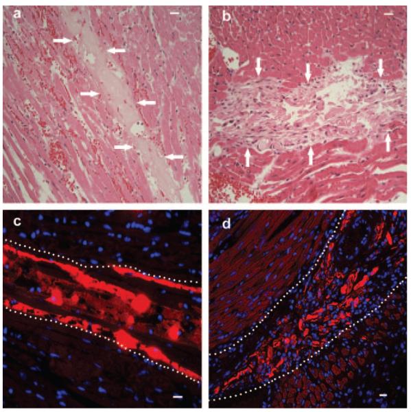
We have previously determined that endothelial cells and myocardial cells survive well within peptide microenvironments made with the RAD16-II peptide in vitro.13 To determine whether microenvironments can be established with injected peptides in vivo, the LV free wall of adult male C57BL6 mice was injected with 1% RAD16-II immediately after suspension of peptides at pH=7.0 and osmolarity of 295 mOsm. The hearts were later excised, fixed in 4% (wt/vol) paraformaldehyde, and embedded in paraffin. When stained with hematoxylin and eosin (H&E), the injected microenvironment was easily distinguishable from the surrounding myocardium (Figure 1a; arrows denote microenvironment borders) and contained few or no nuclei at 3 hours (Figure 1a). In the week after injection, cells had populated the peptide microenvironment (Figure 1b). To confirm the location of the peptide microenvironment and that the cells were within the microenvironment, a biotinylated peptide (0.1% of the total peptide was biotinylated) was injected, and tissue sections were stained with streptavidin—Texas red after fixation. Figure 1c and 1d demonstrate the presence of the microenvironment in the myocardium and the lack of cells immediately after injection and subsequent cell density. As a negative control, we injected biotinylated peptide alone. At concentrations required for normal peptide assembly into fibrils (>0.5% wt/vol), the biotinylated peptide by itself does not assemble when each peptide has the linker and a biotin molecule, most likely because of interference from the linker required for biotin attachment. In contrast, if most of the peptides do not have the linker and biotin (for example, if only 1 of each 100 peptides has the linker and biotin), then the biotinylated peptide is incorporated into the scaffold. We stained sections 7 days after injection of 100% biotinylated peptides at a final concentration of 1% wt/vol with H&E and saw little inflammation in the injection area (data not shown). Furthermore, there was no streptavidin—Texas red staining, indicating that the biotinylated peptide did not remain in the myocardium. Thus, the injected scaffold assembles into a microenvironment in vivo within the myocardium and does not result in a major inflammatory response.
Figure 1.
Cells spontaneously populate cell-free peptide microenvironment after 7 days. a, H&E stain of LV wall of adult male mouse 3 hours after injection of microenvironment. Few nuclei were evident within the microenvironment border, denoted by arrows, although there were some red blood cells. b, H&E of LV wall of adult male mouse 7 days after microenvironment injection shows many nuclei within the microenvironment. c, To confirm microenvironment regions and nuclei content, a biotinylated microenvironment was injected, and tissue was stained with streptavidin—Texas red and DAPI (blue). At 3 hours after injection, there were relatively few nuclei within the microenvironment; borders are denoted by dotted lines as determined by Texas red staining and light microscopy. d, Similarly to b, there were many cells within the microenvironment after 7 days, and the border is defined by streptavidin—Texas red staining. Bars represent 20 μm.
Endothelial Cells Populate Injected Peptide Gel
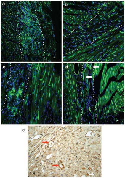
We next sought to determine the identity of the cells within the peptide microenvironment. Because angiogenic cells can invade other 3-dimensional scaffolds after implantation with and without factors,14,15 we hypothesized that endothelial progenitor cells were the dominant cell type initially in the peptide microenvironment. To test this hypothesis, we injected the LV wall of adult mice with 1% RAD16-II, harvested hearts 7, 14, 21, or 28 days later, and stained sections with isolectin, an endothelial cell marker.16 To quantify cell invasion, isolectin-positive cells were counted in 2 sections for each heart and normalized to the microenvironment area (n=4 mice per time point). At 7 days after injection, there were 33.5±1.9 endothelial cells/mm2 of microenvironment (Figure 2a). After 14 days, however, there were significantly fewer endothelial cells (17.3±0.2 endothelial cells/mm2; P<0.001) in the microenvironment (Figure 2b). The cell number remained constant at 21 and 28 days (14.5±0.6 and 14.8±2.7 endothelial cells/mm2 microenvironment, respectively), with the cells appearing more clustered (Figure 2c; 21 days), and capillary-like structures were observed within the microenvironment at 28 days after injection (Figure 2d; arrows). These data demonstrate the presence, organization, and maturation of endothelial cells invading the peptide microenvironment after injection.
Figure 2.
Endothelial cells spontaneously populate cell-free peptide microenvironment after 7 days and organize at later time points. a, Many cells (nuclei=blue, DAPI) within the peptide microenvironment (dotted lines as determined by light microscopy) stained positively with the endothelial cell marker isolectin-FITC (green) 7 days after injection. b, Endothelial cells were still present within the microenvironment 14 days after injection and appeared to be elongated in shape. c, Twenty-one days after injection, the endothelial cells appeared to be clustered within sections of the peptide microenvironment. d, Distinct capillary-like structures (arrows) were seen within the microenvironment 28 days after injection. e, Endothelial cell phenotype was confirmed by immunohistochemical staining for PECAM-1 (CD31, brown staining) within the peptide microenvironment at 28 days. Note the vessels within the gel highlighted by arrows that contain red blood cells. Bars represent 20 μm.
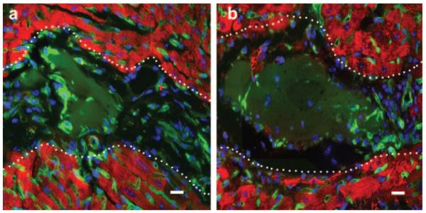
Because some nonendothelial cells can stain positively with isolectin,17 we stained hearts 28 days after injection for the endothelial cell marker CD31 (platelet—endothelial cell adhesion molecule-1 [PECAM-1]). Cells within the peptide microenvironment were positive for CD31 (Figure 2e). Cells in the peptide microenvironment also stained positively for CD144 (VE-cadherin; data not shown). It is important to determine whether the newly formed vascular structures anastamose with the host vasculature. As seen in Figure 2e (arrows), some of the newly formed vessels contained red blood cells, suggesting a connection to the host vasculature. At the 3-hour time point, we also observed occasional red blood cells within the microenvironment; however, we believe that this may be due to the initial trauma of the injection because we did not see red blood cells in the microenvironment outside vascular structures at later time points. Although this is an indirect method, as red blood cells could move during harvesting or tissue processing, these data suggest that functional vessels form in the microenvironment.
Smooth Muscle Cells Populate the Peptide Microenvironment
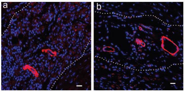
The peptide microenvironment appears to recruit endothelial cells, suggesting the potential of neovascularization. Because smooth muscle cells are also important in cardiac assembly, we stained microenvironment-injected samples with an antibody to α-smooth muscle actin. We did not see significant staining at 7 days after injection (data not shown). However, at 14 days there were aggregates of α-smooth muscle actin—positive cells within the microenvironment (Figure 3a). By 28 days after injection, we found formed arterioles within the microenvironment in all sections (Figure 3b). Furthermore, as shown in Figure 2e, we saw red blood cells within the arterioles, suggesting that these newly formed vascular structures may anastamose with the host vasculature.
Figure 3.
Smooth muscle cells populate the peptide microenvironment and form mature, vessel-like structures. a, At 14 days after injection, the cells (blue=DAPI) in the microenvironment stained positively with an α-smooth muscle actin antibody (red) and showed features of organization. b, At 28 days after injection, several arteriole-like structures were seen within the microenvironment in all samples. Bars represent 10 μm.
Identification of Nonvascular Cells
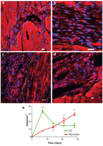
Interestingly, as many as 70% of the cells within the peptide microenvironments were not isolectin positive or smooth muscle actin positive, suggesting that at least 1 other cell type spontaneously invaded the microenvironment after injection. Recent studies indicate that there are cardiac progenitor cells and circulating progenitor cells that may differentiate into adult myocytes after myocardial infarction.18-20 We hypothesized that these nonendothelial cells may be similar to the previously described putative immature myocytes. To explore this, we stained sections from 7-, 14-, 21-, and 28-day peptide microenvironment-injected hearts with the cardiac-specific markers α-sarcomeric actin, as well as α-sarcomeric actinin (data not shown), and determined cell density. There were fewer α-sarcomeric actin—positive cells within the microenvironment at 7 days (8.1±0.7 myocytes/mm2 microenvironment; Figure 4a) compared with endothelial cells (P<0.001). However, at 14 days, there was a 60% increase in α-sarcomeric actin—positive cell density (13.0±1.2 myocytes/mm2 microenvironment; Figure 4b). Increases in α-sarcomeric actin—positive cell density continued at 21 days (21.3±3.4 myocytes/mm2 microenvironment; P<0.01; Figure 4c) and 28 days (29.6±1.5 myocytes/mm2 microenvironment; P<0.01; Figure 4d). These data, summarized graphically in Figure 4e, show that, in addition to endothelial cells, potential myocyte progenitors also populate the microenvironment with a delayed time course. It is important to note, however, that this cannot determine the origin or eventual fate of these cells.
Figure 4.
Putative myocyte precursors spontaneously populate the peptide microenvironment with a later time course than endothelial cell presence. a, There were few cells (blue=DAPI) within the peptide microenvironment that stain positively for α-sarcomeric actin (red) 7 days after injection. b, However, there was an increase in myocyte staining after 14 days, which continued to 21 days (c) and 28 days (d) after injection. Note that many of the myocytes were small cells and remained so at all time points. Bars represent 20 μm. e, Differences in endothelial and myocyte density time courses. ***P<0.001 vs 14-, 21-, and 28-day endothelial cells (EC); **P<0.01, 14 days vs 21 days; 21 days vs 7 and 28 days; 28 days vs 7, 14, and 21 days.
Implantation of Matrigel Recruits Few Cells
To explore a selective advantage of the self-assembling peptide microenvironments, Matrigel was injected intramyocardially, and hearts were harvested and stained after 7 or 28 days. Matrigel has been used in vascularization studies and has also been used for culturing myocytes.21 As Figure 5a shows, there were few endothelial cells (green) in the Matrigel at 7 days, and they were localized around the edges. Similarly, at 28 days there were few endothelial cells (green) within the Matrigel and some capillary formation around the edges (Figure 5b). Additionally, there was no staining for α-sarcomeric actin (red) in any of the Matrigel sections. These data demonstrate an advantage for the self-assembling microenvironments over Matrigel in vascularization.
Figure 5.
Endothelial cells do not populate Matrigel to the same degree as the self-assembling peptide microenvironment. Isolectin (green) and α-sarcomeric actin (red) staining of Matrigel sections 7 days (a) and 28 days (b) after injection. Note that much of the gel area is unpopulated. Blue=DAPI; bars represent 20 μm.
Implantation of Neonatal Myocytes
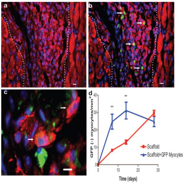
Several investigators have injected various types of myocytes into the myocardium.2,3,5 A shortcoming of cell transplantation, however, is the poor survival of implanted myocytes, possibly because of inadequate vascularization or lack of necessary survival/growth factors.5 One possible effect of transplanted cells is to recruit progenitor cells, even if the transplanted cells themselves do not survive. To explore this, we isolated neonatal cardiac myocytes from 2-day-old mice expressing GFP driven by the actin promoter.22 Using flow cytometry and antibodies to α-sarcomeric actin, we determined that 95% to 99% of the isolated cells were cardiomyocytes (data not shown). These cells were injected together with the self-assembling peptide (100 000 cells per injection), and the hearts were harvested at 7, 14, or 28 days after injection. Surprisingly, very few GFP-positive myocytes remained in the peptide microenvironment 7 days after injection. However, injection of exogenous GFP-positive myocytes increased the density of non-GFP cells that stained positively for the cardiac marker α-sarcomeric actin (24.7±4.3 myocytes/mm2 microenvironment; P<0.01 versus microenvironment alone; Figure 6a). Figure 6b shows the merged image and demonstrates that very few α-sarcomeric actin—positive cells were also GFP positive (arrows denote double positive cells). At 14 days, there were 31.1±4.8 myocytes/mm2 microenvironment (P=NS versus microenvironment alone), and the cells appeared larger and elongated (data not shown). This trend continued at day 28, when there were the same numbers of α-sarcomeric actin—positive cells (24.8±3.3 myocytes/mm2 microenvironment) as the microenvironment alone, but there were several larger, binucleated α-sarcomeric actin—positive cells within the cellembedded microenvironment (Figure 6c; arrows). The data summarized in Figure 6d show a significant increase in α-sarcomeric actin—positive cell density at 7 and 14 days in the group with implanted myocytes within the microenvironment. These data confirm that implantation of exogenous neonatal myocytes increases the density of endogenous potential putative cardiac progenitors in the microenvironment.
Figure 6.
Implantation of GFP myocytes results in increased endogenous myocyte density within the peptide microenvironment. a, α-Sarcomeric actin (red) staining of the peptide microenvironment region 7 days after injection. There were many endogenous putative myocyte precursors within the microenvironment. b, Merged image from 7 days after injection showing that relatively few α-sarcomeric actin—positive cells are GFP positive, as denoted by arrows (green=GFP, merged=yellow). c, Larger, doublenucleated endogenous myocytes (red, arrows) were visible within the microenvironment 28 days after injection, while there were still small endogenous myocytes. d, Difference in myocyte recruitment in GFP myocyte—injected samples and microenvironment alone. **P<0.01 vs microenvironment alone at same time point.
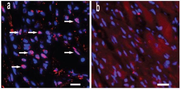
To further define the phenotype of these recruited α-sarcomeric actin—positive cells in the peptide microenvironments, we stained tissues with an antibody against the transcription factor Nkx2.5. Nkx2.5 has been used to stain developing myocytes; we confirmed this by performing immunofluorescence on isolated neonatal myocytes (data not shown). At 7 days after injection with GFP myocytes in peptide microenvironments, many cells stained positive for Nkx2.5 (Figure 7a; arrows), while there was virtually no staining with the secondary alone control (Figure 7b). Similar to stains of isolated cells, Nkx2.5 staining in peptide microenvironments was observed primarily in the nucleus. These data, taken with the α-sarcomeric actin results, further indicate that potential myocyte progenitors populate the peptide microenvironments after injection with GFP-positive cardiomyocytes.
Figure 7.
Potential cardiomyocyte phenotype. a, High-magnification image of cells within the GFP myocyte—embedded microenvironment 7 days after injection, demonstrating positive staining for Nkx2.5 (red; arrows) within the nucleus (DAPI; blue). b, Different peptide microenvironment section of same mouse stained with DAPI (blue) and secondary antibody alone. Note the absence of background staining within the peptide microenvironment. Bars represent 10 μm.
Implantation of Undifferentiated Embryonic Stem Cells
Because the clinical utilization of transplanted neonatal cardiac myocytes is limited, we also examined undifferentiated embryonic stem cell implantation within the microenvironment in vivo. These cells have eGFP under the control of the α-myosin heavy chain promoter and therefore express eGFP when they become cardiac myocytes.12 We injected 100 000 undifferentiated cells embedded within the peptide microenvironment and saw eGFP-positive cells after 14 days (Figure 8). This demonstrates that spontaneous differentiation of embryonic stem cells into cardiac myocytes can occur within the microenvironment in vivo and displays a potential application for optimization of differentiating stem cells in treating cardiovascular disease.
Figure 8.
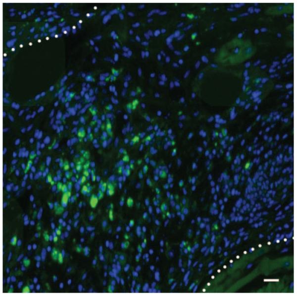
Spontaneous embryonic stem cell differentiation in the injectable microenvironment. Image of a mouse heart section injected with myosin heavy chain—eGFP embryonic stem cells embedded within the peptide microenvironment after 14 days. Green=eGFP, blue=DAPI. Bars represent 20 μm. The bright green cells show that some embryonic stem cells differentiate into cardiac myocytes and survive in the microenvironment.
Discussion
Myocardial regeneration is an emerging field, with several clinical studies already under way to examine the effects of cell implantation. A shortcoming of the cell injection approach is that cell survival may be poor. Our studies demonstrate the feasibility of creating an injectable microenvironment for exogenous cell therapy or recruiting progenitor cells within the myocardium. Both endothelial and smooth muscle cells infiltrated the microenvironments, suggesting the potential for proper vascularization. Furthermore, we demonstrated that injection of exogenous cells within the peptide microenvironment resulted in recruitment of α-sarcomeric actin/Nkx2.5—positive cells.
Although the peptide microenvironments were easily identified with routine histological methods, we also used a biotinylated peptide to identify the microenvironments by staining with streptavidin conjugates. The peptide microenvironments were identified up to 28 days after injection. Thus far, the survival of the peptides within tissues and the mechanisms of degradation are unknown, and further studies are under way. The use of biotinylated peptides should enable studies of microenvironment degradation. An additional application of the biotinylated self-assembling peptides could be to tether or target growth factors or compounds to the microenvironment. Recent studies have shown that injected streptavidin-FITC can be targeted to the biotinylated endothelium of the kidney in rabbits, and this approach may be exploited for the delivery of therapeutic agents.23
Within a week after injection, cells were seen within the peptide microenvironments that stained positively for endothelial cell markers. The time course of cell population was unique, with endothelial cells constituting the major cell type at earlier days and then increasing population by cells expressing α-sarcomeric actin. The unique time course of population suggests that there is a specific pattern to cell assembly within the microenvironment and that the presence of endothelial cells may promote the population of other cell types. These data are consistent with the fact that we have recently demonstrated that the presence of endothelial cells improves survival and organization of cardiomyocytes in vitro.13 The data in the present study suggest that endothelial cell presence within the peptide microenvironments may be important in cell survival and recruitment. Furthermore, we saw development of functional α-smooth muscle actin—positive vessels within the myocardium, including some with red blood cells within, suggesting that angiogenesis also occurs within the microenvironment with a specific time course and that new vessels are capable of connection to the host vasculature.
There were few surviving implanted GFP myocytes at 7, 14, and 28 days, but, interestingly, there was a significant increase in endogenous α-sarcomeric actin—positive cells compared with peptide microenvironment without exogenous cells. This result may be due to nonsurviving implanted cardiomyocytes releasing a signal that recruits endogenous progenitors. Our findings are consistent with previous reports of smaller, immature myocytes that express markers such as c-kit and Nkx2.5 being mobilized after injury.19,20,24 There is a possibility that α-sarcomeric actin—positive cells may have differentiated from non-GFP implanted cells; however, using flow cytometry, we determined that there were no non-GFP cells, and therefore this is improbable.
It is interesting to note that these α-sarcomeric actin—positive cells are not typical cardiac myocytes; they are smaller and have no clear sarcomeres. It has been shown that cells migrating to zones of injury in the heart are smaller cells but can possess molecular markers for immature cardiac myocyte progenitors.19,24 Microenvironment cells stain positively for α-sarcomeric actin as well as Nkx2.5, suggesting that they are similar in nature to published reports of cardiac myocyte precursors. Whether these putative myocyte precursors are capable of commitment and differentiation to adult cardiomyocytes is unclear. Currently, it is challenging to image individual cells over time in vivo in the same mouse. The best tool for evaluating commitment and differentiation is the use of molecular markers. We have shown that these immature myocytes stain positive for the early cardiac marker Nkx2.5, as well as α-sarcomeric actin and actinin. Further studies to determine the fate of these potential putative cardiac progenitors are needed.
A particularly interesting aspect of these experiments is that they demonstrate that endothelial and other progenitor cells migrate into a self-assembling peptide microenvironment environment even though these peptides have no known biological signal. This could be due to diffusion and selective binding of chemotactic factors in the peptide microenvironment. However, our results also raise the intriguing possibility that progenitor cells in normal myocardium are poised to migrate away from the tissue should local injury occur, in a manner similar to cells migrating away from explanted tissue in culture. In this case, the injection itself may represent an acute injury, and the signals released thereafter may contribute to the selective recruitment or differentiation of progenitor cells. It has been shown that there are populations of cells within the myocardium capable of migration and differentiation into cardiac precursors.24 Although this could be a likely source of the α-sarcomeric actin—positive cells within the microenvironment, it is not the only possibility. Cells of bone marrow origin may also have potential to migrate and differentiate into cardiac precursors.19,20 Our data cannot address the origin of cells, and further studies are warranted to elucidate the origin of these putative cardiac progenitors.
Many strategies are being explored for vascularization, and each has advantages and potential pitfalls. As a control for comparison, we injected Matrigel and found little penetration in vivo by endothelial cells and no staining for putative cardiac myocyte precursors. Much of the injected Matrigel remained unpopulated and nonvascularized, suggesting that cardiac assembly in vivo with Matrigel may not be feasible. Here we propose that injectable microenvironments may provide local regions that promote endothelial cell survival and organization. Further work will be required to determine whether progenitor cells mature into functioning myocardial regions, particularly after injury with ischemia or infarction. Furthermore, establishing that these microenvironments are not proarrhythmic is a crucial future goal, as regions with slow or unidirectional conduction could promote reentry dysrhythmias. Additionally, there is usually a profound inflammatory response after myocardial infarction, and careful studies will have to be undertaken to minimize the recruitment/survival of inflammatory cells. However, a powerful advantage of self-assembling peptides is that they can be engineered to incorporate growth factors and other signals that could provide signals for cardiomyocyte recruitment or maturation. Since these factors could be tethered to the peptide microenvironment or even released in a controllable fashion, timing of signals to control the regenerative process could be regulated. Thus, these initial experiments with a self-assembling peptide may provide the technology to modify the regenerative response in beneficial ways.
Acknowledgments
This work was supported by grants from the National Institutes of Health and the Center for the Integration of Medicine and Innovative Technology.
References
- 1.Mann BK, West JL. Tissue engineering in the cardiovascular system: progress toward a tissue engineered heart. Anat Rec. 2001;263:367–371. doi: 10.1002/ar.1116. [DOI] [PubMed] [Google Scholar]
- 2.Li RK, Jia ZQ, Weisel RD, Mickle DA, Zhang J, Mohabeer MK, Rao V, Ivanov J. Cardiomyocyte transplantation improves heart function. Ann Thorac Surg. 1996;62:654–660. doi: 10.1016/s0003-4975(96)00389-x. discussion 660–661. [DOI] [PubMed] [Google Scholar]
- 3.Sakai T, Li RK, Weisel RD, Mickle DA, Kim EJ, Tomita S, Jia ZQ, Yau TM. Autologous heart cell transplantation improves cardiac function after myocardial injury. Ann Thorac Surg. 1999;68:2074–2080. doi: 10.1016/s0003-4975(99)01148-0. discussion 2080–2081. [DOI] [PubMed] [Google Scholar]
- 4.Papadaki M, Bursac N, Langer R, Merok J, Vunjak-Novakovic G, Freed LE. Tissue engineering of functional cardiac muscle: molecular, structural, and electrophysiological studies. Am J Physiol. 2001;280:H168–H178. doi: 10.1152/ajpheart.2001.280.1.H168. [DOI] [PubMed] [Google Scholar]
- 5.Zimmermann WH, Schneiderbanger K, Schubert P, Didie M, Munzel F, Heubach JF, Kostin S, Neuhuber WL, Eschenhagen T. Tissue engineering of a differentiated cardiac muscle construct. Circ Res. 2002;90:223–230. doi: 10.1161/hh0202.103644. [DOI] [PubMed] [Google Scholar]
- 6.Zhang M, Methot D, Poppa V, Fujio Y, Walsh K, Murry CE. Cardiomyocyte grafting for cardiac repair: graft cell death and anti-death strategies. J Mol Cell Cardiol. 2001;33:907–921. doi: 10.1006/jmcc.2001.1367. [DOI] [PubMed] [Google Scholar]
- 7.Reinecke H, Zhang M, Bartosek T, Murry CE. Survival, integration, and differentiation of cardiomyocyte grafts: a study in normal and injured rat hearts. Circulation. 1999;100:193–202. doi: 10.1161/01.cir.100.2.193. [DOI] [PubMed] [Google Scholar]
- 8.Zhang S, Holmes TC, DiPersio CM, Hynes RO, Su X, Rich A. Selfcomplementary oligopeptide matrices support mammalian cell attachment. Biomaterials. 1995;16:1385–1393. doi: 10.1016/0142-9612(95)96874-y. [DOI] [PubMed] [Google Scholar]
- 9.Zhang S. Fabrication of novel biomaterials through molecular selfassembly. Nat Biotechnol. 2003;21:1171–1178. doi: 10.1038/nbt874. [DOI] [PubMed] [Google Scholar]
- 10.Semino CE, Merok JR, Crane GG, Panagiotakos G, Zhang S. Functional differentiation of hepatocyte-like spheroid structures from putative liver progenitor cells in three-dimensional peptide scaffolds. Differentiation. 2003;71:262–270. doi: 10.1046/j.1432-0436.2003.7104503.x. [DOI] [PubMed] [Google Scholar]
- 11.Holmes TC, de Lacalle S, Su X, Liu G, Rich A, Zhang S. Extensive neurite outgrowth and active synapse formation on self-assembling peptide scaffolds. Proc Natl Acad Sci U S A. 2000;97:6728–6733. doi: 10.1073/pnas.97.12.6728. [DOI] [PMC free article] [PubMed] [Google Scholar]
- 12.Takahashi T, Lord B, Schulze PC, Fryer RM, Sarang SS, Gullans SR, Lee RT. Ascorbic acid enhances differentiation of embryonic stem cells into cardiac myocytes. Circulation. 2003;107:1912–1916. doi: 10.1161/01.CIR.0000064899.53876.A3. [DOI] [PubMed] [Google Scholar]
- 13.Narmoneva DA, Vukmirovic R, Davis ME, Kamm RD, Lee RT. Endothelial cells promote cardiac myocyte survival and spatial reorganization: implications for cardiac regeneration. Circulation. 2004;110:962–968. doi: 10.1161/01.CIR.0000140667.37070.07. [DOI] [PMC free article] [PubMed] [Google Scholar]
- 14.Richardson TP, Peters MC, Ennett AB, Mooney DJ. Polymeric system for dual growth factor delivery. Nat Biotechnol. 2001;19:1029–1034. doi: 10.1038/nbt1101-1029. [DOI] [PubMed] [Google Scholar]
- 15.Ennett AB, Mooney DJ. Tissue engineering strategies for in vivo neovascularisation. Expert Opin Biol Ther. 2002;2:805–818. doi: 10.1517/14712598.2.8.805. [DOI] [PubMed] [Google Scholar]
- 16.Sahagun G, Moore SA, Fabry Z, Schelper RL, Hart MN. Purification of murine endothelial cell cultures by flow cytometry using fluoresceinlabeled Griffonia simplicifolia agglutinin. Am J Pathol. 1989;134:1227–1232. [PMC free article] [PubMed] [Google Scholar]
- 17.Pennell NA, Hurley SD, Streit WJ. Lectin staining of sheep microglia. Histochemistry. 1994;102:483–486. doi: 10.1007/BF00269580. [DOI] [PubMed] [Google Scholar]
- 18.Hierlihy AM, Seale P, Lobe CG, Rudnicki MA, Megeney LA. The post-natal heart contains a myocardial stem cell population. FEBS Lett. 2002;530:239–243. doi: 10.1016/s0014-5793(02)03477-4. [DOI] [PubMed] [Google Scholar]
- 19.Orlic D, Kajstura J, Chimenti S, Jakoniuk I, Anderson SM, Li B, Pickel J, McKay R, Nadal-Ginard B, Bodine DM, Leri A, Anversa P. Bone marrow cells regenerate infarcted myocardium. Nature. 2001;410:701–705. doi: 10.1038/35070587. [DOI] [PubMed] [Google Scholar]
- 20.Orlic D, Kajstura J, Chimenti S, Limana F, Jakoniuk I, Quaini F, Nadal-Ginard B, Bodine DM, Leri A, Anversa P. Mobilized bone marrow cells repair the infarcted heart, improving function and survival. Proc Natl Acad Sci U S A. 2001;98:10344–10349. doi: 10.1073/pnas.181177898. [DOI] [PMC free article] [PubMed] [Google Scholar]
- 21.Zimmermann WH, Melnychenko I, Eschenhagen T. Engineered heart tissue for regeneration of diseased hearts. Biomaterials. 2004;25:1639–1647. doi: 10.1016/s0142-9612(03)00521-0. [DOI] [PubMed] [Google Scholar]
- 22.Okabe M, Ikawa M, Kominami K, Nakanishi T, Nishimune Y. ‘Green mice’ as a source of ubiquitous green cells. FEBS Lett. 1997;407:313–319. doi: 10.1016/s0014-5793(97)00313-x. [DOI] [PubMed] [Google Scholar]
- 23.Hoya K, Guterman LR, Miskolczi L, Hopkins LN. A novel intravascular drug delivery method using endothelial biotinylation and avidin-biotin binding. Drug Deliv. 2001;8:215–222. doi: 10.1080/107175401317245895. [DOI] [PubMed] [Google Scholar]
- 24.Beltrami AP, Barlucchi L, Torella D, Baker M, Limana F, Chimenti S, Kasahara H, Rota M, Musso E, Urbanek K, Leri A, Kajstura J, Nadal-Ginard B, Anversa P. Adult cardiac stem cells are multipotent and support myocardial regeneration. Cell. 2003;114:763–776. doi: 10.1016/s0092-8674(03)00687-1. [DOI] [PubMed] [Google Scholar]


