Abstract
Protein trafficking to two different types of vacuoles was investigated in tobacco (Nicotiana tabacum cv SR1) mesophyll protoplasts using two different vacuolar green fluorescent proteins (GFPs). One GFP is targeted to a pH-neutral vacuole by the C-terminal vacuolar sorting determinant of tobacco chitinase A, whereas the other GFP is targeted to an acidic lytic vacuole by the N-terminal propeptide of barley aleurain, which contains a sequence-specific vacuolar sorting determinant. The trafficking and final accumulation in the central vacuole (CV) or in smaller peripheral vacuoles differed for the two reporter proteins, depending on the cell type. Within 2 d, evacuolated (mini-) protoplasts regenerate a large CV. Expression of the two vacuolar GFPs in miniprotoplasts indicated that the newly formed CV was a lytic vacuole, whereas neutral vacuoles always remained peripheral. Only later, once the regeneration of the CV was completed, the content of peripheral storage vacuoles could be seen to appear in the CV of a third of the cells, apparently by heterotypic fusion.
In contrast to yeast or mammalian cells, plant cells may contain different vacuoles. Some vacuoles have storage or digestive functions, whereas others may have other not yet defined functions (Leigh and Sanders, 1997). Some plant cells harbor a single large central vacuole (CV) that may occupy more than 90% of the cell volume. Vacuoles can be characterized by the presence of specific tonoplast intrinsic proteins (TIPs): α-TIP has been associated with protein storage vacuoles (Johnson et al., 1989), whereas γ-TIP has been found in lytic or degradative vacuoles (LVs; Paris et al., 1996) and δ-TIP has been found in pigment-containing vacuoles (Jauh et al., 1998). TIPs have also often been found in various combinations in single vacuoles (Jauh et al., 1998, 1999). Vacuole development has been investigated using several approaches: transmission electron microscopy (Buvat, 1982), biochemical studies of the changes accompanying cell enlargement (Maeshima, 1990), and studies of the mechanisms by which proteins are targeted to the vacuole (for review, see Beevers and Raikhel, 1998; Neuhaus and Rogers, 1998; Vitale and Raikhel, 1999). The intracellular sorting of a number of vacuolar proteins has been investigated, and vacuolar sorting determinants (VSDs) have been determined and classified in three types with different properties (Neuhaus and Rogers, 1998; Matsuoka and Neuhaus, 1999).
A complementary approach is the study of vacuole regeneration. Vacuoles can be removed from plant protoplasts by high-speed centrifugation through a continuous density gradient (Lörz et al., 1981). Evacuolated protoplasts (miniprotoplasts) are viable and can regenerate vacuoles and cell walls in culture (Wu and Tsai, 1992), thus providing a convenient in vitro system in which to study synchronous development of vacuoles in a large number of cells. Vacuole regeneration in evacuolated tobacco (Nicotiana tabacum) and petunia protoplasts was shown to occur after culture for 12 to 44 h (Erdmann et al., 1989). Reappearance of the vacuole coincided with increased levels of hydrolytic enzymes and of tonoplast H+-pyrophosphatase activity, with Neutral Red (NR) uptake and with the reappearance of a 41-kD vacuolar protein of unknown function (Hörtensteiner et al., 1992). Inclusion of bafilomycin A, a specific inhibitor of the vacuolar H+-pumping ATPase (Bowman et al., 1988), into the culture medium decreased the uptake of NR, but did not prevent vacuole regeneration (Sze et al., 1992). Vacuole formation in evacuolated petunia protoplasts was associated with the accumulation of flavonoids, followed by the synthesis of vacuole-associated ethylene-forming activity (Erdmann et al., 1989). Protoplasts cultured in the presence of cycloheximide failed to develop vacuoles, showing that protein synthesis is required, but the strong inhibitory effect of cycloheximide was completely reversed when evacuolated protoplasts were washed with inhibitor-free medium (Hörtensteiner et al., 1994).
The formation and evolution of single vacuoles in plant cells is a complex phenomenon. The use of leaf protoplasts reveals this complexity because they represent a complex mixture of cell types (Di Sansebastiano et al., 1998). Furthermore, production of protoplasts is likely to induce changes in function and composition of preexisting vacuoles. Evacuolation reduces complexity by eliminating most of the chloroplast-poor cell types that were generated from leaves and induces the remaining cells to regenerate a large CV to restore their original volume. To study the biogenesis of vacuoles we thus chose to study the regeneration of vacuoles in these miniprotoplasts.
We had previously described a fluorescent marker for a neutral vacuolar compartment in tobacco protoplasts, a green fluorescent protein (GFP) fused to a C-terminal VSD from tobacco chitinase A (Di Sansebastiano et al., 1998). Since this marker was excluded from acidic vacuoles, we first needed to develop an equivalent reporter protein for this acidic, LV. We fused a sequence-specific VSD from barley aleurain, a protease from the LV, to an enhanced variant of GFP. This allowed the visualization of acidic LVs in protoplasts and miniprotoplasts transiently expressing the hybrid GFPs. With these two GFP markers, specific for a neutral vacuole or an LV, we were able to address the nature of the vacuoles regenerated in miniprotoplasts.
RESULTS
Two Different VSDs Target GFP to Different Compartments
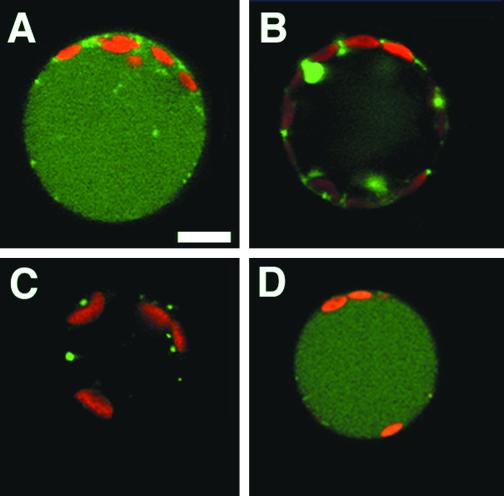
We have shown previously that the C-terminal propeptide of tobacco chitinase A was sufficient to target the reporter protein GFP to pH-neutral vacuoles, but not to acidic vacuoles (Di Sansebastiano et al., 1998). We interpreted this as reflecting the existence of two different types of vacuoles where the pH-neutral vacuole would correspond to a storage vacuole, whereas the acidic vacuole would correspond to a LV. We also observed that the size of the two vacuoles differed in different protoplasts: about three-quarters of chloroplast-rich protoplasts accumulated the GFP (SGFP5T, renamed here GFP5-Chi) in the large CV, as shown in Figure 1A, whereas one-quarter accumulated it in peripheral vacuoles (PVs; Fig. 1B). In the latter cells the CV could be stained with NR and was thus acidic. In chloroplast-poor protoplasts the proportions of the two patterns were reversed, as they mostly accumulated GFP5-Chi in PVs.
Figure 1.
Fluorescence patterns of GFPs targeted to a neutral vacuole (GFP5-Chi, A and B) and to an LV (Aleu-GFP6, C and D) 24 h after protoplast transformation. Confocal images (1-μm section) of chloroplast-rich tobacco mesophyll protoplasts. A and C, More frequent patterns; B and D, less frequent patterns (see Table I). Bar = 10 μm.
To label LVs we fused the same GFP5 variant (Siemering et al., 1996) with the first 143 amino acids of the precursor of barley aleurain, a thiol protease targeted to the LV (Holwerda et al., 1992). This sequence includes the signal sequence and the whole N-terminal propeptide of aleurain. The resulting GFP (Aleu-GFP5) was only visible in compartments smaller than 2 μm and often clustered around the nucleus (not shown). No vacuoles were visible in comparison with GFP5-Chi, which is targeted to the storage vacuole. Since the fluorescence intensity was much lower for Aleu-GFP5 than for GFP5-Chi, we quantified by immunoblots the GFP in the medium and in total protoplast extracts. Aleu-GFP5 was never found in quantities comparable with vacuolar GFP5-Chi or secreted GFP5 (not shown). We, therefore, suspected that Aleu-GFP5 was rapidly degraded in its final target compartment and was thus only visible in the small, unidentified compartments described above, which could be intermediates in the transport pathway. To increase the detection limit of our reporter protein we introduced into Aleu-GFP5 the mutations F64L/S65T (Cormack et al., 1996), producing the brighter and more stable Aleu-GFP6. In addition to the same small fluorescent compartments (Fig. 1C) seen previously with Aleu-GFP5, many protoplasts showed a green CV (Fig. 1D). In contrast to GFP5-Chi, there was no apparent difference in pattern distribution between chloroplast-rich and chloroplast-poor protoplasts. We quantified the distribution of large vacuoles in cells expressing Aleu-GFP6 and found that they are present in 18% of fluorescent chloroplast-rich protoplasts (Table I). This percentage was approximately complementary to the 75% of large vacuoles observed in protoplasts transformed with GFP5-Chi under the same conditions. This indicates that this particular population of chloroplast-rich protoplasts harbors (predominantly) two possible types of CVs, targeted by either of our vacuolar markers. In contrast, in chloroplast-poor protoplasts there was no such complementarity, but we could see large vacuoles accumulating Aleu-GFP6 in 15% of these cells, more than twice the frequency observed with GFP5-Chi. To ascertain that the use in the latter construct of a less bright and possibly less stable form of GFP was not misleading, we introduced the same mutations into GFP5-Chi (making GFP6-Chi). In comparison with GFP5-Chi, we observed a 20% increase of the total number of fluorescent protoplasts. The vacuolar GFP pattern was conserved in the same proportions in the two cell types, although the ER labeling was more visible, even after a long expression time. We concluded that the mutations lowered the detection limit of GFP so that residual GFP6-Chi in the ER was now more often visible. After treatment with cycloheximide, this ER fluorescence strongly decreased (not shown). The apparent increase in transformation efficiency reflected thus the increase in fluorescence intensity.
Table I.
GFP patterns in two protoplast subpopulations expressing GFP5-Chi or Aleu-GFP6
| Vacuole Type | Chloroplast-Rich Protoplasts
|
Chloroplast-Poor Protoplasts
|
||
|---|---|---|---|---|
| Aleu-GFP6n = 154 | GFP5-Chi n = 227 | Aleu-GFP6n = 192 | GFP5-Chi n = 110 | |
| Central | 18% (12–25) | 75% (58–88) | 15% (14–17) | 6% (0–15) |
| Peripheral | 82% (75–88) | 25% (12–42) | 85% (83–86) | 94% (100–85) |
After 24 h of expression, GFP localization was observed in the CV or PV. GFP5-Chi was also often observed in the endoplasmic reticulum (ER). Results are expressed as a percentage within each population analyzed, 100% corresponding to the total no. of fluorescent cells observed in chloroplast-rich or chloroplast-poor protoplasts for three independent transformations. In parentheses we report the observed minimal and maximal percentage in the independent experiments (minimum–maximum).
Because we used the same vector and the same transformation protocol for all three vacuolar GFPs, we expected a similar rate of GFP synthesis. In fact, we found by immunoblotting at early times of expression a 3-fold higher level of GFP6-Chi than Aleu-GFP6, GFP5-Chi being intermediate (data not shown). We chose to keep using GFP5-Chi and Aleu-GFP6 since they gave more comparable fluorescence intensities.
In contrast to GFP5-Chi or to the secreted GFP5, we were never able to visualize Aleu-GFP6 in the ER under our normal experimental conditions. To slow down transit through the ER we incubated protoplasts at low temperature. After an 8-h incubation at 14°C, the nuclear envelope became partially fluorescent and smaller compartments became visible in the neighboring cytoplasm in a pattern very similar to the pattern observed for GFP5-Chi under the same conditions. This indicated that at normal temperatures, Aleu-GFP6 leaves the ER much faster than GFP5-Chi, possibly too fast for the fluorophore to form. This could be due to the presence of an N-terminal domain in Aleu-GFP6, which could accelerate folding of GFP, as reported for other N-terminal fusions (Sacchetti and Alberti, 1999). Pulse-chase analysis indicated that Aleu-GFP6 was processed to a smaller size with a half-time of 1 h or less, whereas for GFP5-Chi the half-time was more than 6 h (not shown).
Aleu-GFP6 Accumulates in Acidic Vacuoles
Despite the use of an enhanced GFP, vacuoles containing Aleu-GFP6 showed a much fainter fluorescence compared with vacuoles containing GFP5-Chi. Because the fluorescence yield depends on GFP stability, we hypothesize that Aleu-GFP6 is accumulated in a more lytic vacuole than GFP5-Chi. The proportion of fluorescent protoplasts in a population expressing Aleu-GFP6 decreased from 20% after 24 h to only 3% after 48 h, whereas a large proportion of GFP5-Chi-expressing protoplasts were still fluorescent. This is an indication that the reporter protein is more rapidly degraded in the compartments that accumulate Aleu-GFP6. After cycloheximide treatment, the protein level of Aleu-GFP6 decreased somewhat faster than GFP5-Chi (by 50% versus 38% within 24 h). These differences seem too small to explain the fast disappearance of the Aleu-GFP6 fluorescence. It is, however, known that the loss of more than one amino acid at the N terminus can cause the loss of GFP fluorescence (Phillips, 1997). Such a short truncation cannot be excluded when Aleu-GFP6 is processed to the apparently normal size in the protoplasts, as detected by protein blotting.
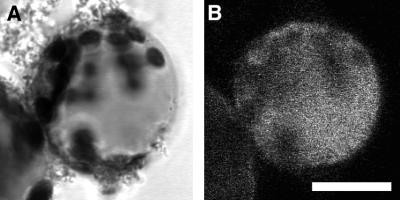
A lower pH can also influence the quantum yield of Aleu-GFP6 since a GFP variant with the same enhancing mutations F64L/S65T was shown to be pH sensitive (Cormack et al., 1996). To assess for the acidity of the vacuoles, a usual property of lytic compartments, we stained the protoplasts with NR, a dye specifically accumulated in acidic compartments. As shown in Figure 2, the green fluorescence of Aleu-GFP6 was visible in many cases in the same compartment as NR staining, a situation never observed in cells expressing GFP5-Chi (Di Sansebastiano et al., 1998) or in cells expressing SGFP6T, the brighter variant (not shown). Thus, it is evident that Aleu-GFP6 can accumulate in acidic vacuoles. We were surprised to observe that the proportion of NR-stained CVs decreased from 70% to 48% compared with control protoplasts or to those expressing GFP5-Chi. In chloroplast-poor protoplasts, this proportion dropped from 95% to 74%. Thus, overexpression of Aleu-GFP6 seems to affect the acidification of the LVs. This significant interference between NR and Aleu-GFP6 also indicated that both were accumulated in the same subcompartment, which is an additional evidence for the accumulation of Aleu-GFP6 in an acidic vacuole.
Figure 2.
NR staining of a protoplast expressing Aleu-GFP6. A, Image in transmitted light. The black spots are chloroplasts and NR appears as a gray stain of the CV. B, Confocal image of the green fluorescence, which is partially quenched by NR. Bar = 20 μm.
From the following three points we conclude that Aleu-GFP6 is targeted to a lytic or acidic vacuole: first, the ssVSD derives from barley aleurain, a protease targeted to LVs; second, Aleu-GFP6 undergoes a faster inactivation; and third, the targeted vacuole has a lower pH than the medium or the neutral vacuole targeted by GFP5-Chi. Furthermore, in chloroplast-rich protoplasts, our two different vacuolar GFPs appear to account for all the CVs, which are lytic or neutral.
Vacuole Regeneration in Transformed Miniprotoplasts
We now had in our hands the tools to study the de novo formation of a CV in the simplified system of evacuolated protoplasts. Because the evacuolation technique is based on the large density difference between chloroplasts and nucleus on one side and vacuoles on the other side, it works best with chloroplast-rich protoplasts, whereas other protoplasts are mostly lost in the procedure.
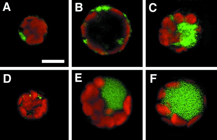
Three to 4 h after evacuolating protoplasts our marker for the neutral vacuoles was found in discrete areas of the ER, where it was still evenly distributed after 6 to 8 h. At this early stage there is no visible CV, but peripheral neutral vacuoles could now be detected (Fig. 3A), and they were similar in size to those observed in whole protoplasts (Fig. 1B). After the same incubation time, the marker for LVs was exclusively detected in small peripheral compartments (Fig. 3D), again similar in size to those observed in whole protoplasts (Fig. 1C).
Figure 3.
Regeneration of vacuoles by miniprotoplasts. Confocal images (1-μm section) of tobacco miniprotoplasts at different times after evacuolation. A through C, Miniprotoplasts expressing the marker for neutral vacuoles 8, 36, or 52 h after evacuolation; D through F, miniprotoplasts expressing the marker for LVs 8, 36, or 52 h after transformation. Bar = 10 μm.
Large CVs only appeared 24 to 36 h after evacuolation, giving to the protoplasts their original size. At this time point our GFP markers allowed us to address the nature of the newly formed vacuoles. As shown in Figure 3E, the CV accumulated the marker for LVs. In contrast, in the protoplasts expressing the reporter for the neutral compartment the CV remained strictly non-fluorescent, whereas peripheral neutral vacuoles still could be seen (Fig. 3B). It is important to note that the difference in transport speed by which Aleu-GFP6 and GFP5-Chi appear to transit through the secretory pathway cannot account for the existence of two separate compartments. In fact we showed previously that 24 h of expression were largely sufficient for vacuolar localization of the slower marker in normal protoplasts (Di Sansebastiano et al., 1998). We counted the proportion of newly formed CVs that were fluorescent in miniprotoplasts expressing either one of our vacuolar markers (Table II). This confirmed that the marker for LVs was almost always transported to the CV, whereas the marker for neutral vacuoles never was. We conclude that evacuolated tobacco protoplasts regenerated a CV that is exclusively lytic in nature. What is most interesting is that these protoplasts regenerated PVs of the neutral type at the same time. It thus appears that the miniprotoplasts strictly separate the two transport pathways to the two separate types of vacuoles at this early stage of vacuole regeneration.
Table II.
GFP accumulation in regenerated central vacuoles of miniprotoplasts
| Reporter Protein | Time after Evacuolation
|
|
|---|---|---|
| 36 | 48–52 | |
| h | ||
| GFP5-Chi | 0% (n = 144) | 36% (n = 245) |
| Aleu-GFP6 | 97% (n = 161) | 84% (n = 208) |
Large CV started to become visible after 24 to 36 h. The percentage of fluorescent CV was calculated for all cells showing any green fluorescence.
Forty-eight to 52 h after evacuolation, the size of the protoplasts remained constant and the marker for the LV was still present in the CV (Fig. 3F), but lost much of its fluorescence intensity, reducing the proportion of protoplasts with a detectable green vacuole to 84% (Table II, 52 h). In the remaining 16% of protoplasts there was residual fluorescence limited to small peripheral compartments similar to those described above (Figs. 1C and 3B), but the CV was no longer fluorescent, which we interpret as an indication of the increasingly lytic character of the CV. Immediately after vacuole regeneration, when the concentration of vacuolar proteases is expected to be low, the fluorescence of the LVs was stronger than ever observed with normal protoplasts, but it decreased rapidly within the time of the experiment, as vacuoles steadily accumulate proteases (Hörtensteiner et al., 1992).
After 2 d, the marker for neutral vacuoles started to appear in some CVs (Fig. 3C). This happened in approximately one-third of the protoplasts after 48 to 52 h (Table II). Since we never observed neutral vacuoles with intermediate sizes in regenerated miniprotoplasts, we assume that the appearance of the marker in the CV occurred by fusion of the peripheral neutral vacuoles with the lytic CV rather than by formation of a new neutral CV and the reduction and/or disappearance of the regenerated LV.
DISCUSSION
In this paper we describe the sorting of two chimeric proteins obtained by the fusion of GFP to two VSDs, a C-terminal propeptide derived from tobacco chitinase A and an N-terminal propeptide derived from barley aleurain. These two proteins have been selected because they represent two types of VSDs that have been shown to follow clearly different pathways (Matsuoka et al., 1995) to two different vacuolar compartments, at least in some cells (Paris et al., 1996).
Confocal laser scanning microscopy was used to visualize the different steps of the two sorting systems. This revealed an interesting difference between the patterns of GFP accumulation for the two constructs. The pattern of GFP5-Chi accumulation described in a previous paper (Di Sansebastiano et al., 1998) led us to conclude that this GFP fusion protein resided some time in the ER and was accumulated in (small) PVs or in the (large) CV. On the contrary, Aleu-GFP6, described here for the first time, resided a very short time in the ER and accumulated in most cells in small compartments, which we chose to call peripheral LVs because they appeared to be the final destination of our vacuolar marker in this experimental system. In some cells Aleu-GFP6 reached the CV. The frequency of fluorescent CVs in different protoplast subpopulations differed compared with GFP5-Chi. For chloroplast-rich protoplasts we could see a clear complementarity in the percentage of cells in which Aleu-GFP6 or GFP5-Chi occupied the CV rather than being limited to PVs and ER. The two markers could label essentially all large vacuoles of this cell type.
An important difference between the two vacuoles was revealed by the difference in NR accumulation. The GFP5-Chi never accumulated in the same vacuoles as NR, which was only found in large vacuoles, when the reporter protein accumulated in small compartments, or in small vacuoles, when the reporter accumulated in the CV. To the contrary, we could observe NR staining of vacuoles containing the reporter Aleu-GFP6. The reduced accumulation of NR in these cells in comparison with the level of NR accumulation in GFP5-Chi -transformed protoplasts and in control protoplasts indicates that Aleu-GFP6 influenced the vacuolar acidification, a further indication of the acidic nature of the labeled vacuoles. We postulate that the pathway used by lytic enzymes and Aleu-GFP6 to the LV is also responsible for acidification. Aleu-GFP6 only becomes visible after leaving the ER. Fluorophore formation is linked to the production of H2O2 (Tsien, 1998), which could affect the function of proton pumps in a post-ER compartment. The overload of this pathway with the chimeric protein could also have increased the volume or the buffering capacity of the LVs without a corresponding increased rate of acidification.
Having shown that GFP5-Chi and Aleu-GFP6 are transported to different vacuoles, presumably through different pathways, we used these proteins to monitor vacuole regeneration in evacuolated protoplasts. We were able to assess the lytic (Aleu-GFP6) or neutral (GFP5-Chi) nature of the vacuoles that appeared during regeneration. We observed that before the formation of a CV, miniprotoplasts regenerate at the same time several PVs, some neutral, others lytic. The large CV of the cell was regenerated after 36 h and almost always accumulated our marker for LVs, with a very bright fluorescence compared with nonevacuolated protoplasts. In contrast, the marker for neutral vacuoles remained limited to PVs. Our results indicate that the CV originates exclusively as an LV. This may correspond to the type of vacuole able to provide the required fast volume increase to restore the original cell volume. Only after the restoration of the cell volume was achieved could we start to see the marker for neutral vacuoles in the CV. Since we did not observe any intermediate compartment with a volume between the PVs and the CV or a parallel decrease and increase of fluorescence in the CV and PVs, respectively, it is probable that the CV became fluorescent by fusion with PVs containing a large amount of GFP5-Chi. The accumulation of NR in a regenerated vacuole has been reported as an indication of its acidic nature (Hörtensteiner et al., 1992), but the authors reported that it only became visible at least 2 d after evacuolation. Since this is the time point at which we start to see GFP5-Chi accumulation in a minority of CVs, NR accumulation and the reappearance of lytic enzymes could not unambiguously reveal the lytic nature of the first regenerated CV.
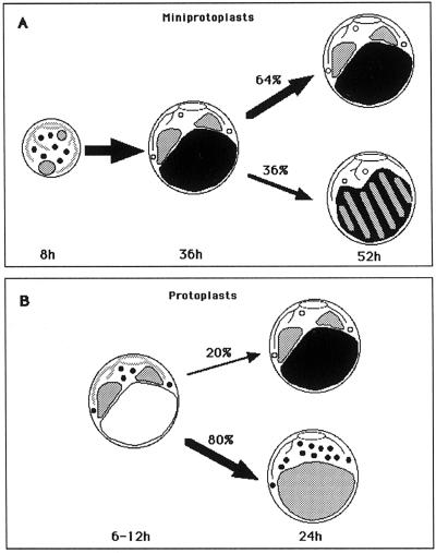
From the comparison of GFP sorting in protoplasts and miniprotoplasts, we propose the model depicted in Figure 4. Miniprotoplasts first regenerate separate small (pre-) vacuoles (Fig. 4A, 8 h), which differ in size for the lytic (shown in black) and neutral compartments (shown in gray). Since they have a similar aspect, they may correspond to the respective PVs seen in whole protoplasts (Fig. 1, B and C). The miniprotoplasts take from 36 to 48 h to regenerate a normal sized CV, but as soon as it becomes visible (after 24 h) it clearly contains the marker for LVs. We suggest that this vacuole forms by maturation and homotypic fusion of the peripheral lytic (pre-)vacuoles and thus receives all of the marker. Meanwhile, the marker for neutral vacuoles continues to accumulate in peripheral neutral vacuoles (Fig. 4, 36 h). After 52 h, due to the progressive accumulation of proteases and to a decreasing pH, the concentration of GFP in the LV decreased, sometimes below the detection level, reducing the proportion of visible fluorescent cells.
Figure 4.
Model of vacuole biogenesis in miniprotoplasts and (chloroplast-rich) protoplasts. The marker for neutral vacuoles (GFP5-Chi) is indicated in gray and the marker for LVs (Aleu-GFP6) is indicated in black. The percentage of cells developing each pattern is indicated next to the arrows. A, Miniprotoplasts: There is no CV yet 8 h after evacuolation. GFP5-Chi labels ER, nuclear envelope, and peripheral (pre-) vacuoles, whereas Aleu-GFP6 labels smaller peripheral (pre-) vacuoles. Thirty-six hours after evacuolation the large CV is an LV labeled by Aleu-GFP6, whereas GFP5-Chi remains limited to PVs. Fifty-two hours after evacuolation the PVs have fused with the lytic CV in about one-third of the cells. B, Protoplasts: The CV is labeled by neither of our GFP markers 6 to 12 h after transformation. GFP5-Chi labels ER, nuclear envelope, and peripheral (pre-) vacuoles, whereas Aleu-GFP6 labels smaller peripheral (pre-) vacuoles. Then, within 24 h there are two possibilities depending on the nature of the pre-existing CV. Top, New peripheral LVs fuse with a lytic CV, whereas the neutral vacuoles remain peripheral. Bottom, The new peripheral neutral vacuoles fuse with a neutral CV, whereas the LVs remain peripheral.
Peripheral neutral vacuoles persist in many protoplasts as final compartments for the accumulation of GFP5-Chi (Fig. 4A, 52 h, top). However, in about 36% of the cells they eventually appear to fuse with the lytic CV. This leads to a hybrid CV with lytic and storage characteristics (Fig. 4A, 52 h, bottom). The proportion of CVs, which acquired GFP5-Chi after 52 h, is more than twice the proportion of CVs that lost Aleu-GFP6 fluorescence in the parallel experiments. Even though overexpression of one marker at a time could have affected vacuole biogenesis, this big difference strongly favors a secondary modification of the regenerated CV of miniprotoplasts. Definite proof for the formation of hybrid vacuoles will require the simultaneous use of two distinct vacuolar markers, such as spectral variants of GFP.
The model for miniprotoplasts can be extended to normal chloroplast-rich protoplasts. GFP5-Chi initially accumulates in peripheral neutral (pre-) vacuoles, whereas Aleu-GFP6 accumulates in peripheral lytic (pre-) vacuoles (Fig. 4B, 6–12 h). Further transport to the pre-existing CV depends on its nature: if it is a neutral vacuole (75%–80% of the protoplasts) it will obtain GFP5-Chi from the peripheral neutral prevacuoles, whereas LVs remain peripheral (Fig. 4B, 24 h, bottom). Similarly, peripheral lytic prevacuoles will give Aleu-GFP6 to a central LV (in 15%–20% of the protoplasts; Fig. 4B, 24 h, top).
Our results obtained with chloroplast-rich protoplasts and with miniprotoplasts (Tables I and II) can be adequately explained with only two types of vacuoles. It is clear that we cannot exclude the existence of vacuoles that accumulate neither type of GFP, especially in chloroplast-poor protoplasts. In the absence of a third marker we cannot decide between faster degradation, absence of homotypic fusion, or a third type of vacuole. The existence of a third sorting pathway is plausible, since three different classes of VSDs have been described (Neuhaus and Rogers, 1998; Kervinen et al., 1999), three different small vacuoles each harboring a different TIP have been visualized in single pea root cells (Jauh et al., 1999), and three different vacuoles were also distinguished in seeds (Jiang et al., 2000).
The nature of the PVs described here is unclear. Neutral PVs are relatively large, comparable in size with chloroplasts, whereas lytic PVs are much smaller, comparable in size with Golgi compartments. Small vacuoles have been described in various plant tissues (e.g. Jauh et al., 1999, Jiang et al., 2000). Prevacuolar compartments reassembling animal endosomes have also been described (Da Silva Conceiçao et al., 1997). It is not clear how proteins are transported from endosomes (prevacuoles) to lysosomes (vacuoles). In animal cells, no transport vesicles have been detected, and late endosomes possibly just fuse with lysosomes (see Luzio et al., 2000, for different models). In plants, if prevacuoles also fuse with vacuoles after removal of recycling components, then (homotypic) fusion can determine the number and size of vacuoles. Inhibition of fusion would lead to the formation of numerous small PVs. Heterotypic fusion of prevacuoles of different types could lead to hybrid vacuoles. Heterotypic fusion does not appear to occur in our protoplast experiments, but seems to occur in about one-third of the regenerated miniprotoplasts.
Hybrid vacuoles probably exist in many plant cells. Immunoelectron microscopy has indicated colocalization of sporamin and barley lectin proteins, respectively, from the LV and neutral vacuole, in all analyzed tissues of doubly transformed tobacco (Schroeder et al., 1993). In a similar manner, in barley and pea root cells many vacuoles harbor two or even three different TIPs (Paris et al., 1996; Jauh et al., 1999). In this respect it will be interesting to determine which TIPs are associated with the tonoplasts in regenerating miniprotoplasts.
The distribution of our two vacuolar GFPs in different tissues and different cell types in transgenic plants will be very informative about the nature of the CV. Preliminary results indicate that in tobacco stably transformed with our vacuolar markers most cells are non-fluorescent. We suppose that GFP is continuously degraded in most tissues and fails to accumulate to a detectable level in either type of vacuoles or in hybrid CV. Protoplast isolation causes reappearance of fluorescence, indicating in fact a change in the nature of the vacuoles. Preliminary observations indicate that each of our two markers labels the CV or small compartments in different cell types of transgenic Arabidopsis, which may offer a better hospitality than tobacco for GFP in different types of vacuoles.
MATERIALS AND METHODS
Fusion Gene Constructs
The pSGFP5 and pSGFP5T plasmids have been described (Di Sansebastiano et al., 1998). The coding sequence for GFP5 was isolated from the plasmid pBIN-mGFP5-ER (Haseloff et al., 1997) and was cloned into the vector pGY1 (Neuhaus et al., 1991) between 35S promotor and termination sequences. GFP6 was obtained by PCR mutagenesis of F64 to L and S65 to T (ctc act). The pAleuGFP5 plasmid was obtained by substitution of the signal sequence of pSGFP5 with the first 431 bases (143 codons) of the coding sequence of the barley aleurain cDNA (Rogers et al., 1985). We introduced by PCR with appropriate primers a BamHI site 5′ of the start codon of aleurain (ggatccggcgaaacgaa atg, restriction site underlined) and a NheI site at the 3′ end of the N-terminal fragment of aleurain (gcc gcc gct agc, changed bases in bold) and a corresponding NheI site at the beginning of the coding sequence for GFP5 or GFP6, in front of the natural start codon of GFP (gct agc gca atg), and used the PstI site previously introduced 3′ of the stop codon of GFP (Di Sansebastiano et al., 1998). The BamHI/NheI aleurain fragment and the NheI/PstI GFP fragment were cloned into the vector pGY1. Plasmids were isolated by alkaline lysis in presence of SDS (Sambrook et al., 1989) and were purified on an ethidium bromide-CsCl density gradient.
Protoplast and Miniprotoplast Transient Expression
Tobacco (Nicotiana tabacum cv SR1) protoplasts were isolated, transformed, and stained with NR (Fluka, Buchs, Switzerland) for 30 min at room temperature as described (Di Sansebastiano et al., 1998).
Protoplasts were evacuolated 2 h after transformation essentially as described by Newell et al. (1998) and were purified as described by Hörtensteiner et al. (1992) with few modifications. Miniprotoplasts were centrifuged 10 min at 200g in a Percoll step gradient: 1.5 mL of 60% (w/v) Percoll solution (0.5 m mannitol, 1 mm CaCl2, and 10 mm MES [2-(N-morpholino)-ethanesulfonic acid]), 5 mL of 40% (w/v) solution, 1.5 mL of 20% (w/v) solution, and finally, the 2 mL of evacuolation solution containing the evacuolated protoplasts. Miniprotoplasts were washed and incubated in the same conditions as normal protoplasts.
Confocal Laser Scanning Microscopy and Data Collection
Images were obtained with a confocal laser-microscope (DMR, Leica Microsystems, Wetzlar, Germany) using the TCS 4D operating system (Leica). GFP was detected with the filter set for fluorescein isothiocyanate, whereas chlorophyll epifluorescence was detected with the filter set for trimethylrhodamine isothiocyanate.
The stored digital images were pseudocolored as red or green images using Photoshop 4.1 (Adobe Systems, Mountain View, CA) in correspondence to the real red or green colors. Negative cells did not show any green fluorescence for the settings at which images were usually collected.
ACKNOWLEDGMENTS
We thank Ricardo Flückiger for his practical help and Enrico Martinoia for many useful discussions.
Footnotes
This work was supported by the Swiss National Science Foundation (grant no. 31–46926.96).
LITERATURE CITED
- Beevers L, Raikhel NV. Transport to the vacuole: receptors and trans elements. J Exp Bot. 1998;49:1271–1279. [Google Scholar]
- Bowman EJ, Siebers A, Altendorf K. Bafilomycins: a class of inhibitors of membrane ATPases from microorganisms, animal cells and plant cells. Proc Natl Acad Sci USA. 1988;85:7972–7976. doi: 10.1073/pnas.85.21.7972. [DOI] [PMC free article] [PubMed] [Google Scholar]
- Buvat R. Some aspects of the origin and evolution of vacuoles-cytophysiological properties and related consequences. Bull Soc Bot. 1982;129:7–17. [Google Scholar]
- Cormack BP, Valdivia RH, Falkow S. FACS-optimized mutants of the green fluorescent protein (GFP) Gene. 1996;173:33–38. doi: 10.1016/0378-1119(95)00685-0. [DOI] [PubMed] [Google Scholar]
- Da Silva Conceiçao A, Marty-Mazars D, Bassham DC, Sanderfoot AA, Marty F, Raikhel NV. The syntaxin homolog atPEP12p resides on a late post-Golgi compartment in plants. Plant Cell. 1997;9:571–582. [PMC free article] [PubMed] [Google Scholar]
- Di Sansebastiano GP, Paris N, Marc-Martin S, Neuhaus JM. Specific accumulation of GFP in a non-acidic vacuolar compartment via a C-terminal propeptide-mediated sorting pathway. Plant J. 1998;15:449–457. doi: 10.1046/j.1365-313x.1998.00210.x. [DOI] [PubMed] [Google Scholar]
- Erdmann H, Griesbach RJ, Lawson RH, Matto AK. Aminocyclopropane-1-carboxylic acid-dependent ethylene production during the reformation of vacuoles in evacuolated protoplasts of Petunia hybrida. Planta. 1989;179:196–202. doi: 10.1007/BF00393689. [DOI] [PubMed] [Google Scholar]
- Haseloff J, Siemering RK, Prasher DC, Hodge S. Removal of a cryptic intron and subcellular localization of green fluorescent protein are required to mark transgenic Arabidopsis plants brightly. Proc Natl Acad Sci USA. 1997;94:2122–21270. doi: 10.1073/pnas.94.6.2122. [DOI] [PMC free article] [PubMed] [Google Scholar]
- Holwerda BC, Padgett HS, Rogers JC. Proaleurain vacuolar targeting is mediated by short contiguous peptide interactions. Plant Cell. 1992;4:307–318. doi: 10.1105/tpc.4.3.307. [DOI] [PMC free article] [PubMed] [Google Scholar]
- Hörtensteiner S, Martinoia E, Amrhein N. Reappearance of hydrolytic activities and tonoplast proteins in the regenerated vacuole of evacuolated protoplasts. Planta. 1992;187:113–121. doi: 10.1007/BF00201632. [DOI] [PubMed] [Google Scholar]
- Hörtensteiner S, Martinoia E, Amrhein N. Factors affecting the re-formation of vacuoles in evacuolated protoplasts and the expression of the two vacuolar proton pumps. Planta. 1994;192:395–403. doi: 10.1007/BF00198576. [DOI] [PubMed] [Google Scholar]
- Jauh G-Y, Fischer AM, Grimes HD, Ryan CAJ, Rogers JC. δ-Tonoplast intrinsic protein defines unique plant vacuole functions. Proc Natl Acad Sci USA. 1998;95:12995–12999. doi: 10.1073/pnas.95.22.12995. [DOI] [PMC free article] [PubMed] [Google Scholar]
- Jauh G-Y, Phillips TE, Rogers JC. Tonoplast intrinsic protein isoforms as markers for vacuolar functions. Plant Cell. 1999;11:1867–1882. doi: 10.1105/tpc.11.10.1867. [DOI] [PMC free article] [PubMed] [Google Scholar]
- Jiang L, Phillips TE, Rogers SW, Rogers JC. Biogenesis of the protein storage vacuole crystalloid. J Cell Biol. 2000;150:755–770. doi: 10.1083/jcb.150.4.755. [DOI] [PMC free article] [PubMed] [Google Scholar]
- Johnson KD, Herman EM, Chrispeels MJ. An abundant, highly conserved tonoplast protein in seeds. Plant Physiol. 1989;91:1006–1013. doi: 10.1104/pp.91.3.1006. [DOI] [PMC free article] [PubMed] [Google Scholar]
- Kervinen J, Tobin GJ, Costa J, Waugh DS, Wlodawer A, Zdanov A. Crystal structure of plant aspartic proteinase prophytepsin: inactivation and vacuolar targeting. EMBO J. 1999;18:3947–3955. doi: 10.1093/emboj/18.14.3947. [DOI] [PMC free article] [PubMed] [Google Scholar]
- Leigh RA, Sanders D. The plant vacuole. In: Callow JA, editor. Advances in Botanical Research. San Diego: Academic Press; 1997. p. 463. [Google Scholar]
- Lörz H, Paszkowski J, Dierks-Ventling D, Potrykus I. Isolation and characterization of cytoplasts and miniprotoplasts derived from protoplasts of cultured cells. Physiol Plant. 1981;53:385–391. [Google Scholar]
- Luzio JP, Rous BA, Bright NA, Pryor PR, Mullock BM, Piper RC. Lysosome-endosome fusion and lysosome biogenesis. J Cell Sci. 2000;113:1515–1524. doi: 10.1242/jcs.113.9.1515. [DOI] [PubMed] [Google Scholar]
- Maeshima M. Development of vacuolar membranes during elongation of cells in mung bean hypocotyls. Plant Cell Physiol. 1990;31:311–317. [Google Scholar]
- Matsuoka K, Bassham DC, Raikhel NV, Nakamura K. Different sensitivity to wortmannin of two vacuolar sorting signals indicates the presence of distinct sorting machineries in tobacco cells. J Cell Biol. 1995;130:1307–1318. doi: 10.1083/jcb.130.6.1307. [DOI] [PMC free article] [PubMed] [Google Scholar]
- Matsuoka K, Neuhaus J-M. Cis-elements of protein transport to the plant vacuoles. J Exp Bot. 1999;50:165–174. [Google Scholar]
- Neuhaus JM, Rogers JC. Sorting of proteins to vacuoles in plant cells. Plant Mol Biol. 1998;38:127–144. [PubMed] [Google Scholar]
- Neuhaus J-M, Sticher L, Meins F, Jr, Boller T. A short C-terminal sequence is necessary and sufficient for the targeting of chitinases to the plant vacuole. Proc Natl Acad Sci USA. 1991;88:10362–10366. doi: 10.1073/pnas.88.22.10362. [DOI] [PMC free article] [PubMed] [Google Scholar]
- Newell JM, Leigh RA, Hall JL. Vacuole development in cultured evacuolated oat mesophyll protoplasts. J Exp Bot. 1998;49:817–827. [Google Scholar]
- Paris N, Stanley CM, Jones RL, Rogers JC. Plant cells contain two functionally distinct vacuolar compartments. Cell. 1996;85:563–572. doi: 10.1016/s0092-8674(00)81256-8. [DOI] [PubMed] [Google Scholar]
- Phillips GN. Structure and dynamics of green fluorescent protein. Curr Opin Struct Biol. 1997;7:821–827. doi: 10.1016/s0959-440x(97)80153-4. [DOI] [PubMed] [Google Scholar]
- Rogers JC, Dean D, Heck GR. Aleurain: a barley thiol protease closely related to mammalian cathepsin H. Proc Natl Acad Sci USA. 1985;82:6512–6516. doi: 10.1073/pnas.82.19.6512. [DOI] [PMC free article] [PubMed] [Google Scholar]
- Sacchetti A, Alberti S. Protein tags enhance GFP folding in eukaryotic cells. Nat Biotechnol. 1999;17:1046. doi: 10.1038/14990. [DOI] [PubMed] [Google Scholar]
- Sambrook J, Fritsch EF, Maniatis T. Molecular Cloning: A Laboratory Manual. Ed 2. Cold Spring Harbor, NY: Cold Spring Harbor Laboratory Press; 1989. [Google Scholar]
- Schroeder MR, Borkhsenious ON, Matsuoka K, Nakamura K, Raikhel NV. Colocalization of barley lectin and sporamin in vacuoles of transgenic tobacco plants. Plant Physiol. 1993;101:451–458. doi: 10.1104/pp.101.2.451. [DOI] [PMC free article] [PubMed] [Google Scholar]
- Siemering KR, Golbik R, Sever R, Haseloff J. Mutations that suppress the thermosensitivity of green fluorescent protein. Curr Biol. 1996;6:1653–1663. doi: 10.1016/s0960-9822(02)70789-6. [DOI] [PubMed] [Google Scholar]
- Sze H, Ward JM, Lai S, Perera I. Vacuolar-type H+-translocating ATPases in plant endomembranes: subunit organization and multigene families. J Exp Biol. 1992;172:123–135. doi: 10.1242/jeb.172.1.123. [DOI] [PubMed] [Google Scholar]
- Tsien RY. The green fluorescent protein. Annu Rev Biochem. 1998;67:509–544. doi: 10.1146/annurev.biochem.67.1.509. [DOI] [PubMed] [Google Scholar]
- Vitale A, Raikhel NV. What do proteins need to reach different vacuoles? Trends Plant Sci. 1999;4:149–155. doi: 10.1016/s1360-1385(99)01389-8. [DOI] [PubMed] [Google Scholar]
- Wu FS, Tsai YZ. Evacuolation and enucleation of mesophyll protoplasts in self-generating Percoll gradients. Plant Cell Environ. 1992;15:685–692. [Google Scholar]


