Abstract
Universal rotavirus (RV) vaccination is expected to reduce hospitalizations for acute gastroenteritis (GE) of children by eliminating most of severe RVGE, but it does not have any effect on norovirus (NV), the second most common causative agent of GE in children. After the introduction of the RV vaccine into the National Immunization Programme (NIP) of Finland in 2009, we conducted a prospective 2-year survey of GE in children seen in Tampere University Hospital either as outpatients or inpatients and compared the results with a similar 2-year survey conducted prior to NIP in the years 2006–2008. Compared with the pre-NIP 2-year period, in 2009–2011, hospitalizations for RVGE were reduced by 76 % and outpatient clinic visits were reduced by 81 %. NVGE showed a slight decreasing trend and accounted for 34 % of all cases of GE seen in hospital in pursuance of RVGE having decreased to 26 % (down from 52 %). In cases admitted to the hospital ward, RV accounted for 28 % and NV accounted for 37 %.The impact of RV vaccination was reflected as a 57 % decrease in all hospital admissions and 62 % decrease in all outpatient clinic visits for GE of any cause. Conclusion: RV vaccination in NIP has led to a major reduction of hospital admissions and clinic visits due to RVGE, but has had no effect on NVGE. After 2 years of NIP, NV has become the leading cause of acute GE in children seen in hospital.
Keywords: Rotavirus, Acute gastroenteritis, Children, Norovirus
Introduction
Rotaviruses (RVs) and noroviruses (NVs) are the two most common causative agents of acute gastroenteritis (GE) in children <5 years of age in Finland [17, 27]. In all resource-rich countries combined, RVs and NVs cause annually an estimated 1,500,000 episodes of GE requiring a hospital visit [22].
A major reduction in severe RVGE has already happened or is expected to happen in countries with extensive use of vaccines against RV [21]. As a consequence, while overall severe GE is expected to decrease, the proportional role of NVs in childhood GE is likely to increase for NV to become the leading cause of GE requiring in-hospital admission [10, 12].
In Finland, RV vaccines were licensed in 2006, and the vaccination coverage rose from 0 to about 30 % between 2006 and 2008. In this pre-National Immunization Programme (NIP) period, we conducted a 2-year prospective study on RVs and NVs as causative agents of GE in children and found RVs to account for 52 % and NVs to account for 25 % of GE seen in hospital [27].
In the season 2008–2009, no prospective surveillance was ongoing. RV vaccination with exclusive use of bovine–human reassortant RV vaccine RotaTeq® (RV5, Merck & Co. Inc.; in Europe, Sanofi Pasteur MSD) was included into the Finnish NIP on 1 September 2009. The coverage of vaccination rose quickly to over 90 % and reached a level 95–97 %, similar to other vaccines in NIP (source: National Institute for Health and Welfare [THL], Finland). The present study, following the same methodology as the pre-NIP study, was started at the same time with the introduction of RotaTeq into the NIP and continued for a 2-year period 2009–2011. This enabled us to compare the absolute numbers and proportions of RVs and NVs in acute GE (AGE) seen in the hospital before and after universal RV vaccination.
Materials and methods
Clinical methods
The prospective study was conducted at Tampere University Hospital from September 2009 to August 2011. The hospital is the pediatric referral center for the Pirkanmaa Hospital District, a mainly urban area with a birth cohort of approximately 6,000 children. The study was approved by the Ethics Committee of Pirkanmaa Hospital District.
All children under 16 years of age seen in the emergency room (ER) or admitted to a pediatric ward with AGE were eligible for enrolment. Prior to enrolment, a parent or legal guardian provided a written informed consent for participation.
Parents were interviewed about their child’s symptoms before the hospital visit and about their child’s RV vaccination. A study nurse confirmed the vaccination status from the records of the respective well baby clinic. A stool specimen was collected during the hospital visit in the ER or at the hospital ward.
If the child had required more than one ER visit or hospitalization due to AGE during the study period and if there were more than seven symptom-free days between them, they were considered as two separate episodes.
Laboratory methods
All stool specimens were tested for the presence of RV and human caliciviruses (including NVs and sapoviruses [SaVs]) using a reverse transcription polymerase chain reaction (RT-PCR) method, as described previously [8, 15, 25–27].
After the detection of RV, the G and P genotypes were determined by nucleotide sequencing of the gene segments encoding for the VP7 and VP4 antigens. The gene segment encoding for VP6 protein was also sequenced to determine the presence of vaccine-derived virus [13].
After the detection of human caliciviruses, RT-PCR typing targeting region C at the beginning of the NV capsid region in open reading frame 2 was done with primers JV21, JV24, and JV24mod [3, 35]. The NV genotypes were defined as the polymerase region A/capsid region C genotype.
RV-positive and NV-positive PCR products were sequenced using the Big Dye Terminator v1.1 Cycle Sequencing Kit and an ABI PRISM 310 Genetic Analyzer (Applied Biosystems, USA).
Nucleotide sequences read from the chromatograms were aligned to published sequences from GenBank (http://www.ncbi.nlm.nih.gov/genbank/) and from the Food-borne Viruses in Europe network (http://www.rivm.nl; National Institute of Public Health and the Environment, The Netherlands).
Statistical analyses
Statistical analyses were performed using the Mann–Whitney U test to compare the age distributions of RVGE and using the chi-square test to calculate the reductions in RVGE between the two study years and two reference years; both tests were performed in SPSS, version 20.0 (SPSS). All tests were two-tailed and a p value <0.05was considered to be statistically significant.
Reference years
The results were compared to reference years of 2006–2007 and 2007–2008 (both seasons from September to August), during which prospective surveillance for RV AGE had been conducted in the same setting using the same methodology [27, 29]. In the second study year in late 2007, an extensive waterborne AGE outbreak occurred in the town of Nokia caused by massive contamination of drinking water by sewage water [27, 28]. We excluded 65 patients associated with this outbreak from the comparative NV analysis to better reflect a normal situation of endemic NVGE [7, 16, 20, 36]. For the RV analysis, those patients were not excluded.
Results
In the 2-year period from 1 September 2009 through 31 August 2011, a total of 495 patients were recruited for the study at Tampere University Hospital. Stool samples were obtained from 330 children (66 % of those recruited)—160 in the first season (September 2009–August 2010) and 170 in the second season (September 2010–August 2011). Of these 330 children, 144 (44 %) were treated in the ER and 186 (56 %) were admitted to a pediatric ward.
For comparison, in the 2-year period of 2006–2008, 1,193 patients were recruited and stool samples were obtained from 809 (68 % of those recruited) children; of whom 434 (54 %) were hospitalized and 375 (46 %) were treated as outpatients.
Rotavirus gastroenteritis
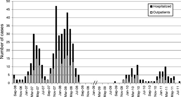
In 2009–2011, of the 330 cases with AGE with a stool specimen, 86 (26 %) were found to have a wild-type RV in stools; 34 of those were treated as outpatients and 52 were hospitalized. Compared to reference years 2006–2007 and 2007–2008 (combined), this means an 81 % reduction of RV AGE in the outpatient clinic (34 vs. 177 cases) and a 76 % reduction in hospital ward admissions (52 vs. 219 cases) (Fig. 1).
Fig. 1.

Timing of the RV seasons between September 2006 and August 2008 and September 2009 and August 2011 and treatment needed in the 507 children seen at Tampere University Hospital because of acute RVGE
The total reduction of all RVGE cases was 80 % during the study years and the proportion of RVGE cases of all AGE cases was decreased from 52 % (421 cases) in the two reference years to 26 % (86 cases) in the two study years combined. The total reduction was statistically significant with p < 0.001.
RV was found in 43 cases each (27 and 25 %, respectively) of 160 and 170 stool samples obtained in the first and the second season after NIP, respectively. In the first RV epidemic season, the majority of RV-positive AGE cases were seen relatively late between March 2010 and May 2010, whereas in the second season, the most active months were between January 2011 and March 2011 (Fig. 1).
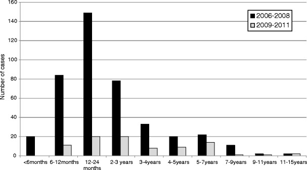
The age distribution of the children with RVGE was from 7 months to 14 years 6 months. The age distribution of RVGE patients shifted towards older children each year (p < 0.001). In 2006–2008, the median age was 19 months, while in 2009–2010, it was 24 months, and in 2010–2011, it was 36 months. Still, the proportion of RVGE cases decreased in every age group, even among children too old for vaccination. The reduction in the age groups eligible for vaccination (patients <1 year of age in 2009–2010 and patients <2 years of age in 2010–2011) was 91 % (16 vs. 178 patients), and in children too old to be vaccinated in NIP, it was 72 % (70 vs. 243 patients). The age distribution of RV-positive cases during 2006–2008 and 2009–2011 are shown in Fig. 2.
Fig. 2.

Reduction of RVGE between 2006–2008 and 2009–2011 in different age groups
The predominant RV types in the two seasons 2009 to 2010 and 2010 to 2011, combined, were G1P[8] (n = 38, 44 %) and G4P[8] (n = 30, 35 %). In the first season, genotype G4P[8] (n = 18, 42 %) was slightly more common than G1P[8] (n = 15, 35 %), but in the second season, genotype G1P[8] was more predominant with 53 % (n = 23) over G4P[8] (n = 13, 30 %). Other common RV genotypes G2P[4], G3P[8], and G9P[8] were all seen to a lesser extent (n = 9, n = 1, and n = 6, respectively, counted from all RV-positive AGE cases in the two seasons combined). In two cases, more than one RV type was found in stools simultaneously: in one case, G1P[8] with G3P[8] and, in the other, G3P[8] with G9P[8]. Other than the predominance of G9P[8] genotype in the season of 2006–2007, no great changes in the genotype distribution were observed during the study years compared to the reference years (data not shown).
Among the 86 wild-type RV-positive cases, there were 4 children who had received at least 1 dose of RotaTeq® and 1 child who had received Rotarix™ before the introduction of NIP. Three of those who had received RotaTeq® were fully vaccinated (two were detected with G4P[8] in the stools and one was detected with G9P[8] in the stools) and one had received only one dose and was detected with G4P[8] RV. The child who had received Rotarix™ was also fully vaccinated with two doses and was detected with the G4P[8] genotype. Two of the four breakthrough cases, 9- and 10-month-old fully vaccinated boys (RotaTeq®), were admitted to the pediatric ward and the other two were seen in the ER only.
We identified three cases of GE in young infants shedding a human–bovine double reassortant G1P[8] vaccine virus. This human–bovine double reassortant was also detected from one patient infected concomitantly with NV. Furthermore, one patient was detected with RotaTeq® vaccine virus G6P[8] and 16 patients were detected to shed the original vaccine virus G1P[5] or just the VP7 G1 part of it separately or detected with several VP4 proteins. No patients were detected with Rotarix™ vaccine strain after 2006–2008. The vaccine-associated cases have been reported separately [13].
Norovirus gastroenteritis
Of all 330 cases of GE in 2009–2011, 111 (33.6 %) were NV-positive. In the first year, NV was found in 52 (33 %) of 160 stool samples and, in the second season, in 59 (35 %) of 170 stool samples (Fig. 3). SaV was found in a total of 23 (7.0 %) cases, 13 (8.1 % of 160) of these in the first season and 10 (5.9 % of 170) in the second season.
Fig. 3.

Seasonality of the NVs seen in AGE in children between 2006–2008 and 2009–2011 in Tampere University Hospital
Of the NV-positive cases, only one was a mixed infection with RV (more specifically with G2P[6]). Three of 23 (13 %) SaV-positive cases were mixed infections with RVs. There were no cases with NV and SaV in the stools at the same time. The reduction of GE positive for NV, SaV, and RV is shown in Fig. 4.
Fig. 4.

Viruses detected in the stools of children admitted to the hospital for AGE in 2006–2008 and 2009–2011
In the reference years 2006–2008, there were 196 cases (excluding the outbreak mentioned in the “Materials and methods” section) of NVGE as compared with 111 cases in the study years 2009–2011. Of the 111 NV-positive cases, 69 (62 %) were admitted to the hospital and 42 (38 %) were treated as outpatients. Even though the absolute number of NV-positive cases decreased slightly (196 vs. 111 cases), the proportion of NVGE of all AGE increased (26 vs. 33.6 %) from the reference years. Moreover, the proportion of NV-positive cases that were admitted to the hospital increased from 47 % (92) in the reference years to 62 % (69) in the study years (Fig. 4).
Compared to the reference years, the proportion and the absolute number of SaV-positive GE increased from 1.6 % (12 cases) to 7.0 % (23 cases). A clear seasonality was seen in the NVGE both in the study years and in the reference years (Fig. 3). The most active months when the majority of NV-positive cases were seen were between January and April in each year.
Of all 111 NV-positive cases, 108 (97 %) were genogroup GII strains and 72 (65 %) were genotype GII.4 (37 (71 % of 52 cases) and 35 (59 % of 59 cases) in the first and the second study years, respectively). In the reference years, genotype GII.4 was even more common with 89 % proportion (175 of 196 cases). The other genotypes detected were GII.b (14 %, n = 15), GII.7 (13 %, n = 14), GII.g (5 %, n = 6), GI.4 (2 %, n = 2), GI.3 (1 %, n = 1), and GII.e (1 %, n = 1). Additional genotypes detected in the reference years included GII.1, GII.c, GI.6, and GII.2, all of which accounted for <1 % each and none of which were detected in the study years 2009–2011.
The age distribution of NV-positive children was similar between the study years and the reference years. The age range was from 7 days to 15 years 7 months (19 days to 13 years 8 months in reference years), with a median age of 12 months (15 months in reference years). Eighty-four of 111 cases (76 %) of children were under 24 months of age, and 41 % was under 12 months of age.
Gastroenteritis with neither RV nor NV
In the study years 2009–2011, of the 330 cases of AGE, 222 (67 %) were positive for RV, NV, or SaV alone or as a mixed infection, whereas in the reference years 2006–2008, 603 (76 %) were positive for RV, NV, or SaV alone or as a mixed infection. The absolute number of GE cases due to other pathogens decreased from 191 cases in the reference years to 108 cases in the study years. Of 108 cases, 53 % (57 cases) detected in the two study years was admitted to a pediatric ward and 47 % (51 cases) was treated as outpatients. RV, NV, or SaV could be detected in the stools in 70 % of all 186 cases admitted to a pediatric ward due to GE and, conversely, 30 % was negative for these viruses (Fig. 4). The absolute number of children who were admitted due to GE and were negative for RV, NV, or SaV decreased from 101 cases in the reference years to 57 cases in the study years. Even though a systematic search for other GE viruses was not performed, some of these patients were found to have other viral agents such as human bocavirus, adenovirus, astrovirus, and coronavirus in their stools [30, 31]. In the emergency department, 35 % (51 of 144 cases) of the children seen for GE symptoms were negative for RV, NV, or SaV. All hospital admissions due to all AGE decreased by 57 % from 434 cases in the reference years to 186 cases in the study years, and the proportion of outpatient clinic visits decreased by 62 % from 375 to 144 cases.
Discussion
In this study, we examined the impact of the National RV Immunization Programme (NIP) on hospitalizations and outpatient clinic visits due to GE in one hospital. The coverage population of the Tampere University Hospital is about one tenth of Finland, and the results may be generalized for the whole country.
We detected a significant reduction in outpatient AGE visits and hospital admissions due to RV (81 and 76 %, respectively) in the 2-year post-NIP period in the entire children population. Similar reductions with the exclusive use of RotaTeq have been observed previously from the USA [32, 33, 37] and from countries using both two available RV vaccines [2, 4, 23].
We did not observe further reduction in RVGE between first (2009–2010) and second (2010–2011) seasons. This might be because the second season post-NIP (2010–2011) might have been a strong epidemic season of RV, resulting in more RV infection pressure; just like in the two reference years, season 2007–2008 was a high epidemic season compared to 2006–2007.
Our study supports the evidence of herd protection in children too old to be vaccinated that have been observed in three studies from the USA after the widespread use of RV vaccines [5, 9, 37]. We observed that the reduction in RVGE cases was statistically significant in every age group. The decrease in hospitalizations in children too old to be vaccinated was 72 %, similar to the findings from the USA (70–79 %) [5, 9, 37]. We found no cases of wild-type RVGE in children <6 months of age. In contrast, in the USA, no reduction in infants <3 months of age, who had been too young to be vaccinated, was observed [5, 9, 37]. Additionally, we observed that the median age distribution of RVGE cases had shifted toward older children.
The high level of herd protection in our study probably resulted from high vaccine coverage. In Austria, no evidence of herd protection was found with vaccine coverage of 57 % in 2007 (RotaTeq) [2]. The reason for herd protection is probably interruption of RV transmission among all children. Exposure of unvaccinated children to vaccine virus shed in the stools of vaccinated infants is possible but unlikely to explain herd protection. Such transmission of vaccine-acquired virus resulting to a symptomatic RVGE has been reported in several countries [24]. In our study, we detected shedding of vaccine virus in a number of children, but all were recently vaccinated and none was unvaccinated.
After the introduction of the RV vaccine into the vaccination program, several studies have detected unusual non-vaccine-included RV strains, such as G8 or G12, or changes in the genotype distribution [14]. However, none of these changes were observed in our study.
In addition, we observed that the impact of RV vaccination was reflected as decreased hospital admissions and outpatient clinic visits for GE of any cause. Compared to the pre-NIP period, there was a 57 % reduction in cases admitted to the hospital ward for all GE. The reduction was higher than the reduction rates observed in previous studies from the USA (29–52 %) [5, 6, 37].
In addition, we observed a reduction of 62 % in all outpatient clinic visits for GE of any cause. Interestingly, such a reduction in all outpatient clinic visits has not been reported from countries where the protective effect of RV vaccination in unvaccinated children has been observed [10, 12].
The important role of NV as a causative agent of endemic (not outbreak-associated) GE in children was first discovered in Finland in connection with an efficacy trial of RotaShield vaccine [18]. In the same study, it was observed that RV vaccine (RotaShield) did not have any effect on NVGE. In that sense, the present findings on the impact of universal RV vaccination on NVGE are (only) confirmatory, and we conclude that the RotaTeq vaccination program does not reduce NVGE. The slight decrease observed in the study vs. reference years may well be explained by natural annual variation. In a decade, there has been considerable year to year variation of NVGE, although the winter epidemic has occurred every year [26].
In reverse, other viruses and notably NVs could theoretically replace RV after its elimination by universal vaccination and fill the available niche as a major causative agent of AGE in children. Our results strongly suggest that such a replacement is not happening. Overall hospitalizations have been reduced according to the share of RV, and NV has become a leading cause of GE only in relative terms, without any increase in absolute numbers.
The reduction of all hospitalizations (57 %) and outpatient clinic visits (62 %) due to GE is well in line with what was observed in the prelicensure efficacy trial (REST) of the RotaTeq vaccine in Finland. RotaTeq reduced all-cause GE requiring medical intervention by 65 % over a period of 3.1 years [34]. To compare the numbers, it should be noted that the present population-based study also includes children who were eligible for vaccination but did not receive it (initially about 10 %, decreasing to 5 % over time).
The present study focused on NV and was not intended as a full etiological examination of GE. Hospitalizations due to GE not associated with RV or NV seemed to decrease somewhat in comparison with the reference years. This observation should be viewed with caution, and a detailed etiological study of GE viruses should be performed before conclusions. However, even though the RV vaccine had no effect on NVGE, it is nevertheless possible that RV vaccination might have a “nonspecific” effect on non-RV-associated GE. Some suggestive evidence of RV vaccine (RotaShield) effect on adenovirus GE was seen in the study in the 1990s [19].
The new leading role of NV as the main causative agent of AGE in children supports the concept of developing an NV vaccine for use in children [11]. Such a vaccine is foreseen and being developed [1].
Conclusion
RV in the NIP of Finland had an immediate and major impact on RVGE cases seen in hospital, i.e., severe RVGE. The age distribution of children with RVGE has shifted upwards at the same time as a statistically significant decrease in every age group was observed as an evidence of herd protection. The impact of RV vaccination was reflected in a decrease of all hospital admissions and outpatient clinic visits for GE, while NV has become the leading cause of AGE in children seen in hospital.
Acknowledgments
We thank the study nurse personnel, especially Marjo Salonen as head study nurse, the staff of the virology laboratory, and the nursing personnel at Tampere University Hospital.
Conflict of interest
Maria Hemming, Sirpa Räsänen, Leena Huhti, Minna Paloniemi, and Marjo Salminen have no conflict of interest to disclose. Timo Vesikari has been the principal investigator of clinical trials of rotavirus vaccines produced by Merck and GlaxoSmithKline and is a member of the advisory boards of Sanofi Pasteur MSD, Merck, Pfizer, Novartis, and GSK.
Source of funding
This study received no funding outside the University of Tampere.
References
- 1.Blazevic V, Lappalainen S, Nurminen K, Huhti L, Vesikari T. Norovirus VLPs and rotavirus VP6 protein as combined vaccine for childhood gastroenteritis. Vaccine. 2011;29:8126–8133. doi: 10.1016/j.vaccine.2011.08.026. [DOI] [PubMed] [Google Scholar]
- 2.Braeckman T, Van Herck K, Raes M, Vergison A, Sabbe M, Van Damme P. Rotavirus vaccines in Belgium: policy and impact. Pediatr Infect Dis J. 2011;30:S21–S24. doi: 10.1097/INF.0b013e3181fefc51. [DOI] [PubMed] [Google Scholar]
- 3.Buesa J, Collado B, Lopez-Andujar P, Abu-Mallouh R, Rodriguez Diaz J, Garcia Diaz A, Prat J, Guix S, Llovet T, Prats G, Bosch A. Molecular epidemiology of caliciviruses causing outbreaks and sporadic cases of acute gastroenteritis in Spain. J Clin Microbiol. 2002;40:2854–2859. doi: 10.1128/JCM.40.8.2854-2859.2002. [DOI] [PMC free article] [PubMed] [Google Scholar]
- 4.Buttery JP, Lambert SB, Grimwood K, Nissen MD, Field EJ, Macartney KK, Akikusa JD, Kelly JJ, Kirkwood CD. Reduction in rotavirus-associated acute gastroenteritis following introduction of rotavirus vaccine into Australia’s National Childhood vaccine schedule. Pediatr Infect Dis J. 2011;30:S25–S29. doi: 10.1097/INF.0b013e3181fefdee. [DOI] [PubMed] [Google Scholar]
- 5.Cortese MM, Tate JE, Simonsen L, Edelman L, Parashar UD. Reduction in gastroenteritis in United States children and correlation with early rotavirus vaccine uptake from national medical claims databases. Pediatr Infect Dis J. 2010;29:489–494. doi: 10.1097/INF.0b013e3181d95b53. [DOI] [PubMed] [Google Scholar]
- 6.Curns AT, Steiner CA, Barrett M, Hunter K, Wilson E, Parashar UD. Reduction in acute gastroenteritis hospitalizations among US children after introduction of rotavirus vaccine: analysis of hospital discharge data from 18 US states. J Infect Dis. 2010;201:1617–1624. doi: 10.1086/652403. [DOI] [PubMed] [Google Scholar]
- 7.Dolin R, Blacklow NR, DuPont H, Buscho RF, Wyatt RG, Kasel JA, Hornick R, Chanock RM. Biological properties of Norwalk agent of acute infectious nonbacterial gastroenteritis. Proc Soc Exp Biol Med. 1972;140:578–583. doi: 10.3181/00379727-140-36508. [DOI] [PubMed] [Google Scholar]
- 8.Farkas T, Zhong WM, Jing Y, Huang PW, Espinosa SM, Martinez N, Morrow AL, Ruiz-Palacios GM, Pickering LK, Jiang X. Genetic diversity among sapoviruses. Arch Virol. 2004;149:1309–1323. doi: 10.1007/s00705-004-0296-9. [DOI] [PubMed] [Google Scholar]
- 9.Giaquinto C, Dominiak-Felden G, Van Damme P, Myint TT, Maldonado YA, Spoulou V, Mast TC, Staat MA. Summary of effectiveness and impact of rotavirus vaccination with the oral pentavalent rotavirus vaccine: a systematic review of the experience in industrialized countries. Hum Vaccin. 2011;7:734–748. doi: 10.4161/hv.7.7.15511. [DOI] [PubMed] [Google Scholar]
- 10.Glass RI. Unexpected benefits of rotavirus vaccination in the United States. J Infect Dis. 2011;204:975–977. doi: 10.1093/infdis/jir477. [DOI] [PubMed] [Google Scholar]
- 11.Glass RI, Parashar UD, Estes MK. Norovirus gastroenteritis. N Engl J Med. 2009;361:1776–1785. doi: 10.1056/NEJMra0804575. [DOI] [PMC free article] [PubMed] [Google Scholar]
- 12.Gray J. Rotavirus vaccines: safety, efficacy and public health impact. J Intern Med. 2011;270:206–214. doi: 10.1111/j.1365-2796.2011.02409.x. [DOI] [PubMed] [Google Scholar]
- 13.Hemming M, Vesikari T. Vaccine derived human–bovine double reassortant rotavirus in infants with acute gastroenteritis. Pediatr Infect Dis J. 2012;31(9):992–994. doi: 10.1097/INF.0b013e31825d611e. [DOI] [PubMed] [Google Scholar]
- 14.Iturriza-Gomara M, Dallman T, Banyai K, Bottiger B, Buesa J, Diedrich S, Fiore L, Johansen K, Koopmans M, Korsun N, Koukou D, Kroneman A, Laszlo B, Lappalainen M, Maunula L, Marques AM, Matthijnssens J, Midgley S, Mladenova Z, Nawaz S, Poljsak-Prijatelj M, Pothier P, Ruggeri FM, Sanchez-Fauquier A, Steyer A, Sidaraviciute-Ivaskeviciene I, Syriopoulou V, Tran AN, Usonis V, VAN Ranst M, DE Rougemont A, Gray J. Rotavirus genotypes co-circulating in Europe between 2006 and 2009 as determined by EuroRotaNet, a pan-European collaborative strain surveillance network. Epidemiol Infect. 2011;139:895–909. doi: 10.1017/S0950268810001810. [DOI] [PubMed] [Google Scholar]
- 15.Jiang X, Huang PW, Zhong WM, Farkas T, Cubitt DW, Matson DO. Design and evaluation of a primer pair that detects both Norwalk- and Sapporo-like caliciviruses by RT-PCR. J Virol Methods. 1999;83:145–154. doi: 10.1016/S0166-0934(99)00114-7. [DOI] [PubMed] [Google Scholar]
- 16.Johnson PC, Mathewson JJ, DuPont HL, Greenberg HB. Multiple-challenge study of host susceptibility to Norwalk gastroenteritis in US adults. J Infect Dis. 1990;161:18–21. doi: 10.1093/infdis/161.1.18. [DOI] [PubMed] [Google Scholar]
- 17.Pang XL, Honma S, Nakata S, Vesikari T. Human caliciviruses in acute gastroenteritis of young children in the community. J Infect Dis. 2000;181(Suppl 2):S288–S294. doi: 10.1086/315590. [DOI] [PubMed] [Google Scholar]
- 18.Pang XL, Joensuu J, Vesikari T. Human calicivirus-associated sporadic gastroenteritis in Finnish children less than two years of age followed prospectively during a rotavirus vaccine trial. Pediatr Infect Dis J. 1999;18:420–426. doi: 10.1097/00006454-199905000-00005. [DOI] [PubMed] [Google Scholar]
- 19.Pang XL, Koskenniemi E, Joensuu J, Vesikari T. Effect of rhesus rotavirus vaccine on enteric adenovirus-associated diarrhea in children. J Pediatr Gastroenterol Nutr. 1999;29:366–369. doi: 10.1097/00005176-199909000-00026. [DOI] [PubMed] [Google Scholar]
- 20.Parrino TA, Schreiber DS, Trier JS, Kapikian AZ, Blacklow NR. Clinical immunity in acute gastroenteritis caused by Norwalk agent. N Engl J Med. 1977;297:86–89. doi: 10.1056/NEJM197707142970204. [DOI] [PubMed] [Google Scholar]
- 21.Patel MM, Steele D, Gentsch JR, Wecker J, Glass RI, Parashar UD. Real-world impact of rotavirus vaccination. Pediatr Infect Dis J. 2011;30:S1–S5. doi: 10.1097/INF.0b013e3181fefa1f. [DOI] [PubMed] [Google Scholar]
- 22.Patel MM, Widdowson MA, Glass RI, Akazawa K, Vinje J, Parashar UD. Systematic literature review of role of noroviruses in sporadic gastroenteritis. Emerg Infect Dis. 2008;14:1224–1231. doi: 10.3201/eid1408.071114. [DOI] [PMC free article] [PubMed] [Google Scholar]
- 23.Paulke-Korinek M, Rendi-Wagner P, Kundi M, Kronik R, Kollaritsch H. Universal mass vaccination against rotavirus gastroenteritis: impact on hospitalization rates in Austrian children. Pediatr Infect Dis J. 2010;29:319–323. doi: 10.1097/INF.0b013e3181c18434. [DOI] [PubMed] [Google Scholar]
- 24.Payne DC, Edwards KM, Bowen MD, Keckley E, Peters J, Esona MD, Teel EN, Kent D, Parashar UD, Gentsch JR. Sibling transmission of vaccine-derived rotavirus (RotaTeq) associated with rotavirus gastroenteritis. Pediatrics. 2010;125:e438–e441. doi: 10.1542/peds.2009-1901. [DOI] [PubMed] [Google Scholar]
- 25.Puustinen L, Blazevic V, Huhti L, Szakal ED, Halkosalo A, Salminen M, Vesikari T. Norovirus genotypes in endemic acute gastroenteritis of infants and children in Finland between 1994 and 2007. Epidemiol Infect. 2012;140:268–275. doi: 10.1017/S0950268811000549. [DOI] [PMC free article] [PubMed] [Google Scholar]
- 26.Puustinen L, Blazevic V, Salminen M, Hamalainen M, Rasanen S, Vesikari T. Noroviruses as a major cause of acute gastroenteritis in children in Finland, 2009–2010. Scand J Infect Dis. 2011;43:804–808. doi: 10.3109/00365548.2011.588610. [DOI] [PubMed] [Google Scholar]
- 27.Rasanen S, Lappalainen S, Halkosalo A, Salminen M, Vesikari T. Rotavirus gastroenteritis in Finnish children in 2006–2008, at the introduction of rotavirus vaccination. Scand J Infect Dis. 2011;43:58–63. doi: 10.3109/00365548.2010.508462. [DOI] [PubMed] [Google Scholar]
- 28.Rasanen S, Lappalainen S, Kaikkonen S, Hamalainen M, Salminen M, Vesikari T. Mixed viral infections causing acute gastroenteritis in children in a waterborne outbreak. Epidemiol Infect. 2010;138:1227–1234. doi: 10.1017/S0950268809991671. [DOI] [PubMed] [Google Scholar]
- 29.Rasanen S, Lappalainen S, Salminen M, Huhti L, Vesikari T. Noroviruses in children seen in a hospital for acute gastroenteritis in Finland. Eur J Pediatr. 2011;170:1413–1418. doi: 10.1007/s00431-011-1443-4. [DOI] [PMC free article] [PubMed] [Google Scholar]
- 30.Risku M, Katka M, Lappalainen S, Rasanen S, Vesikari T. Human bocavirus types 1, 2 and 3 in acute gastroenteritis of childhood. Acta Paediatr. 2012;101:e405–e410. doi: 10.1111/j.1651-2227.2012.02727.x. [DOI] [PMC free article] [PubMed] [Google Scholar]
- 31.Risku M, Lappalainen S, Rasanen S, Vesikari T. Detection of human coronaviruses in children with acute gastroenteritis. J Clin Virol. 2010;48:27–30. doi: 10.1016/j.jcv.2010.02.013. [DOI] [PMC free article] [PubMed] [Google Scholar]
- 32.Tate JE, Mutuc JD, Panozzo CA, Payne DC, Cortese MM, Cortes JE, Yen C, Esposito DH, Lopman BA, Patel MM, Parashar UD. Sustained decline in rotavirus detections in the United States following the introduction of rotavirus vaccine in 2006. Pediatr Infect Dis J. 2011;30:S30–S34. doi: 10.1097/INF.0b013e3181ffe3eb. [DOI] [PubMed] [Google Scholar]
- 33.Tate JE, Panozzo CA, Payne DC, Patel MM, Cortese MM, Fowlkes AL, Parashar UD. Decline and change in seasonality of US rotavirus activity after the introduction of rotavirus vaccine. Pediatrics. 2009;124:465–471. doi: 10.1542/peds.2008-3528. [DOI] [PubMed] [Google Scholar]
- 34.Vesikari T, Karvonen A, Ferrante SA, Ciarlet M. Efficacy of the pentavalent rotavirus vaccine, RotaTeq(R), in Finnish infants up to 3 years of age: the Finnish Extension Study. Eur J Pediatr. 2010;169:1379–1386. doi: 10.1007/s00431-010-1242-3. [DOI] [PMC free article] [PubMed] [Google Scholar]
- 35.Vinje J, Hamidjaja RA, Sobsey MD. Development and application of a capsid VP1 (region D) based reverse transcription PCR assay for genotyping of genogroup I and II noroviruses. J Virol Methods. 2004;116:109–117. doi: 10.1016/j.jviromet.2003.11.001. [DOI] [PubMed] [Google Scholar]
- 36.Wyatt RG, Dolin R, Blacklow NR, DuPont HL, Buscho RF, Thornhill TS, Kapikian AZ, Chanock RM. Comparison of three agents of acute infectious nonbacterial gastroenteritis by cross-challenge in volunteers. J Infect Dis. 1974;129:709–714. doi: 10.1093/infdis/129.6.709. [DOI] [PubMed] [Google Scholar]
- 37.Yen C, Tate JE, Wenk JD, Harris JM, 2nd, Parashar UD. Diarrhea-associated hospitalizations among US children over 2 rotavirus seasons after vaccine introduction. Pediatrics. 2011;127:e9–e15. doi: 10.1542/peds.2010-1393. [DOI] [PubMed] [Google Scholar]


