

Abstract
Melorheostosis is a rare disease (0.9/million habitants), characterized by linear hyperostosis along the cortex bone. It can affect any bone, being more frequent in long bones. The lesions tend to be segmental and unilateral. The etiology remains unknown although several theories proposed over the past year (vascular, inflammatory processes, embryonic defects or genetic). Show no significant difference between sexes or heredity. Clinical manifestations are mainly pain, deformity and joint stiffness. The diagnosis is obtained by combining the clinical findings with imaging studies (mainly radiography with typical image in “candle wax”). There is no definitive or specific treatment, being always palliative. We describe a case of a patient of twenty-four years, followed in Orthopedic consultation since age eight, with a deformity of the right side of the body. X-rays showed hyperostosis of the bones of the limbs in the right side of the body (image in “candle wax”). The patient is in physical therapy program and has a positive response to analgesia with ibuprofen.
Keywords: Melorheostosis Osteosclerosis Hyperostosis
1. Introduction
Melorheostosis is characterized by linear hyperostosis along the bone cortex. It is more common in the long bones, although it can affect any bone of any segment of the human body and tends to be segmental and unilateral.1
The incidence is 0.9 cases per million inhabitants,2 and there are approximately 300 cases described in the literature. Melorheostosis affects men and women in equal proportions, and cases have been described in children and adults.
The etiology is unknown, despite several theories proposed over the past several years (vascular alterations, inflammatory processes and embryonic or genetic defects).3
Clinically, the main symptom is local pain aggravated by mobilization of the affected segment. Melorheostosis can also be characterized by recurrent joint effusions, bone deformity and joint stiffness.4
The diagnosis, in most cases, can be attained by radiographic assessment (“dripping candle wax” sign)1 and can be aided by analytical (normal serum calcium, phosphorus and alkaline phosphatase) and anatomopathological tests (the histological findings are nonspecific and often show a mixture of mature and immature bone in a dense formation with increased trabecular bone)1 or by scintigraphy (higher uptake).
There is no specific treatment for melorheostosis; rather, treatment is symptomatic and involves the use of analgesic or anti-inflammatory drugs.
2. Case Report
We report the clinical case of a 24-year-old patient without any known toxic habits who was also not addicted to any drugs. He had been followed as an outpatient at the Orthopedics Service since the age of 8 due to insidious onset pain in the limbs of the right hemibody (more marked in the lower limb). During the follow-up, he showed a gradual increase in dysmorphia in these limbs, which manifested at that time as a 4-cm increase in the upper limb (compared with the contralateral upper limb) and a 5-cm increase in the lower limb (compared with the contralateral lower limb).
Clinically, the patient had diffuse swelling in the affected limbs, with increased local temperature and hyperpigmentation. All the joints of the affected segments showed decreased range of motion compared with the healthy limbs.
Radiographs of the affected segments showed bone hyperostosis of the limbs in the right hemisphere (except the radius, the 1st and 5th rays of the hand and the 5th ray of the foot), with the typical “dripping candle wax” sign (Fig. 1, Fig. 2, Fig. 3, Fig. 4, Fig. 5).
Fig. 1.
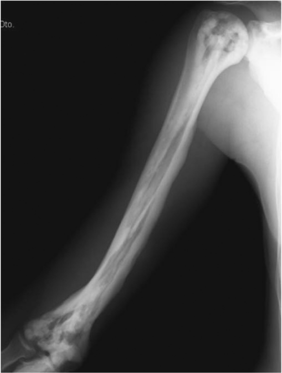
Upper limb X-ray.
Fig. 2.
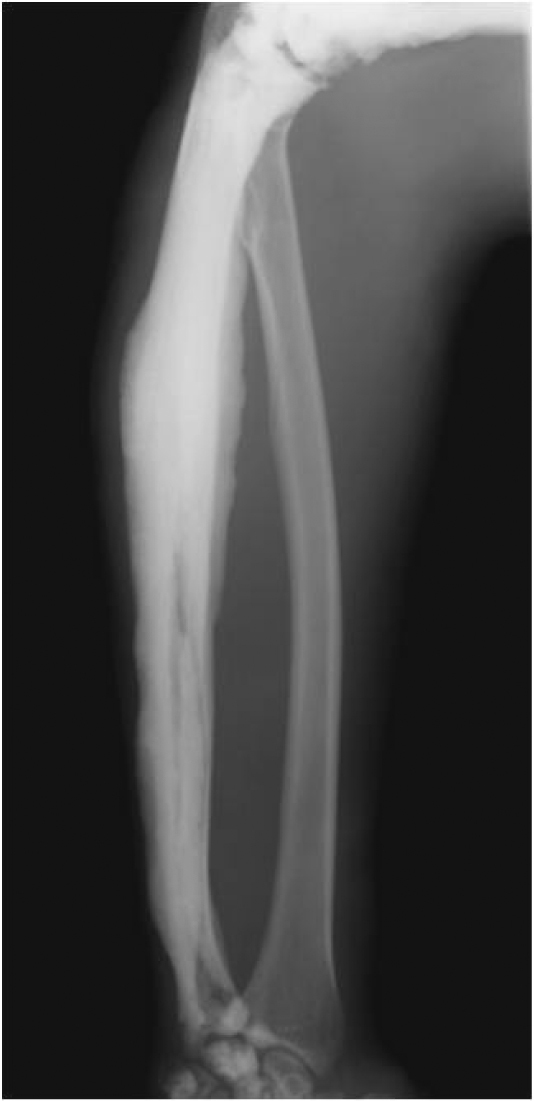
Forearm X-ray.
Fig. 3.
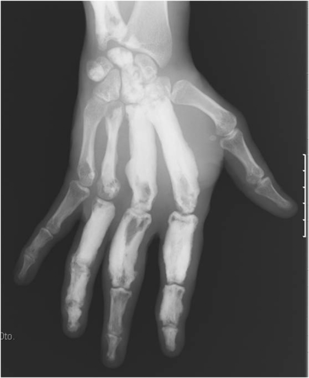
Hand X-ray.
Fig. 4.
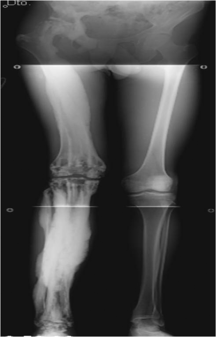
Lower limb X-ray.
Fig. 5.
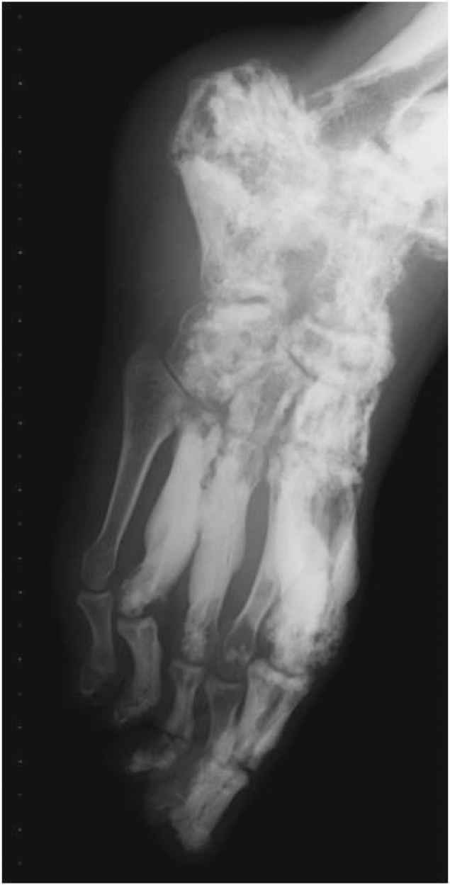
Foot X-ray.
In the laboratory assessment, no alterations were found in any of the parameters, notably serum calcium, alkaline phosphatase, C-reactive protein or erythrocyte sedimentation rate.
The histopathology report of the femur biopsy disclosed: “Large areas of bone tissue with cortical appearance and haversian systems of different calibers combined with areas of spongy bone. The joint injury suggests sclerosing bone dysplasia.”
The patient is currently in a physical therapy program, receives analgesia with ibuprofen (400 mg) and has shown a good response to treatment. He is being clinically followed.
3. Discussion
Melorheostosis is a rare disease first described in 1922 by Leri and Joanny.5 It can occur at any age and affects both sex equally.6
The etiology is unknown, although several theories have been proposed, such as a mesenchymal cell differentiation defect (genetic mutation in LEMD3), vascular disorders or inflammatory processes.3 None of these theories have been completely proven.
The symptoms vary considerably from asymptomatic to severe pain with associated deformity. The onset of complaints is usually insidious, with pain, joint stiffness, skin alterations and bone deformity.4 Our patient began to experience insidious pain at 8 years old, with progressive deformity that has continued to the present.
Regarding the distribution of lesions, they can affect one limb (monomelic, which is the most common) or both upper and lower limbs (hemimelic). The condition can be either monostotic or polyostotic if it affects one or many bones, respectively.7 The distribution in our patient was hemimelic and polyostotic, which is rare. The condition mainly affects the long bones, but rarely the axial skeleton,1 as observed in the aforementioned patient.
The diagnosis is based on radiographic findings (typical “dripping candle wax” sign) characterized by areas of hyperostosis with increased cortical bone thickness and linear density areas in the cortex that can extend into cancellous bone.1 Our patient had clear radiographic findings that were typical of melorheostosis. The laboratory assessment revealed no alterations, as often occurs in this type of pathology.
The histological findings are nonspecific and often show a mixture of mature and immature bone in a dense formation with increased trabecular bone. Osteoclastic activity is unusual, although marginal osteoblast formation is common.1 These characteristics are comparable to those of our case.
Treatment is symptomatic in most cases. Surgical treatment is reserved for contractures and deformities.1 Our patient has been treated with a nonsteroidal anti-inflammatory drug (ibuprofen 400 mg) with good symptomatic response.
Melorheostosis is an extremely rare disease characterized by linear hyperostosis along the bone cortex. Clinically, it progresses with episodes of local pain and deformity. Diagnosis is achieved by a combination of clinical assessment and imaging tests.
There is no specific treatment, and therapy is essentially palliative.
Conflicts of interest
The authors declare no conflict of interest.
Footnotes
The study was carried out at the Service of Orthopedics and Traumatology of Unidade Local de Saúde do Alto Minho, Portugal.
References
- 1.Long H.T., Li K.H., Zhu Y. Case report: severe melorheostosis involving the ipsilateral extremities. Clin Orthop Relat Res. 2009;467(10):2738–2743. doi: 10.1007/s11999-009-0890-y. [DOI] [PMC free article] [PubMed] [Google Scholar]
- 2.Clifford P.D., Jose J. Melorheostosis Am J Orthop (Belle Mead NJ) 2009;38(7):360–361. [PubMed] [Google Scholar]
- 3.Hellemans J., Preobrazhenska O., Willaert A., Debeer P., Verdonk P.C., Costa T. Loss-of-function mutations in LEMD3 result in osteopoikilosis. Buschke-Ollendorff syndrome and melorheostosis Nat Genet. 2004;36(11):1213–1218. doi: 10.1038/ng1453. [DOI] [PubMed] [Google Scholar]
- 4.Biaou O., Avimadje M., Guira O., Adjagba A., Zannou M., Hauzeur J.P. Melorheostosis with bilateral involvement in a black African patient. Joint Bone Spine. 2004;71(1):70–72. doi: 10.1016/S1297-319X(03)00103-9. [DOI] [PubMed] [Google Scholar]
- 5.Leri A., Joanny J. Une affection non décrite des os hyperostose “en coulée” sur toute la longeur d’un member ou “melorhéostose.”. Bull Mem Soc Med Hosp Paris. 1922;46:1141–1145. [Google Scholar]
- 6.Kalbermatten N.T., Vock P., Rüfenacht D., Anderson S.E. Progressive melorheostosis in the peripheral and axial skeleton with associated vascular malformations: imaging findings over three decades. Skeletal Radiol. 2001;30(1):48–52. doi: 10.1007/s002560000283. [DOI] [PubMed] [Google Scholar]
- 7.Nuño C., Heili S., Alonso J., Alcalde M., López P., Villacastín B. Meloreostosis: presentación de un caso y revisión de la literatura. Rev Esp Enferm Metab Oseas. 2001;10(1):50–55. [Google Scholar]