Abstract
Late-stage CCR5 tropic human immunodeficiency virus type 1 (HIV-1) isolates (R5 HIV-1) can deplete nearly all CD4+ thymocytes from human thymus/liver grafts, despite the fact that fewer than 5% of these cells express CCR5. To resolve this paradox, we studied the replication and cytopathic effects (CPE) of late-stage R5 HIV-1 biological clones from two progressors and two long-term nonprogressors (LTNP) in fetal thymic organ culture (FTOC) with and without added cytokines. We found that R5 HIV-1 clones from progressors but not LTNP were cytopathic in untreated FTOC. Moreover, R5 HIV-1 clones from progressors replicated to higher levels than LTNP-derived R5 HIV-1 clones in this system. In contrast, when FTOC was maintained in the presence of interleukin 2 (IL-2), IL-4, and IL-7, both progressor and LTNP clones exhibited similar replication and CPE, which were equal to or greater than the levels achieved by progressor-derived R5 HIV-1 clones in untreated FTOC. This finding was likely due to IL-2-induced CCR5 expression on CD4+ thymocytes in FTOC. R5 HIV-1 clones showed greater pathogenesis for CCR5+ cells but also showed evidence of CPE on CCR5− cells. Furthermore, infection of FTOC by R5 HIV-1 induced IL-10 and transforming growth factor β (TGF-β) expression. Both IL-10 and TGF-β in turn induced CCR5 expression in FTOC. Induction of CCR5 expression via cytokine induction by R5 HIV-1 infection of CCR5+ thymocytes likely permitted further viral replication in newly CCR5+ thymocytes. CCR5 expression, therefore, is a key determinant of pathogenesis of R5 HIV-1 in FTOC.
Human immunodeficiency virus type 1 (HIV-1) infection of humans is characterized by the progressive loss of CD4+ T cells. Infection by most strains of HIV-1 requires interaction with CD4 and a chemokine receptor, either CXCR4 or CCR5 (reviewed in reference 3). During early stages of HIV-1 infection, viral isolates most often use CCR5 to enter cells and are known as R5 HIV-1 (11, 16, 21). Viral isolates that utilize CXCR4 for entry usually arise later during the course of infection and are known as X4 HIV-1 (13, 23). X4 HIV-1 species are rarely transmitted and infrequently detected during the asymptomatic stage of HIV-1 infection for reasons that are not well understood (74). X4 HIV-1 becomes prevalent just before or during the symptomatic stages of HIV-1 infection in approximately 50% of individuals infected with clade B HIV-1 (12, 13, 60, 65, 70). In vivo and in vitro studies have shown that X4 HIV-1 is often more cytopathic than R5 HIV-1 (10, 13, 27, 37, 48, 49, 65). The greater cytopathic effects (CPE) of X4 strains are likely due to the greater fraction of thymocytes and T cells that express CXCR4 than that expressing CCR5 (4, 6, 48, 53, 56, 69, 79). CXCR4 is expressed on a majority of CD4+ T cells and thymocytes, whereas only about 5 to 25% of mature T cells and 1 to 5% of thymocytes express detectable levels of CCR5 on the cell surface. Nonetheless, only about 50% of AIDS patients acquire X4 HIV-1, so it is clear that R5 HIV-1 causes AIDS. In vitro, late-stage AIDS-associated R5 HIV-1 clones replicate more rapidly and exhibit greater CPE than pre-AIDS R5 HIV-1 clones from the same patients, indicating that viral evolution to a more pathogenic phenotype occurs within the CCR5 tropic species (18, 47, 66).
The mechanism by which R5 HIV-1 depletes CD4+ T cells in infected individuals remains poorly characterized. Both decreased thymic production and increased destruction of CD4+ T cells by a number of mechanisms have been proposed to play a role in CD4+-T-cell depletion (28, 50). Infection of the thymus may be an important step in the development of AIDS (36, 45). By infecting and destroying the thymus, HIV-1 blocks the development of new CD4+ T cells, leading to more rapid progression to AIDS and death. Stimulation of naïve T cells by HIV-1, however, also plays a role in T-cell replacement (29, 30). Nevertheless, HIV-1 infection of the thymus has been documented in children and adults and has been shown to correlate with more rapid progression to AIDS and death in children (19, 20, 45, 54).
We and others have studied HIV-1 infection of the thymus by using severe combined immune-deficient mice implanted with human fetal thymus and liver tissue (SCID-hu mice), originally developed by McCune and colleagues (1, 7, 10, 34, 46, 51, 66). Late-stage AIDS-associated R5 HIV-1 clones are cytopathic and replicate well in SCID-hu mice, despite the fact that few thymocytes express CCR5 (43, 66, 69, 79). This paradox was one impetus for the studies described in this paper. We infected human fetal thymic organ culture (FTOC) with R5 HIV-1 from progressors and long-term nonprogressors (LTNP) to study their replication and CPE. We also measured the induction of cytokines by HIV-1 infection in FTOC since cytokines have multiple effects on HIV-1 infection of T lymphocytes and macrophages. The expression of CXCL10, transforming growth factor β1 (TGF-β1), interleukin 10 (IL-10), and other cytokines are increased in HIV-1-infected individuals (24, 40, 44, 55). IL-10 and TGF-β1 have been implicated in the up-regulation of CCR5 on human monocytes, monocyte-derived macrophages, and monocyte-derived dendritic cells (8, 33, 61, 67, 76). HIV-1-induced expression of IL-10 and IFN-α also leads to higher expression of major histocompatibility complex class I (MHC-I) on double positive (DP) thymocytes (39, 46). The pattern of expression of these cytokines and their effects on chemokine receptor and MHC-I expression have important implications for HIV-1 pathogenesis. Our results suggest a mechanism to explain the nearly complete depletion of CD4+ thymocytes by late-stage R5 HIV-1 despite infrequent expression of CCR5 on thymocytes.
MATERIALS AND METHODS
Antibodies and cytokines.
CD3-fluorescein isothiocyanate (FITC), CD4-phycoerythrin (PE), CD7-FITC, CD8-allophycocyanin (APC), CD16-PE, CD11b-APC, and appropriate isotype control monoclonal antibodies (MAb) were obtained from Caltag Laboratories (Burlingame, Calif.). Anti-CCR5 MAb 2D7-APC, CD90-APC, and CMRF-56-FITC were purchased from Pharmingen (San Diego, Calif.). CD8-peridinin chlorophyll protein (PerCP) was obtained from Becton Dickinson Immunocytometry Systems (San Jose, Calif.). To identify HIV-1-infected cells, anti-p24 MAb KC57-FITC from Coulter (Miami, Fla.) was used. IL-2, IL-4, IL-7, IL-10, and TGF-β1 were obtained from R&D Systems, Inc., Minneapolis, Minn.
Preparation and titration of HIV-1 stocks.
HIV-1 biological clones were obtained from the Amsterdam cohort study. HIV-1 was cloned from patients by limiting dilution of patient peripheral blood mononuclear cells (PBMC) cocultured with stimulated normal donor PBMC as previously described (18). Viral stocks were amplified by infection of 2-day phytohemagglutinin- and IL-2-stimulated healthy donor PBMC. One-half of the virus-containing supernatants were removed every 2 days and replaced with fresh medium containing IL-2. Fresh stimulated PBMC were added 7 days postinfection if viral titers of the collected supernatants had not peaked. Virus-containing supernatants were aliquoted and frozen at −80°C until needed. The titer of virus in each supernatant was assayed by limiting dilution infection of 2-day phytohemagglutinin- and IL-2-stimulated healthy donor PBMC, followed 1 week later by assay of supernatant reverse transcriptase activity.
Human FTOC, HIV-1 infection, and cell isolation.
Human fetal thymic tissue of 20 to 24 weeks' gestation was obtained from Advanced Bioscience Resources (Alameda, Calif.). The tissue was washed, dissected into pieces of approximately 8 mm3 in size under a low-power stereomicroscope, and placed into Iscove's medium supplemented with 10% fetal bovine serum, minimal essential medium vitamin solution, 50 μg of gentamicin/ml (all from Life Technologies, Rockville, Md.), and insulin-transferrin-sodium selenite medium supplement (Sigma, St. Louis, Mo.). Tissue pieces were immediately infected with an 8 × 10³ 50% tissue culture infectious dose (TCID50) per tissue by rocking for 2 h at 37°C and compared to uninfected control tissue. Following infection, the tissue pieces were transferred to sterile organ culture membranes (Millipore) floating on the same medium as used for infection in a six-well tissue culture plate (Corning). FTOC was carried with or without the addition of IL-2 (20 IU/ml), IL-4 (0.5 ng/ml), and IL-7 (1 ng/ml). FTOC was maintained for 12 to 20 days with daily medium changes. Tissue was harvested every 3 days beginning 6 days postinfection or as stated in the text, and a single cell suspension was made by mincing the tissue with two scalpels in Iscove's medium supplemented with 2% fetal bovine serum and gentamicin. The cells were filtered through 70-μm nylon mesh and used for subsequent assays. In some cases, uninfected FTOC was cultured with IL-10 (2 ng/ml), TGF-β1 (2 ng/ml), or both cytokines.
Thymic aggregate culture.
Human fetal thymic tissue of 20 to 24 weeks' gestation was obtained from Advanced Bioscience Resources. The tissue was washed, and cells were liberated by using two scalpels, filtered through 70-μm nylon mesh to remove large clumps, and seeded at 2.5 × 10⁶ per well in a U-bottom 96-well plate in Iscove's medium supplemented with 10% fetal bovine serum, minimal essential vitamin solution, 50 μg of gentamicin/ml (all from Life Technologies), insulin-transferrin-sodium selenite medium supplement (Sigma), and IL-7 (1 ng/ml). Sixty percent of the medium was removed and replaced with fresh medium every second day without dispersing the thymic aggregates. To characterize the cell types which constitute thymic aggregate culture (TAC), TAC was prepared as described above and compared with cells liberated from fetal thymic tissue by treatment with 0.2 mg of collagenase B (Roche, Indianapolis, Ind.)/ml and 100 U of DNase (Sigma)/ml in Hanks balanced salt solution without phenol red for 45 min. These cells were also filtered through a 70-μm nylon mesh, and both cell populations were stained with a variety of MAb to detect minor cell types by flow cytometry on a FACSCalibur instrument.
Flow cytometry.
Flow cytometry was used to characterize cell surface expression of CD3, CD4, CD7, CD8, CCR5, HLA-A2, and internal p24 in human thymocytes derived from FTOC. For surface staining, 2 × 10⁶ cells were incubated with each MAb for 30 to 60 min at 4° in the dark. Cells were washed twice with phosphate-buffered saline (PBS) plus 0.02% sodium azide and then subjected to internal staining if desired. Subsequently, the cells were resuspended in 200 μl of PBS with 2% formaldehyde and incubated overnight at 4° in the dark prior to flow cytometry. For internal staining following surface staining, cells were resuspended in 100 μl of PBS containing 2% fetal bovine serum (FBS), 1% paraformaldehyde, 1 mg of human IgG (technical grade; Sigma)/ml, and 1% Tween 20 and incubated for 1 h at room temperature. Following two washes in PBS containing 2% FBS, cells were incubated with KC57-FITC anti-p24 MAb (1:50 dilution in PBS containing 2% FBS and 1% paraformaldehyde) for 30 min at room temperature in the dark. Cells were then washed two times in PBS containing 2% FBS and fixed in 200 μl of PBS with 2% formaldehyde as mentioned above prior to flow cytometry. Cells were analyzed by using a FACSCalibur flow cytometer and Cellquest Pro software (Becton Dickinson Immunocytometry Systems). Cell populations analyzed were defined based on their low angle and 90° light scattering properties. Unstained or isotype control MAb-stained cells were used to set markers defining positive reactivity.
Viral replication and cytokine production.
FTOC medium was collected 4, 6, 9, and 12 days postinfection and centrifuged for 3 min at 1,000 × g to remove cells. Acellular FTOC medium was assayed for HIV-1 capsid protein p24 by enzyme-linked immunosorbent assay (ELISA; NEN Life Science Products, Boston, Mass.) to detect viral replication. FTOC medium was also assayed for IL-10 and TGF-β1 by ELISA as described by the manufacturer (R&D Systems).
Statistical methods.
We used analysis of variance to determine the statistical significance of the results and adjust for intraexperimental variation. In addition, we adjusted for multiple comparisons by using Dunnett's test when we compared groups to a control and the Tukey test for all pairwise comparisons. Statistical tests were done with SAS statistical software (versions 6.12 and 8.2; SAS Institute, Cary, N.C.).
RESULTS
R5 HIV-1 biological clones are cytopathic in FTOC.
HIV-1 biological clones were isolated late in the course of infection from two individuals who progressed to AIDS and two LTNP; all four had normal CCR5 and CCR2b genotypes (Table 1). Of the two individuals who progressed to AIDS, one was a rapid progressor (RP1), and the other progressed at a near average pace (P1) Both LTNP were asymptomatic for more than 12 years and had viral loads below 5,012 HIV-1 RNA copies per ml of plasma at the time of virus isolation. In contrast, both progressors had viral loads greater than 39,810 HIV-1 RNA copies per ml of plasma at the time of virus isolation. All HIV-1 clones were restricted to CCR5 use, as no replication was detected in peripheral blood mononuclear cells from CCR5 Δ32/Δ32 donors (18). We studied the replication and CPE of each R5 HIV-1 clone in FTOC in comparison to uninfected control FTOC. The X4 HIV-1 molecular clone NL4-3 was used as a positive control for viral replication and virally induced CPE. In a preliminary experiment, we compared two methods of gating on live thymocytes: low-angle versus 90° light scatter gating and 7-amino-actinomycin D exclusion. We found that both methods gave very similar results in terms of the populations defined by CD4 and CD8 double staining for uninfected and HIV-1-infected and depleted-cell populations (see Fig. S1 in the supplemental material). We therefore used light scatter gating to study the effects of HIV-1 infection on thymocytes in all subsequent experiments, since it allowed us to use all available fluorescence channels to study specific cell surface and internal proteins.
TABLE 1.
Progressor- and nonprogressor-derived HIV-1 biological clonesa
| Patient (phenotype) | Diagnosis (MASCOSE) | CCR5 Type | CCR2 Type | Clone | MASCOSE | RNA load (log copies/ml of serum) |
|---|---|---|---|---|---|---|
| ACH 424 (RP) | EC (38) | +/+ | +/+ | 424.18.c2 (RP1) | 43 | 4.7 |
| ACH 142 (P) | KS (109) | +/+ | +/+ | 142.*E11 (P1) | 93 | 4.6 |
| ACH 441 (LTNP) | AS (152) | +/+ | +/+ | 441.39.2g10 (LTNP1) | 111 | 3 |
| ACH 583 (LTNP) | AS (149) | +/+ | +/+ | 583.38.1a5 (LTNP2) | 109 | 3.7 |
AS, asymptomatic; EC, esophageal candidiasis; KS, Kaposi's sarcoma; MASCOSE, months after seroconversion or study entry.
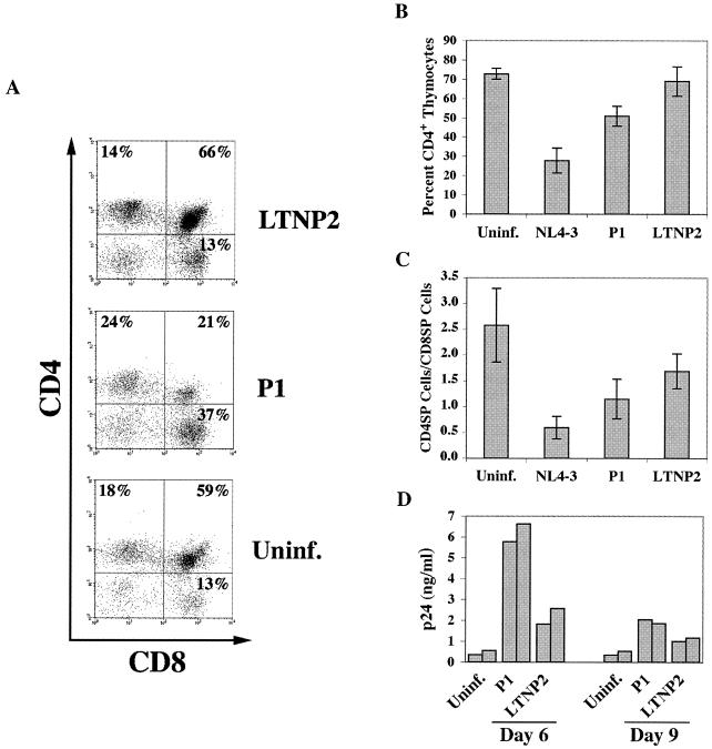
FTOC was infected with 8 × 10³ TCID50 of HIV-1. Culture was initiated immediately after infection. Cells were recovered from FTOC 2 weeks postinfection and analyzed for CD4+ thymocyte depletion as an indicator of viral CPE. In uninfected FTOC 2 weeks after the initiation of culture, approximately 70% of light scatter-gated thymocytes expressed CD4 (Fig. 1A and B), including both mature CD4+ CD8− single-positive cells (CD4SP) and immature CD4+ CD8+ double-positive thymocytes (DP). In FTOC infected with the long-term nonprogressor-derived HIV-1 clone LTNP2, no change was seen in the average percentage of thymocytes that expressed CD4 2 weeks postinfection (Fig. 1B). In contrast, R5 HIV-1 progressor clone P1-infected FTOC had an average of 51% of light scatter-gated thymocytes expressing CD4. In NL4-3-infected FTOC, 28% of the thymocytes remaining 2 weeks postinfection expressed CD4. The depletion of CD4+ thymocytes by P1 was significant compared to uninfected FTOC (P < 0.002) and LTNP2 (P < 0.01) in three separate experiments. The depletion of mature CD4SP cells was also apparent from changes in the ratio of CD4SP to CD8SP cells (Fig. 1C). In three experiments, the average CD4SP-to-CD8SP ratio in uninfected FTOC was 2.5. This ratio was significantly reduced by infection with the R5 HIV-1 LTNP2 clone (CD4SP-to-CD8SP ratio, 1.7; P < 0.01), by infection with the R5 HIV-1 progressor clone P1 (CD4SP-to-CD8SP ratio, 1.2; P < 0.0001), and by infection with the X4 HIV-1 molecular clone NL4-3 (CD4SP-to-CD8SP ratio, 0.6; P < 0.0001). These results show that the progressor-derived R5 HIV-1 clone, P1, was more cytopathic in FTOC than the LTNP2 clone, but not as cytopathic as NL4-3. Moreover, LTNP2 was more efficient in depleting mature CD4 SP thymocytes since it significantly altered the ratio of CD4SP to CD8SP cells but did not significantly deplete the total CD4+ thymocyte population. To study whether the differences in CPE mediated by the R5 HIV-1 clones P1 and LTNP2 were due to differences in the levels or kinetics of viral replication, virus production in FTOC was measured. This measurement was done by assaying the accumulation of HIV-1 capsid antigen p24 in FTOC supernatant by ELISA. P1 produced two- to threefold more p24 than LTNP2 at the peak of viral replication in a representative experiment (Fig. 1D).
FIG. 1.
R5 HIV-1 progressor clone depletes CD4+ thymocytes in untreated FTOC. FTOC was prepared and maintained without added cytokines, as described in Materials and Methods, and was then infected with the X4 HIV-1 molecular clone NL4-3 (n = 4), R5 HIV-1 progressor clone P1 (n = 6), R5 HIV-1 long-term nonprogressor clone LTNP2 (n = 6), or was not infected (n = 6). NL4-3 was used in two experiments done in duplicate. P1 and LTNP2 were used in three experiments done in duplicate along with the uninfected control FTOC (Uninf.). Two weeks postinfection, cells were isolated, incubated with CD4-PE and CD8 PerCP, and analyzed with a FACSCalibur flow cytometer. (A) Representative dot plots showing CD4 and CD8 expression on thymocytes recovered from FTOC 2 weeks postinfection with the indicated HIV-1 clone or from the uninfected control. (B) The average percentages of CD4+ thymocytes (CD4SP and DP) remaining 2 weeks postinfection with the indicated HIV-1 clone or in uninfected control FTOC are shown, with error bars indicating the standard errors of the means (SEM) for two to three independent experiments done in duplicate. (C) Bars indicate the average ratios of the percentages of CD4SP cells to CD8SP cells remaining 2 weeks postinfection with the indicated HIV-1 clone or in uninfected control FTOC. The error bars indicate the SEM for two to three independent experiments done in duplicate. (D) Viral replication in FTOC was quantified by measuring HIV-1 capsid antigen (p24) concentration in FTOC medium with a commercial ELISA kit (NEN Life Science Products). The bars represent viral replication on days 6 and 9 postinfection in a representative experiment.
The dichotomy in CPE mediated by progressor- and LTNP-derived R5 HIV-1 clones was lost in cytokine-treated FTOC.
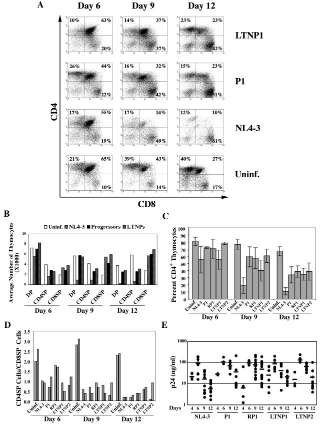
FTOC was infected with 8 × 10³ TCID50 of HIV-1 or not infected and then cultured in the presence of IL-2, IL-4, and IL-7. Thymocytes were recovered 6, 9, and 12 days postinfection, stained with CD4 and CD8 MAb, and analyzed by flow cytometry for depletion of CD4+ thymocytes. Representative dot plots show the percentages of DP, CD4SP, and CD8SP thymocytes in uninfected FTOC and the depletion of CD4SP and DP thymocytes in HIV-1-infected FTOC (Fig. 2A). We also determined the total numbers of DP, CD4SP, and CD8SP cells remaining in FTOC 6, 9, and 12 days postinfection with HIV-1 compared with uninfected FTOC. CD4+ thymocyte depletion mediated by R5 HIV-1 infection of FTOC was evident from the reduced numbers of CD4+ thymocytes remaining compared to uninfected FTOC (Fig. 2B). An average of half as many CD4SP thymocytes remained 12 days following R5 HIV-1 infection compared to uninfected FTOC. The depletion of CD4SP thymocytes by progressor- and LTNP-derived R5 HIV-1 was significant on day 9 (P < 0.001 for progressor clones and P < 0.002 for LTNP clones) and day 12 (P < 0.0001 for both clones) postinfection. R5 HIV-1 infection also significantly depleted DP thymocytes from FTOC 12 days postinfection (P < 0.0002 for progressor clones and P < 0.01 for LTNP clones), although the magnitude of this effect was smaller than the depletion of CD4SP thymocytes. In contrast, a concomitant increase in the number of CD8SP thymocytes was observed with R5 HIV-1- and NL4-3-infected FTOC. Two- to threefold more CD8SP cells were found in HIV-1-infected FTOC than in uninfected FTOC on days 9 (P < 0.0001) and 12 (P < 0.0001) postinfection. These observations strongly suggested that depletion of CD4+ thymocytes in FTOC was specific and mediated directly by HIV-1 infection, since HIV-1 depleted CD4+ thymocytes and spared CD8+ thymocytes. Moreover, HIV-1-infected FTOC was always compared with uninfected FTOC in order to control for the background of cell death in FTOC. Our results clearly validate FTOC as a model system to study HIV-1 pathogenesis.
FIG.2.
HIV-1 replication and cytopathic effects in cytokine-treated FTOC infected ex vivo. FTOC was infected with the X4 HIV-1 molecular clone NL4-3, R5 HIV-1 progressor clone P1, R5 HIV-1 rapid progressor clone RP1, R5 HIV-1 long-term nonprogressor clones LTNP1 and LTNP2, or not infected. FTOC was maintained with IL-2, IL-4, and IL-7 for 12 days with daily medium changes. At 6, 9, and 12 days postinfection, cells were isolated and incubated with CD4-PE and CD8-PerCP. CD4+ T-cell depletion was assessed by flow cytometry with a FACSCalibur instrument. (A) Representative dot plots showing two-color staining of thymocytes in FTOC. Thymocyte expression of CD4 and CD8 are represented in the dot plot for each time point; the percentages of all gated cells found in each positive quadrant are shown. (B) The data shown are the average numbers of light scatter-gated DP, CD4SP, and CD8SP thymocytes analyzed in cytokine-treated FTOC on day 6, 9, and 12 postinfection with NL4-3, progressor-derived R5 HIV-1, LTNP-derived R5 HIV-1 or uninfected control. The numbers of samples used to obtain the data on days 6, 9, and 12 postinfection, respectively, are as follows: n = 10, 20, and 20 for the uninfected control; n = 10, 14, and 14 for NL4-3; n = 4, 9, and 9 for P1; n = 8, 19, and 19 for RP1; n = 5, 8, and 7 for LTNP1; and n = 4, 8, and 10 for LTNP2. Data from infections by the two progressor-derived R5 HIV-1 clones were averaged, as were data from infections by the two LTNP-derived R5 HIV-1 clones. (C) The results shown are the average percentages of CD4+ thymocytes (CD4SP and DP), with error bars indicating the SEM for four to seven independent experiments repeated two, three, or five times. The number of experiments averaged was the same as for panel B. (D) The ratio of CD4SP-to-CD8SP thymocytes is shown for HIV-1-infected or uninfected FTOC. Data from one representative experiment of seven separate FTOC experiments are shown. Data for duplicate cultures are shown with adjacent bars. (E) Viral replication was quantified by measuring HIV-1 capsid antigen (p24) concentration in FTOC medium on day 4, 6, 9, and 12 postinfection with a commercial ELISA kit (NEN Life Science Products). Each dot represents measurement of the medium from an individual FTOC; the bars show the average values. Uninf., uninfected control.
Since our data suggest that some DP thymocytes in FTOC mature to CD4SP and CD8SP cells but are not replaced, we included both DP and CD4SP thymocytes to determine the percentage of thymocytes lost due to HIV-1 infection. In uninfected FTOC 6 and 9 days after the initiation of culture, an average of approximately 80% of light scatter-gated thymocytes expressed CD4 (Fig. 2C). By day 12 of culture, however, a slight decline in the percentage of CD4+ thymocytes had occurred so that an average of 70% of the light scatter-gated thymocytes expressed CD4. In contrast, FTOC infected by progressor- or LTNP-derived R5 HIV-1 clones had a markedly smaller percentage of CD4+ thymocytes: 40 to 60% of the total cells on day 9 and 35 to 40% on day 12 postinfection (Fig. 2B). This CD4+ thymocyte depletion was significant overall in seven separate experiments on day 9 (P < 0.0001) and day 12 (P < 0.0001) postinfection for FTOC infected with progressor clones P1 and RP1 and LTNP clones LTNP1 and LTNP2. There was no significant difference, however, in the CPE mediated by progressor- and LTNP-derived R5 HIV-1 clones in cytokine-treated FTOC on day 9 or 12 postinfection. The X4 HIV-1 clone NL4-3 was more cytopathic than any of the primary R5 HIV-1 clones we tested. NL4-3 infection depleted CD4+ thymocytes more severely both 9 (P < 0.0001) and 12 (P < 0.0001) days postinfection than any of the R5 HIV-1 clones. By 12 days postinfection, an average of only 10% of the remaining thymocytes in NL4-3-infected FTOC were CD4+.
A significant drop in the ratio of CD4SP to CD8SP thymocytes also occurred by 9 (P < 0.002) and 12 (P < 0.002) days postinfection with each of the R5 HIV-1 clones and NL4-3 in comparison to uninfected FTOC, as shown in a representative experiment (Fig. 2D). In uninfected FTOC, the ratio of CD4SP to CD8SP thymocytes was between 2 and 3.2 in duplicate FTOC during the entire course of the culture. In contrast, this ratio was significantly reduced 6 days postinfection with all but one (RP1) of the HIV-1 clones used. On days 9 and 12 of infection, the CD4SP-to-CD8SP ratio was less than one for all R5 HIV-1-infected FTOC and control X4 HIV-1-infected FTOC. Moreover, the depletion of mature CD4SP thymocytes mediated by the R5 HIV-1 clones, indicated by the change in the CD4SP-to-CD8SP ratio, was more similar to the CD4SP cell depletion mediated by NL4-3 than was the overall depletion of CD4+ thymocytes. This finding confirms what we have previously seen with SCID-hu mice: mature CD4+ thymocytes are more susceptible to depletion by R5 HIV-1 infection than immature DP thymocytes (66, 69). Altogether, these observations clearly showed that R5 HIV-1 depleted CD4+ thymocytes in FTOC. Moreover, since the LTNP2-derived R5 HIV-1 clone did not deplete thymocytes in FTOC when cultured in the absence of cytokines IL-2, IL-4, and IL-7, we can rule out the possibility that other factors present in the virus stocks were a cause of thymocyte depletion.
To study whether the differences in CPE mediated by the R5 HIV-1 clones and NL4-3 were due to differences in the levels or kinetics of viral replication, virus production in cytokine-treated FTOC was measured. We observed similar replication levels and kinetics for NL4-3 and R5 HIV-1 clones until day 6, when replication peaked in most cases, except for R5 HIV-1 clone RP1, which exhibited more variability (Fig. 2E). After day 6, a gradual decline in viral load was observed for FTOC infected with R5 HIV-1 biological clones. The decline in viral load, however, was more rapid in NL4-3-infected FTOC than in R5 HIV-1-infected FTOC. This result was likely due to the fact that NL4-3 was more cytopathic in FTOC than the R5 HIV-1 clones we used (Fig. 1B and C; Fig. 2A to D). Nevertheless, the R5 HIV-1 clones replicated to levels as high as those for NL4-3, when measured by the accumulation of p24 in FTOC medium. Since fewer CD4+ thymocytes express CCR5 than CXCR4, however, the R5 HIV-1 clones likely produced more virus per infected cell in cytokine-treated FTOC than the X4 HIV-1 molecular clone, NL4-3.
To investigate whether cytokine treatment led to an increase in the numbers of target cells for R5 HIV-1 clones, the expression of CCR5 was checked in untreated and cytokine-treated FTOC on day 7 of culture. We observed a more-than-fourfold increase in CCR5 expression on CD4+ thymocytes induced by IL-2 treatment alone (Fig. 3A). Little effect on CCR5 expression on CD4+ thymocytes in FTOC was mediated by IL-4 or IL-7 alone or both cytokines together. We also measured CCR5 expression on two CD4+ thymocyte subpopulations in uninfected control FTOC and found that a greater percentage of CD4SP thymocytes express CCR5 than DP thymocytes (Fig. 3B and C; Fig. 4A). IL-2 increased CCR5 expression primarily on mature CD4SP thymocytes, and the addition of IL-4 or IL-4 and IL-7 further increased the fraction of mature thymocytes that expressed CCR5 (Fig. 3B and C). These findings suggested that CD4SP thymocytes are better targets of R5 HIV-1 infection than DP thymocytes and explained why CD4SP thymocytes were more significantly depleted by R5 HIV-1 infection in cytokine-treated FTOC.
FIG. 3.
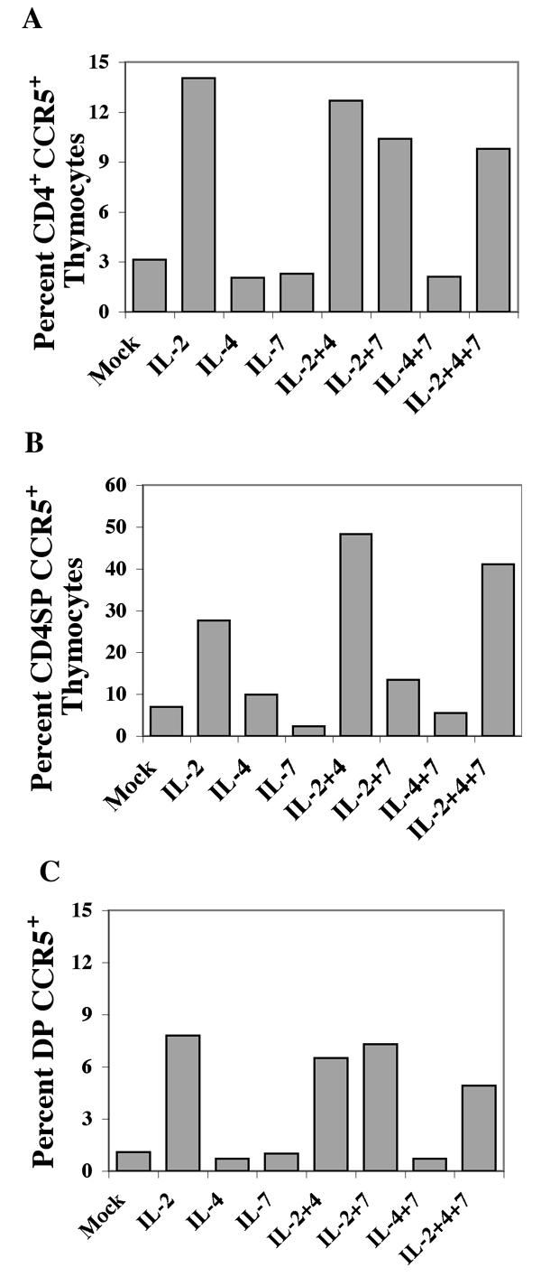
Cytokine treatment induces CCR5 expression on CD4+ thymocytes. FTOC was maintained with or without the cytokines IL-2, IL-4, and IL-7, alone or in combination, as indicated and as described in Materials and Methods. Thymocytes were recovered on day 7 and incubated with CD3-FITC, CD4-PE, CD8-PerCP, and anti-CCR5 (2D7-APC). The cells were gated on CD3, and CCR5 expression was measured on (A) total CD4+ thymocytes, (B) CD4SP thymocytes, or (C) DP thymocytes. Mock, mock-infected control.
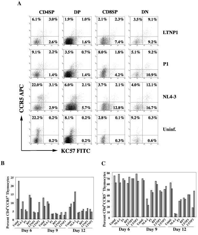
FIG. 4.
Depletion of CCR5+ and CCR5− thymocytes and down-modulation of CD4 and CCR5 in HIV-1-infected, cytokine-treated FTOC. FTOC was infected with the X4 HIV-1 molecular clone NL4-3, R5 HIV-1 biological clones P1 and LTNP2 or not infected. FTOC was cultured with cytokines as described for Fig. 2, and thymocytes were recovered 6, 9, and 12 days postinfection and incubated with CD4-PE, CD8-PerCP, CCR5-APC and then fixed, permeabilized, and incubated with anti-HIV-1 p24 (KC57-FITC) as described in Materials and Methods. (A) Data are from one representative experiment showing cell surface CCR5 expression and internal HIV-1 capsid antigen (p24) expression among the four major subsets of light scatter-gated thymocytes defined by CD4 and CD8 expression 6 days postinfection as indicated. (B) The bars represent the percentage of light scatter-gated thymocytes remaining in representative FTOC that were CD4 positive and CCR5 positive or (C) CD4 positive and CCR5 negative on the indicated day postinfection with NL4-3, the indicated R5 HIV-1 clone or in the uninfected control (Uninf.).
R5 HIV-1 biological clones preferentially depleted CCR5+ CD4SP and DP thymocytes.
Internal staining of HIV-1 p24, along with surface staining of CD4, CD8, and CCR5, showed that R5 HIV-1 clones preferentially infected and depleted the CD4SP CCR5+ and DPCCR5+ thymocyte subset (Fig. 4). The percentages of CD4SP cells expressing CCR5 in FTOC were substantially reduced by P1 or LTNP1 infection (11.3% and 9.1%, respectively) compared to NL4-3 or uninfected FTOC (25.1% and 22.4%, respectively) 6 days postinfection (Fig. 4A). Similarly, the percentages of DP thymocytes expressing CCR5 were reduced approximately twofold by P1 infection and fourfold by LTNP1 infection compared to NL4-3 or uninfected FTOC. Twelve days postinfection, 8 to 10% of the remaining thymocytes in uninfected FTOC were CD4+ CCR5+ (Fig. 4B). In NL4-3-infected FTOC, however, the cells that remained 12 days postinfection were enriched for CCR5 expression, with 10 to 16% expressing CD4 and CCR5. In contrast, for any of four R5 HIV-1 biological clones derived from progressors or LTNP at 12 days after FTOC infection, approximately 5% of the remaining thymocytes were CD4+ CCR5+, showing preferential depletion of CD4+ CCR5+ thymocytes by R5 HIV-1. The preferential depletion of CCR5+ thymocytes by R5 HIV and their preservation following infection by X4 HIV-1 suggested that CPE were primarily mediated directly by HIV-1 infection. Infection by R5 HIV-1 clones had a smaller effect on the percentage of thymocytes remaining that were CD4+CCR5−, while infection with the X4 HIV-1 molecular clone NL4-3 profoundly depleted this thymocyte population by days 9 and 12 postinfection (Fig. 4C). Nevertheless, the partial depletion of these cells 12 days postinfection with the R5 HIV-1 clones P1, RP1, and LTNP2 was statistically significant (P = 0.03, 0.02, and 0.02, respectively) suggesting that CPE was in part due to indirect or bystander killing of uninfected cells. Up-regulation of MHC-1 expression on thymocytes by HIV-1 infection and induction of inflammatory cytokines CXCL10 and oncostatin M may have contributed to this indirect CPE (data not shown).
A small percentage of the CD4SP and DP cells in the HIV-1-infected cultures expressed HIV-1 p24 internally, indicating that they were infected. The fraction of DN and CD8SP thymocytes that expressed p24 internally, however, was usually greater than the percentage of CD4SP or DP thymocytes that had internal p24. This result was likely due to CD4 down-modulation in infected cells that were CD4+ prior to infection. Furthermore, in R5 HIV-1-infected FTOC a substantial portion of the internal p24 was found in CCR5− cells, indicating that CCR5 was also down-modulated by infection. Nevertheless, the contribution of CD4 down-modulation to our measurement of CD4+ thymocyte depletion was relatively minor, as shown in Fig. 5.
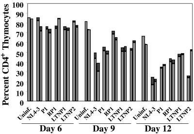
FIG. 5.
Down-modulation of CD4 on HIV-1-infected thymocytes is a minor component of the CD4+ thymocyte depletion seen following HIV-1 infection of cytokine-treated FTOC. The bars represent the percentages of thymocytes that were CD4+ remaining in FTOC following HIV-1 infection or in the uninfected control FTOC (light portions of the bars), plus the percentage that down-modulated CD4 (dark portions of the bars). The total height of each bar, therefore, represents the percentage of CD4+ or formerly CD4+ thymocytes remaining on the day shown, following FTOC infection with each HIV-1 clone depicted or in the uninfected control (Uninf.).
CD4+ thymocyte depletion following R5 HIV-1 infection is not due to CD4 down-modulation by HIV-1.
We considered the possibility that the observed depletion of CD4+ thymocytes was in part due to HIV-1-mediated down-modulation of CD4 on infected thymocytes. To test this hypothesis, cells were recovered 6, 9, and 12 days postinfection and stained for surface expression of CD4 and CD8 and then for internal expression of HIV-1 capsid p24. CD4SP, DP, CD8SP, and DN thymocytes were separately gated and analyzed for HIV-1 infection by internal p24 staining. If HIV-1 infection caused enough CD4 down-modulation for thymocytes, which were previously CD4SP or DP, to be negative for surface CD4, then we would expect to see internal p24+ cells in the DN and CD8SP quadrants, respectively. The sum of the p24+ DN and p24+ CD8SP cell populations constitutes the number of previously CD4+ thymocytes, which lost CD4 expression due to down-modulation by HIV-1. This population of cells, however, constituted only a minor fraction of the total light scatter-gated thymocytes (always less than 10%). In fact, the percentage of CD4 down-modulated thymocytes exceeded 5% of the total for NL4-3-infected FTOC only on days 9 (9%) and 12 (8%) postinfection and was always less than 5% of thymocytes for R5 HIV-1-infected FTOC. Therefore, the percentage of thymocytes that had down-modulated CD4 was far less than the percentage of CD4+ thymocyte depletion we observed. Figure 5 shows the percentage of thymocytes which were CD4+ remaining in FTOC following HIV-1 infection (light portions of the bars) plus the percentage that down-modulated CD4 due to HIV-1 infection (dark portions of the bars). The total height of each bar in Fig. 5 depicts the percentage of CD4+ thymocytes remaining in FTOC plus the percentage of CD4− HIV-1 p24+ cells. The differences in the overall heights of the bars in Fig. 5 between uninfected and HIV-1-infected FTOC represent the fractions of CD4+ thymocytes depleted, corrected for CD4 down-modulation.
IL-10 and TGF-β1 were induced by HIV-I infection of FTOC.
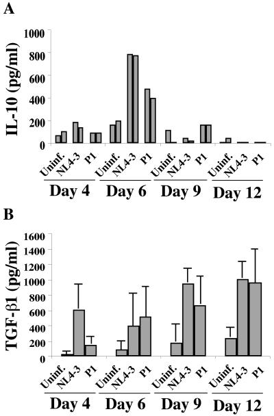
A preliminary cDNA array experiment, which we performed in collaboration with Millennium Pharmaceuticals, indicated that many cytokines were induced by HIV-1 infection of SCID-hu thymus/liver grafts (data not shown). We used commercial ELISA kits to measure IL-10 and TGF-β1 induction in FTOC since these cytokines have been implicated in CCR5 up-regulation on human monocyte-derived macrophages and dendritic cells (77). The cytokines were measured 4, 6, 9, and 12 days postinfection with FTOC for the X4 HIV-1 clone NL4-3 and the R5 HIV-1 clone P1 (Fig. 6). Despite different levels of infection by NL4-3 and P1, we saw patterns of cytokine induction that were consistent in their magnitudes and temporal patterns following infection with either virus in the presence of IL-2, IL-4, and IL-7. IL-10 was induced at the peak of viral replication, on day 6 postinfection, with both viruses. In contrast, TGF-β1 was induced on day 4 postinfection with P1 or NL4-3 and was elevated to even higher levels for the remaining period of culture. The increase in TGF-β1 in FTOC medium was significant following both NL4-3 and P1 infection on day 9 (P < 0.005) and day 12 (P < 0.01).
FIG. 6.
Induction of IL-10 and TGF-β1 expression in HIV-1-infected, cytokine-treated FTOC. FTOC was infected with the X4 HIV-1 molecular clone, NL4-3, the R5 HIV-1 biological clone, P1, or not infected. FTOC was maintained with IL-2, IL-4, and IL-7 for 12 days with daily medium changes as described in Materials and Methods. Cytokine production in FTOC medium was measured on days 4, 6, 9, and 12 postinfection by commercial ELISA (R&D Systems). (A) The results from a representative experiment are shown with adjacent bars showing duplicate cultures. (B) The results shown are the averages for three experiments, performed in duplicate, along with error bars depicting the standard errors of the means. Uninf., uninfected control.
IL-10 and TGF-β1 induced CCR5 expression on CD4+ thymocytes.
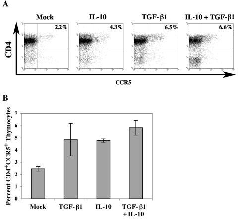
We checked whether IL-10 and TGF-β1 could increase the expression of CCR5 on CD4+ thymocytes in FTOC when added at a concentration similar to those detected in R5 HIV-1-infected FTOC medium. FTOC was carried out in the absence or presence of IL-10 (2 ng/ml), TGF-β1 (2 ng/ml), or both cytokines and without other cytokines. CCR5 expression on CD4+ thymocytes was determined on day 7 by flow cytometry. IL-10 or TGF-β1 treatment of FTOC, or treatment with both cytokines, caused a two- to threefold increase in the percentages of CCR5 expressing CD4+ thymocytes in a representative experiment (Fig. 7A). We averaged results from three experiments in which CCR5 expression was measured on day 7 (Fig. 7B). The average increase in FTOC cells expressing CCR5 following IL-10 or TGF-β1 treatment was twofold, while for FTOC-treated with IL-10 and TGF-β1 together, a 2.3-fold increase in CCR5 expression was observed, which was statistically significant (P = 0.01) (Fig. 7B).
FIG. 7.
Effect of IL-10 and TGF-β1 on CCR5 expression on CD4+ thymocytes in FTOC. FTOC was maintained in the absence or presence of IL-10 (2 ng/ml), TGF-β1 (2 ng/ml), or both cytokines with medium changes every second day. No other cytokines were used. CCR5 expression on CD4+ thymocytes was determined on day 7 by flow cytometry. (A) Representative dot plots for one of three independent experiments are shown. (B) The average values obtained in three independent experiments are shown along with the standard errors of the means for each measure. Mock, mock-infected control.
We also measured the time course of CCR5 expression on CD4+ thymocytes in TAC, similar to the human lymphoid aggregate culture system described by Eckstein et al. (22). TAC was prepared from fresh fetal thymic tissue by dispersal with scalpels followed by filtration to remove large clumps. This method gave similar percentages of thymocyte subpopulations, as well as thymic epithelial cells, dendritic cells, and macrophages, as those found in collagenase B-digested thymic tissue (38). Both methods gave 1 to 2% CD90+ thymic epithelial cells, 1 to 3% CMRF-56+ dendritic cells, and 3 to 6% CD11b+ macrophages, although the collagenase method gave higher cell viability (31, 57). TAC allowed us to plate the cells from a small piece of tissue in multiple wells, thereby facilitating multiple culture conditions.
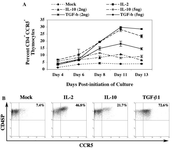
TAC was incubated in complete medium with IL-2, IL-10, or TGF-β1, and CCR5 expression was determined 4, 6, 8, 11, and 13 days postinfection by flow cytometry (Fig. 8A). CCR5 expression on CD4+ thymocytes was maximal on day 11 posttreatment with IL-2 or TGF-β1. IL-10 induction of CCR5 peaked on day 8 at the higher concentration used and on day 11 at the lower concentration. IL-10 and TGF-β1 both increased CCR5 expression, primarily on CD4SP thymocytes, but this increase was more pronounced for TGF-β1 (Fig. 8B). Thus, the cellular response to HIV-1 infection of FTOC includes a loop whereby increased IL-10 and TGF-β1 production induces CCR5 expression, which in turn may support further rounds of HIV-1 infection, CD4+ thymocyte depletion, and new cytokine production. This cycle may allow efficient depletion of CD4+ thymocytes by R5 HIV-1, despite the fact that at any given time few CD4+ thymocytes express CCR5.
FIG. 8.
Effect of IL-10 and TGF-β1 on CCR5 expression on CD4+ thymocytes in TAC. TAC was maintained in the presence of IL-7 (1 ng/ml) and treated with either medium alone, IL-2 (20 IU/ml), or two concentrations of IL-10 and TGF-β1, as indicated. CCR5 expression on CD4+ thymocytes was determined on the indicated days following initiation of TAC by flow cytometry. (A) The average values obtained in a representative experiment done in duplicate are shown with error bars indicating standard errors of the means. (B) Representative dot plots showing expression of CCR5 on CD4SP thymocytes on day 11, when CCR5 expression peaked for cells treated with IL-2 (20 IU/ml), IL-10 (5 ng/ml), or TGF-β1 (5 ng/ml). Mock, mock-infected control.
DISCUSSION
We analyzed the replication and CPE of four diverse, primary R5 HIV-1 clones in FTOC. We used R5 HIV-1 clones from two individuals who progressed rapidly or normally to AIDS and from two long-term nonprogressors (LTNP). FTOC was also infected with the X4 HIV-1 molecular clone NL4-3 or was not infected. When FTOC was maintained without adding cytokines, we found that progressor-derived R5 HIV-1 clones exhibited greater CPE than R5 HIV-1 clones from LTNP. The progressor-derived clones also replicated better than R5 HIV-1 clones from LTNP. Similar results were obtained by Blaak et al. on phytohemagglutinin-stimulated PBMC; the two LTNP clones replicated slower than progressor clones, suggesting that LTNP clones are less fit than progressor clones (5). We and others have previously observed that LTNP-derived HIV-1 clones replicate more slowly than progressor clones in tissue culture and in SCID-hu mice (10, 18, 37, 47, 73). Certain mutations or deletions in the HIV-1 nef gene have been shown to correlate with LTNP status in some cases (14, 41, 42). Sequencing of the HIV-1 nef genes from RP1, LTNP1, and LTNP2, however, showed few mutations and none could be associated with the progressor or LTNP status of the infected individuals (5).
The pattern of CPE was quite different, however, when R5 HIV-1 clones from progressors and LTNP were used to infect FTOC that was maintained in the presence of IL-2, IL-4, and IL-7. Under these conditions, R5 HIV-1 clones from progressors and LTNP were nearly equally cytopathic and replicated to similar levels. The levels of viral replication achieved by progressor- and LTNP-derived R5 HIV-1 clones in cytokine-treated FTOC was approximately 10-fold greater than that achieved by progressor clones in untreated FTOC. Moreover, all R5 HIV-1 clones depleted approximately the same percentages of CD4+ thymocytes, altered the CD4SP-to-CD8SP ratio similarly, and depleted approximately the same numbers of DP, CD4SP, and CD8SP thymocytes in cytokine-treated FTOC. The likely explanation for these differences in the behavior of R5 HIV-1 clones in FTOC with and without the addition of IL-2, IL-4, and IL-7 is that these cytokines induced CCR5 expression on thymocytes. Overall, threefold more thymocytes expressed CCR5 and four- to fivefold more CD4SP cells expressed CCR5 when FTOC was maintained in the presence of the cytokines. Several studies have shown or suggested that one difference between more and less cytopathic R5 HIV-1 clones is their affinity for CCR5 (26, 62). Our results suggest that the LTNP-derived HIV-1 clones we studied had lower affinity for CCR5 than the progressor-derived clones, since increased CCR5 expression may compensate for the lower replication and CPE of the LTNP-derived HIV-1 clones. Other mechanisms may also play a role in cytokine-mediated enhancement of R5 HIV-1 replication and CPE in FTOC. IL-2, IL-4, and IL-7 are important in thymocyte development and may therefore contribute to viral replication by increasing thymocyte activation (reviewed in reference 68). IL-2, IL-4, and IL-7, either alone or in combination, also enhanced HIV-1 replication in isolated thymocytes (58, 59, 63, 71, 72). The effects of these cytokines on viral replication in isolated thymocytes were also explained in part by their positive effects on CCR5 expression on mature thymocytes.
All four R5 HIV-1 clones mediated greater CPE on the CCR5+ subset of CD4+ thymocytes than on CCR5− CD4+ thymocytes. Conversely, the X4 HIV-1 clone NL4-3 was more cytopathic for the much larger CXCR4+ CD4+ thymocyte subset. NL4-3 infection spared some CCR5+ CD4+ thymocytes, presumably since they did not express CXCR4. Nevertheless, the R5 HIV-1 clones significantly depleted CCR5− CD4+ thymocytes, indicating that they may have induced bystander CPE or CCR5 down-modulation. Alternatively, some apparently CCR5− CD4+ thymocytes may express low levels of CCR5, which are not detectable by flow cytometry but are sufficient to allow entry by these viruses. An analysis of CCR5 expression versus internal p24 expression indicated that CCR5 down-modulation likely did occur in some thymocytes following infection with R5 HIV-1 as has been previously reported (78). Taken together, our data indicate that while direct killing of infected cells was likely the predominant mechanism of CPE following R5 HIV-1 infection of FTOC, indirect killing may also have played a role in thymocyte depletion.
Internal p24 staining showed that two- to fivefold more thymocytes were infected with NL4-3 than with any of the four R5 HIV-1 clones. Nevertheless, similar levels of virus were produced by FTOC infected with R5 HIV-1 clones or NL4-3, leading to the conclusion that the R5 HIV-1 clones produced more p24 per infected cell than NL4-3. This may result from infection of a greater diversity of cell types in FTOC by R5 HIV-1 than X4 HIV-1. These cell types may include medullary stromal cells and macrophages, which, while relatively few in number in the thymus, may produce a significant quantity of HIV-1 (4).
HIV-1 infection of FTOC induced the production of IL-10 and TGF-β1, which have previously been implicated in inducing CCR5 expression in monocytes, macrophages, and monocyte-derived dendritic cells (8, 33, 39, 46, 67, 76, 77). PBMC from AIDS patients was shown to produce more IL-10 than PBMC from healthy donors (55). Moreover, IL-10 production by PBMC and monocyte-derived macrophages could be induced in vitro by Tat, Nef, gp120, or Gag (2, 8, 9, 55, 64). Elevated levels of TGF-β proteins with increased expression of TGF-β1 mRNA have been identified in HIV-1-infected individuals and can be induced by HIV-1 in PBMC, macrophages, and astrocytes (25, 32, 40, 75). Both IL-10 and TGF-β1 induced CCR5 expression on thymocytes at a concentration similar to that measured in HIV-1-infected FTOC medium. Thus, HIV-1 infection induces the production of IL-10 and TGF-β1 in FTOC, which then in turn increase CCR5 expression. This increased CCR5 expression may play a crucial role in R5 HIV-1 infection in the thymus by making more cells available for viral entry, replication, and CPE. Our results and those of Jekle et al. suggest that CPE on infected cells is the major mechanism of CD4+ thymocyte and T-cell depletion by R5 HIV-1 strains (35). CCR5 expression may limit R5 HIV-1 in the thymus just as it must in patients, since individuals heterozygous for the normal (CCR5+) and CCR5Δ32 alleles progress to AIDS 1 to 2 years more slowly on average after HIV-1 seroconversion relative to individuals with the CCR5 +/+ genotype (15, 17, 52, 80). Our results suggest that the small fraction of thymocytes that express CCR5 limits the replication and CPE of R5 HIV-1 clones in the thymus. This limitation can be overcome, however, by late-stage R5 HIV-1 clones, which induce the production of IL-10 and TGF-β1 in the thymus, which in turn induce a greater fraction of thymocytes to express CCR5 and thereby create more target cells for R5 HIV-1 infection.
Supplementary Material
Acknowledgments
We thank Victoria Camerini for helpful discussions and review of the manuscript. We also thank Lesley White for help with p24 ELISA.
This work was supported by NIH grants R01 AI 47729 and R21 AI 55385 awarded to D.C.
Footnotes
Supplemental material for this article may be found at http://jvi.asm.org/.
REFERENCES
- 1.Aldrovandi, G. M., G. Feuer, L. Gao, B. Jamieson, M. Kristeva, I. S. Chen, and J. A. Zack. 1993. The SCID-hu mouse as a model for HIV-1 infection. Nature 363:732-736. [DOI] [PubMed] [Google Scholar]
- 2.Badou, A., Y. Bennasser, M. Moreau, C. Leclerc, M. Benkirane, and E. Bahraoui. 2000. Tat protein of human immunodeficiency virus type 1 induces interleukin-10 in human peripheral blood monocytes: implication of protein kinase C-dependent pathway. J. Virol. 74:10551-10562. [DOI] [PMC free article] [PubMed] [Google Scholar]
- 3.Berger, E. A., P. M. Murphy, and J. M. Farber. 1999. Chemokine receptors as HIV-1 coreceptors: roles in viral entry, tropism, and disease. Annu. Rev. Immunol. 17:657-700. [DOI] [PubMed] [Google Scholar]
- 4.Berkowitz, R. D., K. P. Beckerman, T. J. Schall, and J. M. McCune. 1998. CXCR4 and CCR5 expression delineates targets for HIV-1 disruption of T cell differentiation. J. Immunol. 161:3702-3710. [PubMed] [Google Scholar]
- 5.Blaak, H., M. Brouwer, L. J. Ran, F. de Wolf, and H. Schuitemaker. 1998. In vitro replication kinetics of human immunodeficiency virus type 1 (HIV-1) variants in relation to virus load in long-term survivors of HIV-1 infection. J. Infect. Dis. 177:600-610. [DOI] [PubMed] [Google Scholar]
- 6.Bleul, C. C., L. Wu, J. A. Hoxie, T. A. Springer, and C. R. Mackay. 1997. The HIV coreceptors CXCR4 and CCR5 are differentially expressed and regulated on human T lymphocytes. Proc. Natl. Acad. Sci. USA 94:1925-1930. [DOI] [PMC free article] [PubMed] [Google Scholar]
- 7.Bonyhadi, M. L., L. Rabin, S. Salimi, D. A. Brown, J. Kosek, J. M. McCune, and H. Kaneshima. 1993. HIV induces thymus depletion in vivo. Nature 363:728-732. [DOI] [PubMed] [Google Scholar]
- 8.Borghi, P., L. Fantuzzi, B. Varano, S. Gessani, P. Puddu, L. Conti, M. R. Capobianchi, F. Ameglio, and F. Belardelli. 1995. Induction of interleukin-10 by human immunodeficiency virus type 1 and its gp120 protein in human monocytes/macrophages. J. Virol. 69:1284-1287. [DOI] [PMC free article] [PubMed] [Google Scholar]
- 9.Brigino, E., S. Haraguchi, A. Koutsonikolis, G. J. Cianciolo, U. Owens, R. A. Good, and N. K. Day. 1997. Interleukin 10 is induced by recombinant HIV-1 Nef protein involving the calcium/calmodulin-dependent phosphodiesterase signal transduction pathway. Proc. Natl. Acad. Sci. USA 94:3178-3182. [DOI] [PMC free article] [PubMed] [Google Scholar]
- 10.Camerini, D., H. P. Su, G. Gamez-Torre, M. L. Johnson, J. A. Zack, and I. S. Chen. 2000. Human immunodeficiency virus type 1 pathogenesis in SCID-hu mice correlates with syncytium-inducing phenotype and viral replication. J. Virol. 74:3196-3204. [DOI] [PMC free article] [PubMed] [Google Scholar]
- 11.Choe, H., M. Farzan, Y. Sun, N. Sullivan, B. Rollins, P. D. Ponath, L. Wu, C. R. Mackay, G. LaRosa, W. Newman, N. Gerard, C. Gerard, and J. Sodroski. 1996. The beta-chemokine receptors CCR3 and CCR5 facilitate infection by primary HIV-1 isolates. Cell 85:1135-1148. [DOI] [PubMed] [Google Scholar]
- 12.Connor, R. I., and D. D. Ho. 1994. Human immunodeficiency virus type 1 variants with increased replicative capacity develop during the asymptomatic stage before disease progression. J. Virol. 68:4400-4408. [DOI] [PMC free article] [PubMed] [Google Scholar]
- 13.Connor, R. I., K. E. Sheridan, D. Ceradini, S. Choe, and N. R. Landau. 1997. Change in coreceptor use coreceptor use correlates with disease progression in HIV-1-infected individuals. J. Exp. Med. 185:621-628. [DOI] [PMC free article] [PubMed] [Google Scholar]
- 14.Deacon, N. J., A. Tsykin, A. Solomon, K. Smith, M. Ludford-Menting, D. J. Hooker, D. A. McPhee, A. L. Greenway, A. Ellett, C. Chatfield, et al. 1995. Genomic structure of an attenuated quasi species of HIV-1 from a blood transfusion donor and recipients. Science 270:988-991. [DOI] [PubMed] [Google Scholar]
- 15.Dean, M., M. Carrington, C. Winkler, G. A. Huttley, M. W. Smith, R. Allikmets, J. J. Goedert, S. P. Buchbinder, E. Vittinghoff, E. Gomperts, S. Donfield, D. Vlahov, R. Kaslow, A. Saah, C. Rinaldo, R. Detels, and S. J. O'Brien. 1996. Genetic restriction of HIV-1 infection and progression to AIDS by a deletion allele of the CKR5 structural gene. Science 273:1856-1862. [DOI] [PubMed] [Google Scholar]
- 16.Deng, H., R. Liu, W. Ellmeier, S. Choe, D. Unutmaz, M. Burkhart, P. Di Marzio, S. Marmon, R. E. Sutton, C. M. Hill, C. B. Davis, S. C. Peiper, T. J. Schall, D. R. Littman, and N. R. Landau. 1996. Identification of a major co-receptor for primary isolates of HIV-1. Nature 381:661-666. [DOI] [PubMed] [Google Scholar]
- 17.de Roda Husman, A. M., M. Koot, M. Cornelissen, I. P. Keet, M. Brouwer, S. M. Broersen, M. Bakker, M. T. Roos, M. Prins, F. de Wolf, R. A. Coutinho, F. Miedema, J. Goudsmit, and H. Schuitemaker. 1997. Association between CCR5 genotype and the clinical course of HIV-1 infection. Ann. Intern. Med. 127:882-890. [DOI] [PubMed] [Google Scholar]
- 18.de Roda Husman, A. M., R. P. van Rij, H. Blaak, S. Broersen, and H. Schuitemaker. 1999. Adaptation to promiscuous usage of chemokine receptors is not a prerequisite for human immunodeficiency virus type 1 disease progression. J. Infect. Dis. 180:1106-1115. [DOI] [PubMed] [Google Scholar]
- 19.Douek, D. C., R. A. Koup, R. D. McFarland, J. L. Sullivan, and K. Luzuriaga. 2000. Effect of HIV on thymic function before and after antiretroviral therapy in children. J. Infect. Dis. 181:1479-1482. [DOI] [PubMed] [Google Scholar]
- 20.Douek, D. C., R. D. McFarland, P. H. Keiser, E. A. Gage, J. M. Massey, B. F. Haynes, M. A. Polis, A. T. Haase, M. B. Feinberg, J. L. Sullivan, B. D. Jamieson, J. A. Zack, L. J. Picker, and R. A. Koup. 1998. Changes in thymic function with age and during the treatment of HIV infection. Nature 396:690-695. [DOI] [PubMed] [Google Scholar]
- 21.Dragic, T., V. Litwin, G. P. Allaway, S. R. Martin, Y. Huang, K. A. Nagashima, C. Cayanan, P. J. Maddon, R. A. Koup, J. P. Moore, and W. A. Paxton. 1996. HIV-1 entry into CD4+ cells is mediated by the chemokine receptor CC-CKR-5. Nature 381:667-673. [DOI] [PubMed] [Google Scholar]
- 22.Eckstein, D. A., M. L. Penn, Y. D. Korin, D. D. Scripture-Adams, J. A. Zack, J. F. Kreisberg, M. Roederer, M. P. Sherman, P. S. Chin, and M. A. Goldsmith. 2001. HIV-1 actively replicates in naive CD4+ T cells residing within human lymphoid tissues. Immunity 15:671-682. [DOI] [PubMed] [Google Scholar]
- 23.Feng, Y., C. C. Broder, P. E. Kennedy, and E. A. Berger. 1996. HIV-1 entry cofactor: functional cDNA cloning of a seven-transmembrane, G protein-coupled receptor. Science 272:872-877. [DOI] [PubMed] [Google Scholar]
- 24.Finnegan, A., K. A. Roebuck, B. E. Nakai, D. S. Gu, M. F. Rabbi, S. Song, and A. L. Landay. 1996. IL-10 cooperates with TNF-alpha to activate HIV-1 from latently and acutely infected cells of monocyte/macrophage lineage. J. Immunol. 156:841-851. [PubMed] [Google Scholar]
- 25.Garba, M. L., C. D. Pilcher, A. L. Bingham, J. Eron, and J. A. Frelinger. 2002. HIV antigens can induce TGF-β1-producing immunoregulatory CD8+ T cells. J. Immunol. 168:2247-2254. [DOI] [PubMed] [Google Scholar]
- 26.Gorry, P. R., J. Taylor, G. H. Holm, A. Mehle, T. Morgan, M. Cayabyab, M. Farzan, H. Wang, J. E. Bell, K. Kunstman, J. P. Moore, S. M. Wolinsky, and D. Gabuzda. 2002. Increased CCR5 affinity and reduced CCR5/CD4 dependence of a neurovirulent primary human immunodeficiency virus type 1 isolate. J. Virol. 76:6277-6292. [DOI] [PMC free article] [PubMed] [Google Scholar]
- 27.Grivel, J. C., M. L. Penn, D. A. Eckstein, B. Schramm, R. F. Speck, N. W. Abbey, B. Herndier, L. Margolis, and M. A. Goldsmith. 2000. Human immunodeficiency virus type 1 coreceptor preferences determine target T-cell depletion and cellular tropism in human lymphoid tissue. J. Virol. 74:5347-5351. [DOI] [PMC free article] [PubMed] [Google Scholar]
- 28.Hazenberg, M. D., D. Hamann, H. Schuitemaker, and F. Miedema. 2000. T cell depletion in HIV-1 infection: how CD4+ T cells go out of stock. Nat. Immunol. 1:285-289. [DOI] [PubMed] [Google Scholar]
- 29.Hazenberg, M. D., S. A. Otto, J. W. Cohen Stuart, M. C. Verschuren, J. C. Borleffs, C. A. Boucher, R. A. Coutinho, J. M. Lange, T. F. Rinke de Wit, A. Tsegaye, J. J. van Dongen, D. Hamann, R. J. de Boer, and F. Miedema. 2000. Increased cell division but not thymic dysfunction rapidly affects the T-cell receptor excision circle content of the naive T cell population in HIV-1 infection. Nat. Med. 6:1036-1042. [DOI] [PubMed] [Google Scholar]
- 30.Hazenberg, M. D., J. W. Stuart, S. A. Otto, J. C. Borleffs, C. A. Boucher, R. J. de Boer, F. Miedema, and D. Hamann. 2000. T-cell division in human immunodeficiency virus (HIV)-1 infection is mainly due to immune activation: a longitudinal analysis in patients before and during highly active antiretroviral therapy (HAART). Blood 95:249-255. [PubMed] [Google Scholar]
- 31.Hock, B. D., D. B. Fearnley, A. Boyce, A. D. McLellan, R. V. Sorg, K. L. Summers, and D. N. Hart. 1999. Human dendritic cells express a 95 kDa activation/differentiation antigen defined by CMRF-56. Tissue Antigens 53:320-334. [DOI] [PubMed] [Google Scholar]
- 32.Hori, K., P. R. Burd, J. Kutza, K. A. Weih, and K. A. Clouse. 1999. Human astrocytes inhibit HIV-1 expression in monocyte-derived macrophages by secreted factors. AIDS 13:751-758. [DOI] [PubMed] [Google Scholar]
- 33.Houle, M., M. Thivierge, C. Le Gouill, J. Stankova, and M. Rola-Pleszczynski. 1999. IL-10 up-regulates CCR5 gene expression in human monocytes. Inflammation 23:241-251. [DOI] [PubMed] [Google Scholar]
- 34.Jamieson, B. D., and J. A. Zack. 1998. In vivo pathogenesis of a human immunodeficiency virus type 1 reporter virus. J. Virol. 72:6520-6526. [DOI] [PMC free article] [PubMed] [Google Scholar]
- 35.Jekle, A., O. T. Keppler, E. De Clercq, D. Schols, M. Weinstein, and M. A. Goldsmith. 2003. In vivo evolution of human immunodeficiency virus type 1 toward increased pathogenicity through CXCR4-mediated killing of uninfected CD4 T cells. J. Virol. 77:5846-5854. [DOI] [PMC free article] [PubMed] [Google Scholar]
- 36.Joshi, V. V., and J. M. Oleske. 1985. Pathologic appraisal of the thymus gland in acquired immunodeficiency syndrome in children. A study of four cases and a review of the literature. Arch. Pathol. Lab. Med. 109:142-146. [PubMed] [Google Scholar]
- 37.Kaneshima, H., L. Su, M. L. Bonyhadi, R. I. Connor, D. D. Ho, and J. M. McCune. 1994. Rapid-high, syncytium-inducing isolates of human immunodeficiency virus type 1 induce cytopathicity in the human thymus of the SCID-hu mouse. J. Virol. 68:8188-8192. [DOI] [PMC free article] [PubMed] [Google Scholar]
- 38.Keir, M. E., M. G. Rosenberg, J. K. Sandberg, K. A. Jordan, A. Wiznia, D. F. Nixon, C. A. Stoddart, and J. M. McCune. 2002. Generation of CD3+CD8low thymocytes in the HIV type 1-infected thymus. J. Immunol. 169:2788-2796. [DOI] [PubMed] [Google Scholar]
- 39.Keir, M. E., C. A. Stoddart, V. Linquist-Stepps, M. E. Moreno, and J. M. McCune. 2002. IFN-alpha secretion by type 2 predendritic cells up-regulates MHC class I in the HIV-1-infected thymus. J. Immunol. 168:325-331. [DOI] [PubMed] [Google Scholar]
- 40.Kekow, J., W. Wachsman, J. A. McCutchan, M. Cronin, D. A. Carson, and M. Lotz. 1990. Transforming growth factor beta and noncytopathic mechanisms of immunodeficiency in human immunodeficiency virus infection. Proc. Natl. Acad. Sci. USA 87:8321-8325. [DOI] [PMC free article] [PubMed] [Google Scholar]
- 41.Kirchhoff, F., P. J. Easterbrook, N. Douglas, M. Troop, T. C. Greenough, J. Weber, S. Carl, J. L. Sullivan, and R. S. Daniels. 1999. Sequence variations in human immunodeficiency virus type 1 Nef are associated with different stages of disease. J. Virol. 73:5497-5508. [DOI] [PMC free article] [PubMed] [Google Scholar]
- 42.Kirchhoff, F., T. C. Greenough, D. B. Brettler, J. L. Sullivan, and R. C. Desrosiers. 1995. Brief report: absence of intact nef sequences in a long-term survivor with nonprogressive HIV-1 infection. N. Engl. J. Med. 332:228-232. [DOI] [PubMed] [Google Scholar]
- 43.Kitchen, S. G., and J. A. Zack. 1999. Distribution of the human immunodeficiency virus coreceptors CXCR4 and CCR5 in fetal lymphoid organs: implications for pathogenesis in utero. AIDS Res. Hum. Retrovir. 15:143-148. [DOI] [PubMed] [Google Scholar]
- 44.Kolb, S. A., B. Sporer, F. Lahrtz, U. Koedel, H. W. Pfister, and A. Fontana. 1999. Identification of a T cell chemotactic factor in the cerebrospinal fluid of HIV-1-infected individuals as interferon-gamma inducible protein 10. J. Neuroimmunol. 93:172-181. [DOI] [PubMed] [Google Scholar]
- 45.Kourtis, A. P., C. Ibegbu, A. J. Nahmias, F. K. Lee, W. S. Clark, M. K. Sawyer, and S. Nesheim. 1996. Early progression of disease in HIV-infected infants with thymus dysfunction. N. Engl. J. Med. 335:1431-1436. (Erratum, 336:595) [DOI] [PubMed] [Google Scholar]
- 46.Kovalev, G., K. Duus, L. Wang, R. Lee, M. Bonyhadi, D. Ho, J. M. McCune, H. Kaneshima, and L. Su. 1999. Induction of MHC class I expression on immature thymocytes in HIV-1-infected SCID-hu Thy/Liv mice: evidence of indirect mechanisms. J. Immunol. 162:7555-7562. [PMC free article] [PubMed] [Google Scholar]
- 47.Kwa, D., J. Vingerhoed, B. Boeser, and H. Schuitemaker. 2003. Increased in vitro cytopathicity of CC chemokine receptor 5-restricted human immunodeficiency virus type 1 primary isolates correlates with a progressive clinical course of infection. J. Infect. Dis. 187:1397-1403. [DOI] [PubMed] [Google Scholar]
- 48.Kwa, D., J. Vingerhoed, B. Boeser-Nunnink, S. Broersen, and H. Schuitemaker. 2001. Cytopathic effects of non-syncytium-inducing and syncytium-inducing human immunodeficiency virus type 1 variants on different CD4+-T-cell subsets are determined only by coreceptor expression. J. Virol. 75:10455-10459. [DOI] [PMC free article] [PubMed] [Google Scholar]
- 49.Maas, J. J., S. J. Gange, H. Schuitemaker, R. A. Coutinho, R. van Leeuwen, and J. B. Margolick. 2000. Strong association between failure of T cell homeostasis and the syncytium-inducing phenotype among HIV-1-infected men in the Amsterdam Cohort Study. AIDS 14:1155-1161. [DOI] [PubMed] [Google Scholar]
- 50.McCune, J. M. 2001. The dynamics of CD4+ T-cell depletion in HIV disease. Nature 410:974-979. [DOI] [PubMed] [Google Scholar]
- 51.McCune, J. M., R. Namikawa, H. Kaneshima, L. D. Shultz, M. Lieberman, and I. L. Weissman. 1988. The SCID-hu mouse: murine model for the analysis of human hematolymphoid differentiation and function. Science 241:1632-1639. [DOI] [PubMed] [Google Scholar]
- 52.Michael, N. L., G. Chang, L. G. Louie, J. R. Mascola, D. Dondero, D. L. Birx, and H. W. Sheppard. 1997. The role of viral phenotype and CCR-5 gene defects in HIV-1 transmission and disease progression. Nat. Med. 3:338-340. [DOI] [PubMed] [Google Scholar]
- 53.Mo, H., S. Monard, H. Pollack, J. Ip, G. Rochford, L. Wu, J. Hoxie, W. Borkowsky, D. D. Ho, and J. P. Moore. 1998. Expression patterns of the HIV type 1 coreceptors CCR5 and CXCR4 on CD4+ T cells and monocytes from cord and adult blood. AIDS Res. Hum. Retrovir. 14:607-617. [DOI] [PubMed] [Google Scholar]
- 54.Nahmias, A. J., W. S. Clark, A. P. Kourtis, F. K. Lee, G. Cotsonis, C. Ibegbu, D. Thea, P. Palumbo, P. Vink, R. J. Simonds, S. R. Nesheim, et al. 1998. Thymic dysfunction and time of infection predict mortality in human immunodeficiency virus-infected infants. J. Infect. Dis. 178:680-685. [DOI] [PubMed] [Google Scholar]
- 55.Ostrowski, M. A., J. X. Gu, C. Kovacs, J. Freedman, M. A. Luscher, and K. S. MacDonald. 2001. Quantitative and qualitative assessment of human immunodeficiency virus type 1 (HIV-1)-specific CD4+ T cell immunity to gag in HIV-1-infected individuals with differential disease progression: reciprocal interferon-γ and interleukin-10 responses. J. Infect. Dis. 184:1268-1278. [DOI] [PubMed] [Google Scholar]
- 56.Ostrowski, M. A., S. J. Justement, A. Catanzaro, C. A. Hallahan, L. A. Ehler, S. B. Mizell, P. N. Kumar, J. A. Mican, T. W. Chun, and A. S. Fauci. 1998. Expression of chemokine receptors CXCR4 and CCR5 in HIV-1-infected and uninfected individuals. J. Immunol. 161:3195-3201. [PubMed] [Google Scholar]
- 57.Patel, D. D., L. P. Whichard, G. Radcliff, S. M. Denning, and B. F. Haynes. 1995. Characterization of human thymic epithelial cell surface antigens: phenotypic similarity of thymic epithelial cells to epidermal keratinocytes. J. Clin. Immunol. 15:80-92. [DOI] [PubMed] [Google Scholar]
- 58.Pedroza-Martins, L., W. J. Boscardin, D. J. Anisman-Posner, D. Schols, Y. J. Bryson, and C. H. Uittenbogaart. 2002. Impact of cytokines on replication in the thymus of primary human immunodeficiency virus type 1 isolates from infants. J. Virol. 76:6929-6943. [DOI] [PMC free article] [PubMed] [Google Scholar]
- 59.Pedroza-Martins, L., K. B. Gurney, B. E. Torbett, and C. H. Uittenbogaart. 1998. Differential tropism and replication kinetics of human immunodeficiency virus type 1 isolates in thymocytes: coreceptor expression allows viral entry, but productive infection of distinct subsets is determined at the postentry level. J. Virol. 72:9441-9452. [DOI] [PMC free article] [PubMed] [Google Scholar]
- 60.Richman, D. D., and S. A. Bozzette. 1994. The impact of the syncytium-inducing phenotype of human immunodeficiency virus on disease progression. J. Infect. Dis. 169:968-974. [DOI] [PubMed] [Google Scholar]
- 61.Sato, K., H. Kawasaki, H. Nagayama, M. Enomoto, C. Morimoto, K. Tadokoro, T. Juji, and T. A. Takahashi. 2000. TGF-β1 reciprocally controls chemotaxis of human peripheral blood monocyte-derived dendritic cells via chemokine receptors. J. Immunol. 164:2285-2295. [DOI] [PubMed] [Google Scholar]
- 62.Scarlatti, G., E. Tresoldi, A. Bjorndal, R. Fredriksson, C. Colognesi, H. K. Deng, M. S. Malnati, A. Plebani, A. G. Siccardi, D. R. Littman, E. M. Fenyo, and P. Lusso. 1997. In vivo evolution of HIV-1 co-receptor usage and sensitivity to chemokine-mediated suppression. Nat. Med. 3:1259-1265. [DOI] [PubMed] [Google Scholar]
- 63.Schmitt, N., L. Chene, D. Boutolleau, M. T. Nugeyre, E. Guillemard, P. Versmisse, C. Jacquemot, F. Barre-Sinoussi, and N. Israel. 2003. Positive regulation of CXCR4 expression and signaling by interleukin-7 in CD4+ mature thymocytes correlates with their capacity to favor human immunodeficiency X4 virus replication. J. Virol. 77:5784-5793. [DOI] [PMC free article] [PubMed] [Google Scholar]
- 64.Schols, D., and E. De Clercq. 1996. Human immunodeficiency virus type 1 gp120 induces anergy in human peripheral blood lymphocytes by inducing interleukin-10 production. J. Virol. 70:4953-4960. [DOI] [PMC free article] [PubMed] [Google Scholar]
- 65.Schuitemaker, H., M. Koot, N. A. Kootstra, M. W. Dercksen, R. E. de Goede, R. P. van Steenwijk, J. M. Lange, J. K. Schattenkerk, F. Miedema, and M. Tersmette. 1992. Biological phenotype of human immunodeficiency virus type 1 clones at different stages of infection: progression of disease is associated with a shift from monocytotropic to T-cell-tropic virus population. J. Virol. 66:1354-1360. [DOI] [PMC free article] [PubMed] [Google Scholar]
- 66.Scoggins, R. M., J. R. Taylor, Jr., J. Patrie, A. B. van't Wout, H. Schuitemaker, and D. Camerini. 2000. Pathogenesis of primary R5 human immunodeficiency virus type 1 clones in SCID-hu mice. J. Virol. 74:3205-3216. [DOI] [PMC free article] [PubMed] [Google Scholar]
- 67.Sozzani, S., S. Ghezzi, G. Iannolo, W. Luini, A. Borsatti, N. Polentarutti, A. Sica, M. Locati, C. Mackay, T. N. Wells, P. Biswas, E. Vicenzi, G. Poli, and A. Mantovani. 1998. Interleukin 10 increases CCR5 expression and HIV infection in human monocytes. J. Exp. Med. 187:439-444. [DOI] [PMC free article] [PubMed] [Google Scholar]
- 68.Spits, H. 2002. Development of αβ T cells in the human thymus. Nat. Rev. Immunol. 2:760-772. [DOI] [PubMed] [Google Scholar]
- 69.Taylor, J. R., Jr., K. C. Kimbrell, R. Scoggins, M. Delaney, L. Wu, and D. Camerini. 2001. Expression and function of chemokine receptors on human thymocytes: implications for infection by human immunodeficiency virus type 1. J. Virol. 75:8752-8760. [DOI] [PMC free article] [PubMed] [Google Scholar]
- 70.Tersmette, M., R. E. de Goede, B. J. Al, I. N. Winkel, R. A. Gruters, H. T. Cuypers, H. G. Huisman, and F. Miedema. 1988. Differential syncytium-inducing capacity of human immunodeficiency virus isolates: frequent detection of syncytium-inducing isolates in patients with acquired immunodeficiency syndrome (AIDS) and AIDS-related complex. J. Virol. 62:2026-2032. [DOI] [PMC free article] [PubMed] [Google Scholar]
- 71.Uittenbogaart, C. H., D. J. Anisman, J. A. Zack, A. Economides, I. Schmid, and E. F. Hays. 1995. Effects of cytokines on HIV-1 production by thymocytes. Thymus 23:155-175. [PubMed] [Google Scholar]
- 72.Uittenbogaart, C. H., W. J. Boscardin, D. J. Anisman-Posner, P. S. Koka, G. Bristol, and J. A. Zack. 2000. Effect of cytokines on HIV-induced depletion of thymocytes in vivo. AIDS 14:1317-1325. [DOI] [PubMed] [Google Scholar]
- 73.van 't Wout, A. B., H. Blaak, L. J. Ran, M. Brouwer, C. Kuiken, and H. Schuitemaker. 1998. Evolution of syncytium-inducing and non-syncytium-inducing biological virus clones in relation to replication kinetics during the course of human immunodeficiency virus type 1 infection. J. Virol. 72:5099-5107. [DOI] [PMC free article] [PubMed] [Google Scholar]
- 74.van't Wout, A. B., N. A. Kootstra, G. A. Mulder-Kampinga, N. Albrecht-van Lent, H. J. Scherpbier, J. Veenstra, K. Boer, R. A. Coutinho, F. Miedema, and H. Schuitemaker. 1994. Macrophage-tropic variants initiate human immunodeficiency virus type 1 infection after sexual, parenteral, and vertical transmission. J. Clin. Investig. 94:2060-2067. [DOI] [PMC free article] [PubMed] [Google Scholar]
- 75.Wahl, S. M., J. B. Allen, N. McCartney-Francis, M. C. Morganti-Kossmann, T. Kossmann, L. Ellingsworth, U. E. Mai, S. E. Mergenhagen, and J. M. Orenstein. 1991. Macrophage- and astrocyte-derived transforming growth factor beta as a mediator of central nervous system dysfunction in acquired immune deficiency syndrome. J. Exp. Med. 173:981-991. [DOI] [PMC free article] [PubMed] [Google Scholar]
- 76.Wang, J., K. Crawford, M. Yuan, H. Wang, P. R. Gorry, and D. Gabuzda. 2002. Regulation of CC chemokine receptor 5 and CD4 expression and human immunodeficiency virus type 1 replication in human macrophages and microglia by T helper type 2 cytokines. J. Infect. Dis. 185:885-897. [DOI] [PubMed] [Google Scholar]
- 77.Wang, J., G. Roderiquez, T. Oravecz, and M. A. Norcross. 1998. Cytokine regulation of human immunodeficiency virus type 1 entry and replication in human monocytes/macrophages through modulation of CCR5 expression. J. Virol. 72:7642-7647. [DOI] [PMC free article] [PubMed] [Google Scholar]
- 78.Wang, J. M., H. Ueda, O. M. Howard, M. C. Grimm, O. Chertov, X. Gong, W. Gong, J. H. Resau, C. C. Broder, G. Evans, L. O. Arthur, F. W. Ruscetti, and J. J. Oppenheim. 1998. HIV-1 envelope gp120 inhibits the monocyte response to chemokines through CD4 signal-dependent chemokine receptor down-regulation. J. Immunol. 161:4309-4317. [PubMed] [Google Scholar]
- 79.Zaitseva, M. B., S. Lee, R. L. Rabin, H. L. Tiffany, J. M. Farber, K. W. Peden, P. M. Murphy, and H. Golding. 1998. CXCR4 and CCR5 on human thymocytes: biological function and role in HIV-1 infection. J. Immunol. 161:3103-3113. [PubMed] [Google Scholar]
- 80.Zimmerman, P. A., A. Buckler-White, G. Alkhatib, T. Spalding, J. Kubofcik, C. Combadiere, D. Weissman, O. Cohen, A. Rubbert, G. Lam, M. Vaccarezza, P. E. Kennedy, V. Kumaraswami, J. V. Giorgi, R. Detels, J. Hunter, M. Chopek, E. A. Berger, A. S. Fauci, T. B. Nutman, and P. M. Murphy. 1997. Inherited resistance to HIV-1 conferred by an inactivating mutation in CC chemokine receptor 5: studies in populations with contrasting clinical phenotypes, defined racial background, and quantified risk. Mol. Med. 3:23-36. [PMC free article] [PubMed] [Google Scholar]
Associated Data
This section collects any data citations, data availability statements, or supplementary materials included in this article.


