Abstract
The sleeping neocortex shows nested oscillatory activity in different frequency ranges, characterized by fluctuations between “up-states” and “down-states.” High-density neuronal ensemble recordings in rats now reveal the interaction between synchronized activity in the hippocampus and neocortex: Electroencephalographic sharp waves in the hippocampus were more probable during down-states than during up-states, and tended to coincide with transitions from down-states to up-states. The form of cortical activity fluctuations and their interactions with sharp waves depend on sleep depth: In deeper sleep stages, characterized by strong neocortical oscillation in the delta range or slower (∼0.8-4 Hz), sharp-wave-triggered peri-event time histograms (PETH) are consistent with a longer duration for down-states than for up-states. In lighter sleep, the sharp-wave-triggered PETH suggested longer up-states than down-states. These results highlight the interplay in the hippocampal/neocortical loop: Decreased neocortical input during down-states may be a factor in generation of sharp waves. In turn, sharp waves may facilitate down-to-up transitions. This interplay may reflect joint memory trace reactivation in the hippocampus and in the neocortex, possibly contributing to consolidation of long-term memory: Off-line reactivation of recent neural activity patterns in the hippocampus occurs during 50-100-msec electroencephalographic sharp waves, corresponding to pyramidal-cell population bursts. The neocortical up-states starting in correspondence with sharp waves may be influenced by the reactivated information carried by the hippocampal sharp wave.
Brain activity during sleep or idle periods may be crucial for memory processes (Plihal and Born 1999; Stickgold et al. 2000; Sutherland and McNaughton 2000; Maquet 2001). Hypotheses descending from David Marr's theory of memory (Marr 1970) assume that information acquired during a waking period is reactivated in the hippocampus during sleep episodes, leading to a reactivation and consequent consolidation (i.e., reorganization and strengthening) of the neocortical memory trace (Pavlides and Winson 1989; Wilson and McNaughton 1994; Skaggs and McNaughton 1996; Kudrimoti et al. 1999; Louie and Wilson 2001; Hoffman and McNaughton 2002; Ribeiro et al. 2004). The intracortical connections created as the outcome of this trace-reactivation process would provide the link between memory items stored in different cortical areas (Marr 1970) and support long-term memory, in a hippocampus-independent manner.
During slow wave sleep, the neocortex engages in largely synchronized activity patterns, with alternation of periods of generalized elevated activity (“up-states”) and depressed activity (“down-states”) (e.g., Steriade and Buzsaki 1990; Cowan and Wilson 1994; Timofeev et al. 2001; Petersen et al. 2003). This process appears to be a network phenomenon that originates in the neocortex. Up-states are supported by excitatory intracortical connections (Steriade et al. 1993; Sanchez-Vives and McCormick 2000) balanced by an increased activity of inhibitory interneurons (Timofeev et al. 2001). The positive feedback induced by these recurrent connections makes the cortical network capable of sustaining coherent activity fluctuations at several time scales, from the slow (<1 Hz), regular oscillations observed in ketamine-anesthetized animals (Steriade et al. 1993; Destexhe et al. 1999), to the more irregular, slow (∼1 Hz) fluctuations seen in the un-anesthetized animal (Timofeev et al. 2001; Petersen et al. 2003) and also in human surface electroencephalogram (Achermann and Borbely 1997), to the activity in the faster delta frequency range (2-4 Hz, e.g., Ball et al. 1977). The slower end of this frequency range is often referred to as “up/down-state fluctuations.” Such activities in different frequency domains can actually coexist in a coordinate fashion: for example, delta oscillations are especially prominent during up-states, with each up-state encompassing several delta cycles, and the amplitude of delta declining from the beginning to the end of the up-states.
The concomitant hippocampal EEG pattern, termed large irregular activity (LIA), is characterized by short (50-150 msec) episodes of coherent burst firing, accompanied by dendritic sharp wave (SPW) LFPs and high-frequency LFP oscillations in the CA1 pyramidal layer (100-200 Hz, ripple oscillations) (O'Keefe and Nadel 1978; Buzsaki et al. 1992; Csicsvari et al. 2000), amidst relatively suppressed activity. SPWs occur at random intervals with a mean frequency of ∼1 Hz during LIA, and appear to be generated in CA3, the portion of the hippocampus richest in recurrent connections (Ishizuka et al. 1990).
SPWs influence the firing of neurons in the entorhinal cortex (the major cortical efferent of the hippocampus) (Chrobak and Buzsaki 1994, 1996), and are weakly correlated with the occurrence of thalamocortical spindle oscillations (Siapas and Wilson 1998), which are also related to the occurrence of up-states (Steriade 2000). Sirota et al. (2003) showed that cortical oscillations in somatosensory cortex and hippocampal activity are related on the short time scale: Ripple events are associated with cortical discharges occurring 50-100 msec earlier. Moreover, they showed that both spindle and delta oscillations affect hippocampal activity, with hippocampal synaptic inputs phase-locked to the cortical oscillations, possibly influencing the generation of SPWs.
Hippocampal memory trace reactivation is strongest during SPWs (Kudrimoti et al. 1999), indicating that trace reactivation might, indeed, reflect the information stored in the CA3 synaptic matrix (McNaughton 1983; Buzsaki 1989; Shen and McNaughton 1996). Hippocampal SPWs, therefore, are a possible mechanism for the transfer of reactivated memories to the neocortex, and for the organization of coordinated cortical retrieval and consolidation (Hoffman and McNaughton 2002). These considerations motivated the investigation of the relationship between global cortical activity at slow time scales and hippocampal sharp waves, which is the subject of the present work.
Some of these results have been presented in an abstract format (Battaglia et al. 2001).
Results
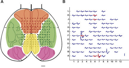
To establish the relationship between SPWs and neocortical activity, multiple single unit activity was recorded from an array of 144 independently positionable electrodes chronically implanted on three rats. The electrode array spanned about two-thirds of the dorsal surface of the neocortex, including anterior cingulate, primary and secondary motor, somatosensory, dorsal parietal, retrosplenial, and visual areas (Fig. 1A). At the same time, the left CA1 pyramidal layer EEG (Fig. 2) was recorded from one electrode.
Figure 1.
Recording location and yield of isolated units from a particularly successful recording session. Average yield per session was approximately 50 neurons. (A) Outline of the cortical structures spanned by the electrode array, as implanted on rats 1 and 2. The array on rat 3 was placed symmetrically over the midline. Electrode locations are represented by black dots. Colors indicate neocortical areas; (purple) visual; (green) somatosensory; (yellow) retrosplenial; (orange) motor and anterior cingulate. Calibration bar, 1 mm. (This figure was adapted from Zilles 1985 with permission from Springer © 1985.) (B) Graph of 96 simultaneously recorded neocortical single unit waveforms, plotted at locations of the corresponding recording electrodes. Calibration bar, 150 μV.
Figure 2.
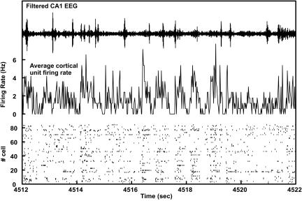
Example of simultaneous recording of hippocampal LFPs and 84 neocortical single units. This is a 10-sec excerpt from a rest period showing: LFP trace from the CA1 pyramidal layer, bandpass (100-300 Hz) filtered, with SPW/ripple complexes (top); the global average cortical firing rate (middle); and raster plot containing the spike trains from the simultaneously recorded cells (bottom). Cortical firing shows oscillations in the delta range (2-3 Hz) synchronized across all the sampled cortical areas. During the troughs of these oscillations, neuronal activity was often completely suppressed.
During recording sessions, rats 1 and 2 slept or rested for two periods (Rest 1 and Rest 2) of 25-40 min. Between these periods, they performed a behavioral task. Rat 3 was only recorded during one rest period of 25-40 min in each session and did not perform a behavioral task. Only the rest period data were analyzed for this report. Up to 96 cells were recorded simultaneously (Fig. 1B; total 2812 cells, in 61 sessions; 2298 from rat 1, 288 from rat 2, 226 from rat 3). Some of the cells may have been recorded from more than once across sessions, given that not all electrodes were moved every day (firing rate: mean: 1.94 ± 2.89 Hz; min: 0.005 Hz, max: 31.06 Hz).
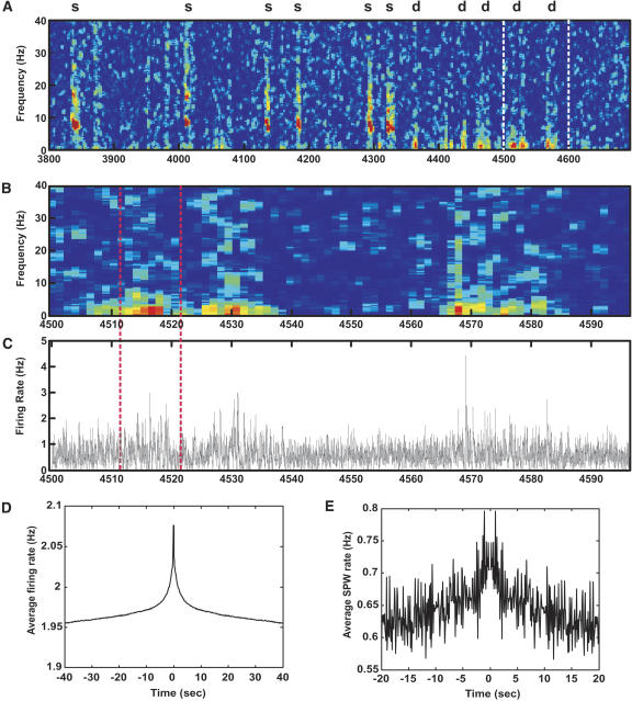
Neocortical neuronal activity exhibited coherent fluctuations spanning all of the recorded areas. Spectral analysis of the total spike activity revealed discrete bouts of oscillations in the spindle range and in the delta/slow range during most of the recorded rest sessions (see, e.g., Fig. 3A). These episodes of oscillations accounted for 35% of the total recording time.
Figure 3.
Global fluctuations of cortical activity and hippocampal sharp waves. (A) Power spectral density analysis of 700 sec of cortical activity. The power spectrum of combined instantaneous firing rate for the 84 recorded cells was estimated using a sliding 5-sec window. Episodes of coordinated oscillations in the spindle range (indicated by the s) and in the delta/slow range (indicated by the d) are visible. (B) Enlargement of the 100-sec period between the white dashed lines in A, showing delta oscillations. (C) Average instantaneous cortical firing rate during the same interval. The interval between the two red lines is the same as depicted in Figure 2. Note that the absolute values of the firing rates are different from Figure 2 because a larger smoothing parameter was used. (D) Autocorrelogram of the total activity of the recorded neocortical population. In each recording session, the spikes from all the recorded cells were pooled into a single binned time series that was used to compute the autocorrelation function. The normalization was chosen such that the asymptotic value of the autocorrelation coincides with the population average firing rate per cell. The data shown are an across-session average. The decline of the autocorrelogram was fit with an exponential function with a time constant of 2.4 ± 0.5 sec. Time bin, 200 msec. (E) Autocorrelogram of the time of occurrence of SPWs. There is a decay from t = 0 to the baseline, that can be approximated with an exponential function with a time constant of 5.5 ± 0.06 sec. This indicates that SPWs do not occur as a Poisson process with a constant rate throughout the recording sessions.
During troughs of slow/delta oscillations, generalized neuronal silence was often observed for a duration of ∼200-400 msec (Figs. 2 and 3B,C). The same degree of synchrony was observed across the sampled recorded areas during spindle oscillations (data not shown). Fluctuations on a longer time scale were also present, and were reflected in the autocorrelogram of the global population activity, which decayed with a time constant of 2.4 ± 0.5 sec (Fig. 3D). “Up-states” were defined arbitrarily as those 1-sec periods in which the number of “active” cells (one or more spikes) exceeded the 95% confidence threshold, computed from the randomly and independently shuffled interval series of all cells (see Materials and Methods). In all, 15.2% ± 4.3% of the intervals in Rest 1 and 13.0% ± 3.0% in Rest 2 exceeded this threshold, with activity in at least 58.3% ± 5.0% and 57.5% ± 5.1% of neurons, respectively. The recording session with the smallest effect yielded 7.6% of above-threshold intervals (p < 0.0001).
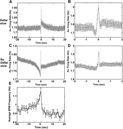
The sharp-wave-triggered Peri-event time histograms (PETH) of the global cortical activity showed that SPW events were accompanied by a brief burst of neocortical firing (Fig. 4). PETHs computed during periods of identified delta/slow oscillations were different from those computed during periods in which no delta/slow oscillations were present. During delta/slow oscillations (Fig. 4A,B), sharp wave events were preceded by a dip in average cortical firing lasting for ∼500 msec. The average cortical firing was at the baseline level immediately before the dip. After the sharp wave event, the average cortical firing was increased, and slowly decayed to baseline with a time constant of the order of 3 sec. In epochs that did not include delta/slow oscillations, the sharp wave-triggered PETH of cortical firing exhibited different features (Fig. 4C,D): Although there was still a transient increase at the time of the sharp wave event (albeit not as strong as during delta/slow oscillations), no “fast” dip was observed preceding sharp waves. Instead, an exponential decline in average activity was observed preceding the sharp wave, with a time constant of the order of seconds. Baseline firing resumed shortly after sharp waves. Conversely, the generation of SPW events was affected by the neocortical state (Fig. 4E): the frequency of sharp wave occurrence was higher during “down-states” (1.12 ± 0.22 Hz; p < 10-6) than during “up-states” (0.77 ± 0.20 Hz). The “down/up” transition-centered PETH of SPW events also revealed an increase in SPW frequency at the transition, which started 1-2 sec ahead of the transition.
Figure 4.
SPW interaction with neocortical neural activity. (A) Peri-event time histogram (PETH) of cortical population activity during the periods of identified global oscillations in the delta/slow range, centered on hippocampal sharp wave events. A transient increase in firing rate was evident at the time of the sharp waves. On the long time scale, the firing rate was larger after the sharp wave event. Firing rate showed a short lasting dip right before (200-400 msec) the SPW, probably related to the in-phase occurrence of delta oscillation, and was at baseline level shortly before that. After the SPW events, average cortical firing rate showed a much longer decay, spanning several seconds (error bars = SEM; bin size, 100 msec). (B) Same PETH, with an expanded scale, illustrating both the fast dip before SPWs and the transient increase at the time of SPWs (bin size, 20 msec). (C) Sharp-wave-triggered PETH (bin size, 100 msec) of cortical firing during periods in which oscillations in the delta/slow range were absent. The transient increase at SPW time was still present, albeit of smaller amplitude, but there was no fast dip preceding SPWs. A prolonged period of increasingly negative modulation was observed for several seconds leading up to the sharp waves. After SPWs, the return to baseline was relatively fast. (D) Same PETH shown on an enlarged scale (bin size, 20 msec). Note that, because the shapes of the PETH functions represent averages over many SPW events, they do not necessarily reflect the shape of individual events (see Fig. 6 for further explanation). (E) PETH of SPW event occurrence, centered on the down-to-up-state transitions in the total cortical spike activity, showing increased SPW probability around such transitions. Note that the baseline value was higher on the left-hand side of the PETH than on the right-hand side, signaling that sharp waves were more frequent during the down-states than during the up-states.
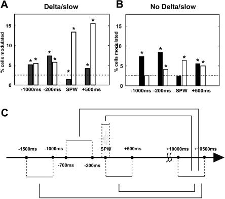
To estimate the proportion of individual cells exhibiting statistically significant modulation around SPWs, the firing rates before, during, and after each SPW event were compared with an estimate of the SPW-independent firing rates (Fig. 5; Table 1), for both delta/slow epochs and non-delta/slow epochs. For all intervals, there was an elevated number of significantly (two-tailed t-test, p < 0.05) up- or down-modulated (or both) cells. The proportion of up- and down-modulated cells was in most cases different from chance; the significance of such differences was tested by a binomial sign test, whose results are reported in Table 1. The delta/slow epochs showed similar numbers of up- and down-modulated cells before the sharp waves (consistent lack of deviation from baseline observed in the PETH prior to sharp waves), and larger numbers of up-modulated cells at the sharp wave times and in the subsequent interval (consistent with the long-lasting increase from baseline registered in the PETH). At the time of the sharp waves, there were significantly fewer down-modulated cells than chance. In non-delta/slow epochs, there was an elevated number of down-modulated cells in the pre-SPW intervals, a prevalence of up-modulated cells at sharp wave times, and similar numbers of up- and down-modulated cells in the interval following the sharp waves. The pattern was similar in each of the experimental animals (Table 1). To assess whether these effects were localized in only some of the recorded cortical areas, the 12 × 12 electrode array was divided into nine square regions (3 × 3). The number of SPW-modulated cells was above chance in all regions studied both in delta/slow epochs and non-delta/slow epochs, although there were regional differences (p < 0.02, χ2 test). The effects seemed to be strongest in the cortical regions spanning the midline, but further study would be required to rule out variables other than region in the generation of this effect. The z-scores for sharp wave-modulation of individual cells in the two rest periods were only moderately correlated (r = 0.48 in the interval corresponding to SPWs, and lower in the other intervals), indicating that different ensembles were activated during different SPW events.
Figure 5.
Single unit activity modulation by SPWs. (A) Percentage of cells with firing rates that were significantly (p < 0.05) up-modulated (white bars) or down-modulated (black bars) in intervals centered on, or surrounding, the SPWs, as they were detected by the thresholding algorithm during the periods of identified global delta/slow cortical oscillations. The firing rates in 500-msec intervals spanning from 1500 to 1000 msec before the SPW (first pair of bars), from 700 to 200 msec before the SPW (second pair), the 500 msec after the SPW (fourth pair) and in the intervals between the beginnings and ends of SPWs (third pair), were compared with intervals of the same length starting 10 sec after each SPW. A large percentage of cells was up-modulated at the time of SPWs and 500 msec thereafter. At the time of the SPW, the number of down-modulated cells was actually significantly lower than the chance value of 2.5%. In the intervals before the SPW there were similar proportions of up-modulated and down-modulated cells. (B) Same comparison for the periods of time without global delta/slow oscillations. A large percentage of cells was down-modulated before the SPWs. A large proportion of cells was up-modulated at the time of the SPW, whereas similar proportions of cells were up- and down-modulated in the +500-msec interval. The dashed line here and in A represents the 2.5% chance level. A fraction of modulated cells close to that chance level would be attained if there were no statistical relationship between sharp waves and cortical firing, as in that case the intervals considered and the controls 10 sec later could be considered random time intervals with respect to the cortical firing (*: fraction of modulated cells different from chance level; p < 0.00001). (C) Schematic representation of the intervals used for the comparisons in A. The intervals used for the -1000 msec (500 msec), -200 msec (500 msec), SPW (beginning and ends detected by thresholding), and +500 msec (500 sec), and the corresponding control intervals are displayed.
Table 1.
Detail of the numbers of up- and down-modulated cells during delta/slow and non-delta/slow epochs
| −1000 msec | −200 msec | SPW | +500 msec | |
|---|---|---|---|---|
| Rat 1, N = 2298 | ||||
| Delta/slow up-modulated | 135 (5.9%, p < 0.00001) | 152 (6.6%, p < 0.00001) | 333 (14.5%, p < 0.00001) | 376 (16.36%, p < 0.00001) |
| Delta/slow down-modulated | 118 (5.1%, p < 0.00001) | 168 (7.3%, p < 0.00001) | 32 (1.4%, p < 0.001) | 97 (4.2%, p < 0.00001) |
| Non-delta/slow up-modulated | 48 (2.0%, n.s.) | 90 (3.9%, p < 0.00005) | 139 (6.0%, p < 0.00001) | 100 (4.45, p < 0.00001) |
| Non-delta/slow down-modulated | 188 (8.1%, p < 0.00001) | 203 (8.8%, p < 0.00001) | 59 (2.6%, n.s.) | 140 (6.0%, p < 0.00001) |
| Rat 2, N = 288 | ||||
| Delta/slow up-modulated | 14 (4.9%, p < 0.02) | 8 (2.8%, n.s.) | 13 (4.5%, p < 0.05) | 28 (9.7%, p < 0.00001) |
| Delta/slow down-modulated | 17 (5.9%, p < 0.0005) | 26 (9.0%, p < 0.00001) | 5 (1.7%, n.s.) | 18 (6.2%, p < 0.00005) |
| Non-delta/slow up-modulated | 18 (6.2%, p < 0.00005) | 12 (4.1%, n.s.) | 19 (6.6%, p < 0.00001) | 16 (5.5%, p < 0.001) |
| Non-delta/slow down-modulated | 8 (2.8%, n.s.) | 27 (9.4%, p < 0.00001) | 7 (2.4%, n.s.) | 11 (3.8%, n.s.) |
| Rat 3, N = 226 | ||||
| Delta/slow up-modulated | 5 (2.2%, n.s.) | 1 (0.04%, p < 0.05) | 30 (13.2%, p < 0.00001) | 34 (15.0%, p < 0.00001) |
| Delta/slow down-modulated | 8 (3.5%) | 12 (5.3%, p < 0.01) | 0 (0.0%, p < 0.02) | 1 (0.04%, p < 0.05) |
| Non-delta/slow up-modulated | 5 (2.2%, n.s.) | 15 (6.6%, p < 0.00001) | 21 (9.3%, p < 0.00001) | 25 (11.0%, p < 0.00001) |
| Non-delta/slow down-modulated | 10 (4.4% n.s., p = 0.06) | 8 (3.5%, n.s.) | 2 (0.09%, n.s.) | 5 (2.2%, n.s.) |
| Total, N = 2812 | ||||
| Delta/slow up-modulated | 154 (5.5%, p < 0.00001) | 161 (5.7%, p < 0.00001) | 376 (13.4%, p < 0.00001) | 438 (15.6%, p < 0.00001) |
| Delta/slow down-modulated | 143 (5.1%, p < 0.00001) | 206 (7.3%, p < 0.00001) | 37 (1.3%, p < 0.00001) | 116 (4.1%, p < 0.00001) |
| Non-delta/slow up-modulated | 71 (2.5%, n.s.) | 117 (4.1%, p < 0.00001) | 179 (6.3%, p < 0.00001) | 141 (5.0%, p < 0.00001) |
| Non-delta/slow down-modulated | 206 (7.3%, p < 0.00001) | 238 (8.4%, p < 0.00001) | 68 (2.4%, n.s.) | 156 (5.5%, p < 0.00001) |
The numbers of cells whose rates were modulated up and down (p < 0.05, two-tailed t-test) and the fractions of the total are shown for each of the three experimental animals and for the totals. The p-values are relative to the significance of the difference between the measured fraction of modulated cells and the chance level of 2.5% (binomial, z-test).
The probability of SPW occurrence also fluctuated weakly on time scales comparable to those of neocortical slow oscillations, as shown by the autocorrelogram of SPW times, which decays with a time constant of 5.51 ± 0.06 sec (Fig. 3E).
Dicussion
The three main results of this study are the observation of deeply synchronized fluctuations in neuronal firing in the delta/slow frequency range across almost the entire neocortex, an observation that extends previously published results (Destexhe et al. 1999), of the existence of longer time-scale fluctuations in nonanesthetized animals, and, most importantly, of the relationship between these cortical phenomena and hippocampal sharp waves. A coupling between cortical delta rhythm and sharp waves was previously demonstrated by Sirota et al. (2003) in somatosensory cortex. It is shown here that longer time-scale coherent fluctuations in cortical activity, namely, the transitions between what have been referred to as “down-states” and “up-states” were correlated with an increased probability of occurrence of sharp waves in the hippocampus (Fig. 4E), and sharp waves were more likely to occur during down-states than during up-states. Because the coupling between cortical activity and hippocampal activity is of a probabilistic nature, there was no one-to-one correspondence between cortical transitions and sharp waves, which explains why the modulation observed in the PETHs is relatively small. The small size of the effect may have prevented Pelletier et al. (2004) from observing a relation between neocortical firing and sharp waves in the entorhinal cortex, which are tightly related to the hippocampal ones (Chrobak and Buzsaki 1996), because of their smaller sample size. The effect was nevertheless observed in a large fraction of the cells, thus it could not be attributed to a modulation of a small subset of cells (Fig. 5). These two facts taken together suggest that different, relatively small, groups of cells are recruited by each sharp wave episode. The small size of this group may reflect the sparse coding of information in neocortical modules, and may be partly caused by the high inhibitory pressure in the entorhinal cortex (Pelletier et al. 2004). The correlation between hippocampal and neocortical dynamics was also evident when the change in sharp wave frequency associated with cortical activity states was considered: sharp wave events were 20%-30% more likely in correspondence with down-to-up transitions compared with baseline (Fig. 4E). Sharp waves were also more likely during down-states than during up-states.
The intracellular physiology literature has introduced the concept of “up” and “down” states as membrane potential fluctuations between two well-defined levels. In fact, in some regions or experimental conditions, the membrane potential fluctuations are bimodal in nature (e.g., as observed in the striatum by Wilson and Groves 1981). Bimodal fluctuations of membrane potential have been observed in intracellular recordings in anesthetized and naturally sleeping animals (Timofeev et al. 2001; Petersen et al. 2003) in the delta range, with frequencies corresponding to those of the population firing rate oscillations shown here. The picture emerging from population firing rates is, however, somewhat different: The population firing rate during delta/slow oscillations did not show bimodality, as the oscillations are characterized by an abrupt upward transition and a slower decline (see, e.g., Fig. 2). This might be owing to a gradual loss in coherence across neocortical cells (perhaps because of different durations of up-states in different neocortical networks) or to firing rate adaptation, which may slow down cell firing as the cortex remains in an up-state. Nevertheless, the data are consistent with the conclusion that sharp waves were correlated with discrete transitions from relatively low to relatively high states of activity. The precise shape of the sharp-wave-triggered PETH depends on the current dynamical state of the neocortex: During delta/slow oscillations, the PETH stays at baseline until few hundreds of milliseconds from the sharp wave, whereas, following the sharp waves, it decays back to baseline in a few seconds, starting from a more elevated value. In periods without delta/slow oscillations, the PETH is basically flat after the sharp wave, but exhibits a slowly increasing depression leading to up the sharp wave (2-3 sec).
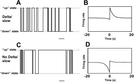
One possible scenario that would produce this pattern is depicted in Figure 6. In this oversimplified scheme, transitions between up-states and down-states are described by a Markov process, so that the state durations follow an exponential distribution, and at least a fraction of the sharp waves coincide with the down-to-up transitions, the others being distributed randomly in time. If the up-state has a much shorter mean duration than the down-state, the sharp wave-triggered PETH of cortical activity has a shape similar to that observed during delta oscillations. If down-states are shorter than up-states, the PETH resembles that observed in no-delta/slow periods. Thus, whereas the abrupt increase in firing at the time of the sharp wave event is observed in both cases (in the model, because of the assumption of coincidence of sharp waves and down/up transitions), the difference in the shape of the PETH in these two dynamical regimes appears to reflect a different relative duration of up- and down-states, as might be expected in deeper or more superficial sleep stages, possibly because of cholinergic modulation of leakage K+ conductances (Compte et al. 2003).
Figure 6.
Qualitative picture of the interaction between up-state and down-state transitions in cortical firing and hippocampal sharp waves and how the apparently gradual trends in the average PETH functions shown in Figure 4 can be accounted for by averaging over fluctuations in discrete states with variable durations. In these numerical simulations, the cortical state transitions occur randomly, according to a two-state Markov process, whose parameters represent the mean life of each state, and cortical firing rates are assumed to be constant within a given state. (A) In this situation, the mean life of the up-states is much shorter (3 sec) than the mean duration of the down-states (20 sec). If at least some proportion of the SPW occurs in correspondence with the down-to-up transition, the SPW-triggered PETH of cortical firing would look like the one in B, which resembles what is observed during the periods dominated by delta/slow oscillations, at least in terms of the long time scale behavior (long decline after the SPW, mostly flat before; see Fig. 4). (C) In the scenario in which the down-state is much shorter (3 sec) than the up-state (20 sec), the SPW-triggered, cortical PETH would look like the one in D, which is reminiscent of what is observed in the periods without delta/slow oscillations (long-lasting exponential trough leading to the SPWs, small modulation after the SPWs). The calibration bar in A and C represents 20 sec.
At present, there is no clear evidence for a causal relationship between these phenomena, even though a modulation in the neocortical EEG has been shown to precede sharp wave events by 50-100 msec (Sirota et al. 2003). Subtle changes in neocortical input, related to the slow fluctuations, may affect the probability of triggering an SPW in the hippocampus. More specifically, a decrease in cortical input might lead to hyperpolarization and low spike rates of CA3 cells, and to a de-inactivation of Ca++ channels (Buzsaki et al. 1992), leading to higher excitability of the hippocampal neurons. A sharp wave event may then be triggered by some fluctuation in the hippocampal input (e.g., by a neocortical transition to an up-state). On the other hand, it has been observed (Lewis and O'Donnell 2000) that stimulation of hippocampus or the ventral tegmental area can trigger up-states. Thus, the appropriate framework to describe the hippocampal/neocortical interactions might well be that of a closed loop, with causal relationships actually going both ways. During down-states, the cortex may have a role in mediating the increase in excitability in the hippocampus, which may lead to sharp waves, which could explain the increased sharp wave frequency during down-states. The burst discharge associated to a sharp wave in the CA3 network may converge on a stored memory state, making that memorized information available to the neocortex (Shen and McNaughton 1996). Both the neocortex, especially at the end of a down-state (Compte et al. 2003), and the hippocampus are in an extremely excitable state, prone to explode in coherent, self-sustained activations. Therefore, they can readily follow the input coming from the other structure: The hippocampal drive during a sharp wave could increase the probability of a transition from a down-state to an up-state. Conversely, as mentioned above, the neocortical down/up transition may provide the triggering input causing the sharp wave. Common inputs from other brain structures may influence the generation of these related events in the hippocampus and neocortex.
The set of network relationships highlighted in the present study might be relevant to the memory consolidation process: Sharp waves, reflecting the stored patterns of hippocampal activity (Kudrimoti et al. 1999), could elicit coordinated retrieval of corresponding activity configurations stored in the neocortex that are indexed by the hippocampal cue (for review, see McNaughton et al. 2003). The neocortical input may, in turn, influence the memory content retrieved by the hippocampus at that time.
Materials and Methods
Electrode array and surgical procedures
The animals were treated according to NIH guidelines and approved IACUC protocols. Rats were anesthetized with sodium pentobarbital (40 mg/kg), and a square craniotomy was opened to accommodate the 8-mm square, 12 × 12 guide cannulae array (Hoffman and McNaughton 2002). The dura was left intact, and the array was isolated from the dura surface by a layer of biocompatible silastic (Dow Corning). The coordinates were bregma AP +2.6 mm (anterior side), AP -5.2 mm (back), DL -2.4 mm (left), DL +5.4 mm (right) (Fig. 1A). The cannulae contained 75-μm stainless steel electrodes, tapered to a fine tip (FHC). The uninsulated rear part of the electrodes made electrical contact with the cannulae, which, in turn, were connected to a circuit board that provided the connections between the cannulae and the buffer preamplifiers (Neuralynx). The implant was anchored to the skull with jeweler's screws and dental cement. After surgery, the electrodes were pushed through the insulation layer and the dura to the brain surface by means of a 0.006-inch wire push-rod, connected to a digital micromanipulator, which was inserted in the top end of the cannulae. Once every few recording sessions, a subset of the electrodes was advanced to new locations, searching for new units in the neocortex, while a custom-written program kept track of the depth of the electrodes. A few electrodes were advanced to the CA1 pyramidal layer for LFP recording.
Electrophysiological recording procedures
For single unit recordings, the signal coming from the buffer preamplifiers was differentially amplified, referred to an electrode in a quiet location in the white matter, and band-pass filtered between 600 Hz and 6 kHz. Whenever the signal exceeded a threshold, a spike waveform was acquired at 25 kHz (duration 1.3 msec) and recorded on the hard disk (Cheetah recording system; Neuralynx, Inc.). LFPs were filtered between 1 Hz and 475 Hz and continuously recorded at 1.6 kHz. The acquired waveforms were sorted off-line using a semiautomated clustering algorithm (BBClust, P. Lipa, unpubl.) based on the waveform amplitude, and the wave-shape principal components, and successively refined manually with custom-written software (MClust, A.D. Redish, unpubl.).
Recording session protocol
For rats 1 and 2, during each recording session, rats were allowed to rest and/or sleep for two sessions of 25/40 min, while electrophysiological data were recorded. In between these sessions, the rats ran for food back and forth on a circular track. The two reward sites were separated by a barrier so that the rat had to alternate directions on the track. Along the track, many multi-modal sensory cues were placed: odors, floor coverings with different textures, rods that hit the whiskers while the rat was passing by, and an earphone playing music. Only the data from the rest sessions were used for this work. Rat 3 was allowed to rest and/or sleep for 25/40 min during electrophysiological recordings, and did not perform any behavioral task.
Up-state detection
For this analysis, only 28 sessions with 40 or more recorded cells were considered. Spike-trains were binned in 1-sec intervals. For each interval, the number of active cells (i.e., cells that fired at least one spike in the interval) was computed. If that number exceeded a threshold, an up-state was detected. The threshold was computed as the 95th percentile of the distribution, computed from randomly and independently shuffled spike trains. The threshold was therefore a value representative of the expected amount of fluctuation in the total activity in spike trains with similar firing rates, but in which the coherence across cells was disrupted.
Sharp wave detection
The CA1 pyramidal layer LFP was filtered in the ripple oscillation frequency domain (100-300 Hz). SPW events were detected by means of a thresholding algorithm. Suprathreshold events closer to each other than 100 msec were merged, and events shorter than 50 msec were discarded. For neocortical activity PETH calculations, SPW timestamps were computed as the times of the highest peaks in the ripple oscillation within the SPW events.
Spectral analysis of cortical activity
The combined spike train from all the recorded cortical cells in each session was transformed into a time series by a 5-msec time window binning. The power spectrum of the resulting time series was estimated by means of multitaper analysis (Mitra and Pesaran 1999; Percival and Walden 2002), in a sliding time window with a width of ∼10 sec. The aggregate power in the 0.8-4 Hz as a function of time was used as an index of oscillatory activity in the delta/slow range. Epochs in which this index exceeded a manually defined threshold area are considered as delta/slow oscillation periods.
Acknowledgments
We thank K. Bohne, K. Poneta, L. Gronenberg, J. Lammers, S. Cowen, M. Tatsuno, and K. Hoffman for help with running the experiments; K. Stengel (Neuralynx, Inc.) for technical assistance; and A. Fuglevand, K. Gothard, and K. Hoffman for suggestions on the manuscript. This work is supported by NIH, MH46823, and MH01565. F.P.B. was partially funded by HFSP LT150/99B. G.R.S. was partially funded by NIH NIGMS 5 T32 GM08400.
Article and publication are at http://www.learnmem.org/cgi/doi/10.1101/lm.73504.
References
- Achermann, P. and Borbely, A.A. 1997. Low-frequency (<1 Hz) oscillations in the human sleep electroencephalogram. Neuroscience 81: 213-222. [DOI] [PubMed] [Google Scholar]
- Ball, G.J., Gloor, P., and Schaul, N. 1977. The cortical electromicrophysiology of pathological delta waves in the electroencephalogram of cats. Electroencephalogr. Clin. Neurophysiol. 43: 346-361. [DOI] [PubMed] [Google Scholar]
- Battaglia, F.P, Sutherland, G.R., and McNaughton, B.L. 2001. Widespread modulation of neocortical cell activity during hippocampal sharp waves. Soc. Neurosci. Abs. 27: 643.16. [Google Scholar]
- Buzsaki, G. 1989. Two-stage model of memory trace formation: A role for “noisy” brain states. Neuroscience 31: 551-570. [DOI] [PubMed] [Google Scholar]
- Buzsaki, G., Horvath, Z., Urioste, R., Hetke, J., and Wise, K. 1992. High-frequency network oscillation in the hippocampus. Science 256: 1025-1027. [DOI] [PubMed] [Google Scholar]
- Chrobak, J.J. and Buzsaki, G. 1994. Selective activation of deep layer (V-VI) retrohippocampal cortical neurons during hippocampal sharp waves in the behaving rat. J. Neurosci. 14: 6160-6170. [DOI] [PMC free article] [PubMed] [Google Scholar]
- ____. 1996. High-frequency oscillations in the output networks of the hippocampal-entorhinal axis of the freely behaving rat. J. Neurosci. 16: 3056-3066. [DOI] [PMC free article] [PubMed] [Google Scholar]
- Compte, A., Sanchez-Vives, M.V., McCormick, D.A., and Wang, X.J. 2003. Cellular and network mechanisms of slow oscillatory activity (<1 Hz) and wave propagations in a cortical network model. J. Neurophysiol. 89: 2707-2725. [DOI] [PubMed] [Google Scholar]
- Cowan, R.L. and Wilson, C.J. 1994. Spontaneous firing patterns and axonal projections of single corticostriatal neurons in the rat medial agranular cortex. J. Neurophysiol. 71: 17-32. [DOI] [PubMed] [Google Scholar]
- Csicsvari, J., Hirase, H., Mamiya, A., and Buzsaki, G. 2000. Ensemble patterns of hippocampal CA3-CA1 neurons during sharp wave-associated population events. Neuron 28: 585-594. [DOI] [PubMed] [Google Scholar]
- Destexhe, A., Contreras, D., and Steriade, M. 1999. Spatiotemporal analysis of local field potentials and unit discharges in cat cerebral cortex during natural wake and sleep states. J. Neurosci. 19: 4595-4608. [DOI] [PMC free article] [PubMed] [Google Scholar]
- Hoffman, K.L. and McNaughton, B.L. 2002. Coordinated reactivation of distributed memory traces in primate neocortex. Science 297: 2070-2073. [DOI] [PubMed] [Google Scholar]
- Ishizuka, N., Weber, J., and Amaral, D.G. 1990. Organization of intrahippocampal projections originating from CA3 pyramidal cells in the rat. J. Comp. Neurol. 295: 580-623. [DOI] [PubMed] [Google Scholar]
- Kudrimoti, H.S., Barnes, C.A., and McNaughton, B.L. 1999. Reactivation of hippocampal cell assemblies: Effects of behavioral state, experience, and EEG dynamics. J. Neurosci. 19: 4090-4101. [DOI] [PMC free article] [PubMed] [Google Scholar]
- Lewis, B.L. and O'Donnell, P. 2000. Ventral tegmental area afferents to the prefrontal cortex maintain membrane potential `up' states in pyramidal neurons via D(1) dopamine receptors. Cereb. Cortex. 10: 1168-1175. [DOI] [PubMed] [Google Scholar]
- Louie, K. and Wilson, M.A. 2001. Temporally structured replay of awake hippocampal ensemble activity during rapid eye movement sleep. Neuron 29: 145-156. [DOI] [PubMed] [Google Scholar]
- Maquet, P. 2001. The role of sleep in learning and memory. Science 294: 1048-1052. [DOI] [PubMed] [Google Scholar]
- Marr, D. 1970. A theory for cerebral neocortex. Proc. R Soc. Lond. B Biol. Sci. 176: 161-234. [DOI] [PubMed] [Google Scholar]
- McNaughton, B.L. 1983. Comments in hippocampus symposium panel discussion. In Neurobiology of the hippocampus (ed. W. Siefert), p. 610. Academic Press, New York.
- McNaughton, B.L., Barnes, C.A., Battaglia, F.P., Bower, M.R., Cowen, S.L., Ekstrom, A.D., Gerrard, J.L., Hoffman, K.L., Houston, F.P., Karten, Y., et al. 2003. Off-line reprocessing of recent memory and its role in memory consolidation: A progress report. In Sleep and brain plasticity (eds. P. Maquet et al.), pp. 225-246. Oxford University Press, Oxford, UK.
- Mitra, P.P. and Pesaran, B. 1999. Analysis of dynamic brain imaging data. Biophys. J. 76: 691-708. [DOI] [PMC free article] [PubMed] [Google Scholar]
- O'Keefe, J. and Nadel, L. 1978. The hippocampus as a cognitive map. Oxford University Press, Oxford.
- Pavlides, C. and Winson, J. 1989. Influences of hippocampal place cell firing in the awake state on the activity of these cells during subsequent sleep episodes. J. Neurosci. 9: 2907-2918. [DOI] [PMC free article] [PubMed] [Google Scholar]
- Pelletier, J.G., Apergis, J., and Paré, D. 2004. Low probability transmission of neocortical and entorhinal impulses through the perirhinal cortex. J. Neurophysiol. 91: 2079-2089. [DOI] [PubMed] [Google Scholar]
- Percival, D.B. and Walden, A.T. 2002. Spectral analysis for physical applications. Cambridge University Press, Cambridge, UK.
- Petersen, C.C., Hahn, T.T., Mehta, M., Grinvald, A., and Sakmann, B. 2003. Interaction of sensory responses with spontaneous depolarization in layer 2/3 barrel cortex. Proc. Natl. Acad. Sci. 100: 13638-13643. [DOI] [PMC free article] [PubMed] [Google Scholar]
- Plihal, W. and Born, J. 1999. Effects of early and late nocturnal sleep on priming and spatial memory. Psychophysiology 36: 571-582. [PubMed] [Google Scholar]
- Ribeiro, S., Gervasoni, D., Soares, E.S., Zhou, Y., Lin, S.C., Pantoja, J., Larine, M., and Nicolelis, M.A. 2004. Long-lasting novelty-inducing neuronal reverberation during slow-wave sleep in multiple forebrain areas. PLoS Biol. 2: E24. [DOI] [PMC free article] [PubMed] [Google Scholar]
- Sanchez-Vives, M.V. and McCormick, D.A. 2000. Cellular and network mechanisms of rhythmic recurrent activity in neocortex. Nat. Neurosci. 3: 1027-1034. [DOI] [PubMed] [Google Scholar]
- Shen, B. and McNaughton, B.L. 1996. Modeling the spontaneous reactivation of experience-specific hippocampal cell assembles during sleep. Hippocampus 6: 685-692. [DOI] [PubMed] [Google Scholar]
- Siapas, A.G. and Wilson, M.A. 1998. Coordinated interactions between hippocampal ripples and cortical spindles during slow-wave sleep. Neuron 21: 1123-1128. [DOI] [PubMed] [Google Scholar]
- Sirota, A., Csicsvari, J., Buhl, D., and Buzsaki, G. 2003. Communication between neocortex and hippocampus during sleep in rodents. Proc. Natl. Acad. Sci. 100: 2065-2069. [DOI] [PMC free article] [PubMed] [Google Scholar]
- Skaggs, W.E. and McNaughton, B.L. 1996. Replay of neuronal firing sequences in rat hippocampus during sleep following spatial experience. Science 271: 1870-1873. [DOI] [PubMed] [Google Scholar]
- Steriade, M. 2000. Corticothalamic resonance, states of vigilance and mentation. Neuroscience 101: 243-276. [DOI] [PubMed] [Google Scholar]
- Steriade, M. and Buzsaki, G. 1990. Parallel activation of thalamic and cortical neurons by brainstem and basal forebrain cholinergic systems. In Brain cholinergic systems (eds. M. Steriade and D. Biesold), pp. 3-64. Oxford University Press, Oxford, UK.
- Steriade, M., Nunez, A., and Amzica, F. 1993. A novel slow (<1 Hz) oscillation of neocortical neurons in vivo: Depolarizing and hyperpolarizing components. J. Neurosci. 13: 3252-3265. [DOI] [PMC free article] [PubMed] [Google Scholar]
- Stickgold, R., James, L., and Hobson, J.A. 2000. Visual discrimination learning requires sleep after training. Nat. Neurosci. 3: 1237-1238. [DOI] [PubMed] [Google Scholar]
- Sutherland, G.R. and McNaughton, B. 2000. Memory trace reactivation in hippocampal and neocortical neuronal ensembles. Curr. Opin. Neurobiol. 10: 180-186. [DOI] [PubMed] [Google Scholar]
- Timofeev, I., Grenier, F., and Steriade, M. 2001. Disfacilitation and active inhibition in the neocortex during the natural sleep-wake cycle: An intracellular study. Proc. Natl. Acad. Sci. 98: 1924-1929. [DOI] [PMC free article] [PubMed] [Google Scholar]
- Wilson, C.J. and Groves, P.M. 1981. Spontaneous firing patterns of identified spiny neurons in the rat neostriatum. Brain Res. 220: 67-80. [DOI] [PubMed] [Google Scholar]
- Wilson, M.A. and McNaughton, B.L. 1994. Reactivation of hippocampal ensemble memories during sleep. Science 265: 676-679. [DOI] [PubMed] [Google Scholar]
- Zilles, K. 1985. The cortex of the rat. Springer-Verlag, New York.


