Abstract
Two articles in this issue of Neuron explore the structural basis of allosteric inhibition in ionotropic glutamate receptors, providing key insights into how iGluRs function in the brain as well as how they might be pharmacologically modulated in neurological disorders and disease.
Ionotropic glutamate receptors (iGluRs) are critical for proper function of the human brain. For that reason, this family of ion channel proteins has garnered a tremendous amount of interest in recent years in their function and dysfunction in neurological diseases and disorders. iGluRs are tetrameric transmembrane proteins which can be divided into four subfamilies, each with distinct functional profiles: N-methyl-D-aspartate receptors (NMDARs), α-amino-3-hydroxy-5-methyl-4-isoxazolepropionic acid receptor (AMPARs), Kainate receptors, and Delta receptors, and several subtypes exist within each of these subfamilies. However, disentangling the various activities of these protein complexes in the brain has been a daunting task. Two subfamilies of iGluRs, NMDA receptors and AMPA receptors, have further been implicated in a number of neurological disorders including schizophrenia and depression (in the case of NMDARs) and epilepsy (both NMDARs and AMPARs) (Traynelis et al., 2010).
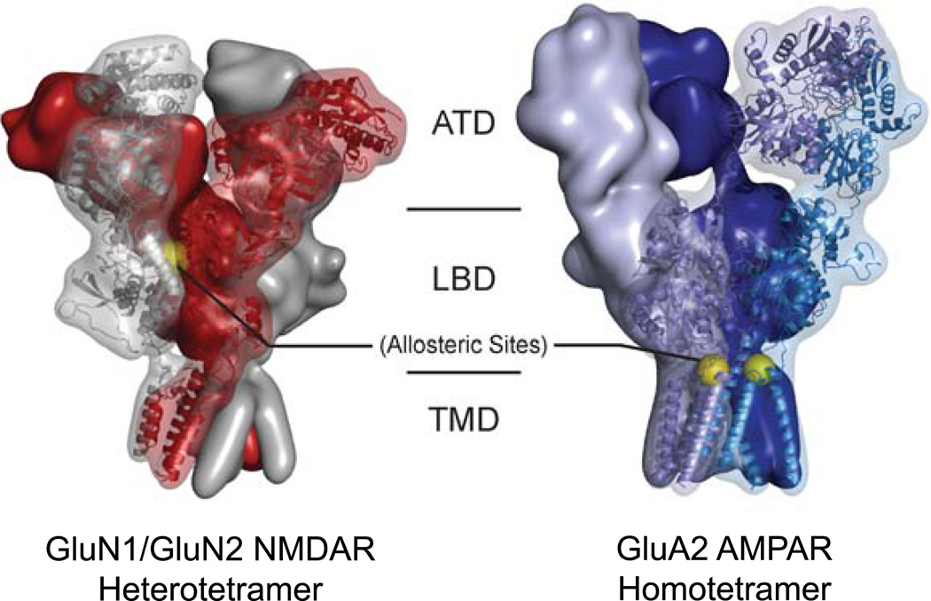
In this issue of Neuron, both AMPARs and NMDARs figure prominently in a pair of articles (Yelshanskaya et al., 2016; Yi et al., 2016, respectively) that begin to dissect the mechanisms underlying allosteric inhibition in iGluRs, which could potentially be further extended to produce novel therapies for the above conditions. Of critical importance to this endeavor, however, is uncovering the means to produce sufficient specificity so as to target only a single subtype in a large family of closely-related proteins. Both NMDARs and non-NMDARs (AMPA, Kainate and Delta receptors) have a similar overall domain organization consisting of an extracellular amino-terminal domain (ATD) and ligand binding domain (LBD), and a transmembrane domain (TMD) which comprises the ion channel, as well as a primarily unstructured carboxyl-terminal domain (CTD) (Karakas and Furukawa, 2014; Sobolevsky et al., 2009) (Figure 1). While AMPA receptors can form functional homotetramers activated by glutamate alone, NMDA receptors are obligate heterotetramers requiring binding of both glycine and glutamate (Traynelis et al., 2010). In both cases, the totality of iGluRs expressed in the brain are not “pure” populations of subtypes, but rather a wide mixture: four different subtypes exist of the AMPA receptor (GluA1-4), while NMDA receptors have seven (GluN1, GluN2A-D, and GluN3A-B) (Regan et al., 2015; Sobolevsky et al., 2009). During development the expression pattern of these iGluRs changes dramatically, and concurrent alterations in channel subtype composition produce receptors with very different functional properties (Traynelis et al., 2010). These features of iGluRs begin to frame two key obstacles to the development of clinically useful small molecules: first, due to the wide range in functional characteristics of heteromeric iGluRs, the most appealing leads will show specificity for only those iGluR subtypes which are involved in the disorder in question. Second, as nearly all iGluRs share a glutamate-binding pocket in LBD, any drug targeting the glutamate-binding site is likely to have significant off-target effects; thus, an allosteric inhibitor would be the most viable candidate for iGluR subfamily- and subtype-specific binding.
Figure 1. Locations of allosteric binding sites in NMDAR and AMPAR.
Allosteric binding sites (yellow spheres) could prove to be critical in the development of novel, highly-specific modulators of iGluRs. Yi, et al. (2016) characterized a site at the subunit interface between the LBDs of the NMDAR GluN1 and GluN2 subunits (left, grays and reds respectively). Yelshanskaya, et al. (2016) present structural data of inhibitor binding at the TMD/LBD interface of the homomeric AMPAR (right).
To this end, Yi et al. (2016) begin by focusing on a class of compounds which shows very good selectivity for NMDARs containing the GluN2A subunit, the most well-known of which is the negative allosteric modulator (NAM) TCN-201 (Bettini et al., 2010) in addition to a pair of related compounds referred to as MPX-004 and MPX-007. Interestingly, earlier work showed that while increased glycine concentrations could overcome the inhibitory effect of TCN-201 (suggestive of a competitive binding mode), mutagenesis studies pointed to a binding site at the subunit interface of the GluN1/GluN2A LBD heterodimer (Hansen et al., 2012). Very recent work by Hackos and colleagues confirmed the latter inter-LBD binding site but, perplexingly, found that both positive allosteric modulators (PAMs) specific to NMDARs and AMPAR modulators bound in very nearly the same site (Hackos et al., 2016) (Figure 1).
To better understand the mechanism through which ligands could bind at this allosteric site and affect channel activity, Yi et al. crystallized the isolated GluN1/GluN2A heterodimer in the presence of the agonists glycine and glutamate, then soaked these crystals into solutions containing either TCN-201, MPX-004, or MPX-007. By solving a series of structures ranging from 1.7 Å – 2.7 Å resolution, Yi and colleagues confirmed the allosteric binding site and, in conjunction with extensive biochemical and electrophysiological data, began to piece together a functional mechanism.
Similar to the findings of Hackos et al. (2016), Yi et al. observed convincing electron density for each of the NAMs in a pocket at the subunit interface of the GluN1/GluN2A LBD heterodimer near the hinge region of the GluN1 subunit, folded into a hairpin conformation, and partially overlapping with the PAM binding site previously identified (Hackos et al., 2016). Notably, even in the presence of allosteric modulators, the structures were all highly similar to earlier examples of agonist-bound LBD heterodimers (Jespersen et al., 2014). Upon careful examination of the binding site, Yi and colleagues observed two conformational alterations that they believed to be instrumental to NAM activity. First, they noted a subtle shift in the positioning of the sidechain of Val783 in the GluN2A LBD. While the absolute motion was small, only on the order of 0.5 Å– 1.0 Å, this residue had previously been shown to be critical for TCN-201 binding (Hackos et al., 2016; Hansen et al., 2012) and apparently engaged in a hydrophobic interaction with Phe754 of GluN1. This phenylalanine residue was seen to undergo a much larger conformational shift, leading these researchers to posit that perhaps the interaction between GluN1/Phe754 and GluN2A/Val783 was itself the “molecular switch” whereby NAM binding induced allosteric inhibition by shifting the GluN1 LBD towards a conformation less favorable to glycine binding. This was confirmed by mutagenesis experiments that demonstrated substantially altered glycine potency upon mutating these residues. (Yi et al., 2016). The authors are also able to tie the interaction between GluN1 Phe754 and GluN2A Val783 into the specificity of this class of NAMs’ binding to GluN2A, as the residues corresponding to GluN2A Val783 in GluN2B and GluN2C/D are phenylalanine and leucine, respectively, suggesting a steric basis for this specificity (Yi et al., 2016).
A final crystal structure provided further evidence of an interplay between glycine binding and NAM binding: that of the GluN1/GluN2A LBD soaked against a combination of MPX-007 and the GluN1 competitive antagonist 5,7-dichlorokyurenic acid (DCKA). DCKA binding to the glycine site of the GluN1 LBD stabilizes an open cleft conformation of the bilobe architecture, which mimics the apo state (Jespersen et al., 2014). In this structure, Yi et al. found that GluN1/Phe754 is rotated by nearly 90° away from GluN2A/Val783, which strongly hinted that this sidechain interaction is the molecular switch, but due to the constraints of the crystal itself they were unable to observe a true open cleft conformation of the GluN1 LBD, even in the presence of DCKA (Yi et al., 2016).
Based on this information, Yi and colleagues put forth a mechanism of NAM binding and allosteric inhibition, wherein the GluN1 Phe754/GluN2A Val783 interaction serves as the lynchpin. Initially, the apo-state NMDAR is closed, until binding by glycine to the GluN1 LBD and glutamate to the GluN2A LBD induces a conformational shift to activate the channel. Binding of a NAM to the allosteric pocket between the LBD subunits interrupts the interaction between GluN1 Phe754 and GluN2A Val783, stabilizing an open cleft of the GluN1 LBD which leads to the release of the bound glycine molecule -- indeed, these researchers’ mutagenesis studies showed that mutating these two residues had a striking effect on glycine potency. At this point, the receptor would deactivate, the NAM would eventually be released, and residues of the GluN1 LBD hinge region would be allowed to relax back to their apo conformation (Yi et al., 2016).
A distinct group of allosteric inhibitors that have been predicted to bind at the interface between LBD and TMD of the AMPA receptor (Balannik et al., 2005). The binding site for those allosteric compounds cannot be captured unless one works on the intact AMPAR ion channel. This is where the work by Yelshanskaya and colleagues begins.
Building upon earlier work which elucidated the structure of the intact AMPA receptor (Sobolevsky et al., 2009), Yelshanskaya et al. modified their initial crystal construct to produce a homotetrameric GluA2 receptor with functional properties similar to the wild type which they could employ for crystallization studies. Of particular interest to Yelshanskaya and colleagues were a group of structurally dissimilar compounds which they refer to as GYKI, CP, and PMP. While a considerable number of AMPAR inhibitors have been published, only PMP is available for clinical use, and the mechanism of inhibition was largely unknown (Balannik et al., 2005; Yelshanskaya et al., 2016).
The authors were able to co-crystallize each of these compounds bound to their AMPAR construct and solved structures with resolutions between 3.8 Å – 4.4 Å. They found that the receptor has generally the same architecture as that which had been previously published for the apo state of the protein (Sobolevsky et al., 2009) but, importantly, Yelshanskaya et al. were able to identify electron density of high enough quality to be able to place the inhibitor molecules, although, as the authors point out, due to the limitations in resolution the specific protein/ligand interactions are somewhat ambiguous. Interestingly, all three inhibitors appeared to bind in the same site in their respective structures: between the LBD/TMD linkers and the TMD itself, for a total of four binding sites per each intact tetrameric receptor (Figure 1). Notably, this binding site agreed relatively well with previous mutagenesis studies predicting the binding site to be located at the LBD/TMD interface (Balannik et al., 2005). However, this earlier work had suggested these inhibitors bound between the individual subunits; in their new crystal structures, Yelshanskaya et al. instead show the binding sites to consist almost entirely of a single subunit, with the lone exception being Ser615 from the neighboring subunit forming a critical hydrogen bond with the inhibitor in question (Balannik et al., 2005; Yelshanskaya et al., 2016).
Given that CP, GYKI, and PMP all appear to bind the same site, the question arises as to how this site can accommodate these quite different small molecules. While the authors are able to verify the binding site structurally with the use of a brominated GYKI analog and in silico docking studies, they turned to electrophysiology as a means to functionally verify binding at this site through extensive mutagenesis experiments. Mutation of two residues at the allosteric site, Ser615 and Phe623, showed remarkable effects on IC50 values for GYKI and PMP, but a number of residues had more subtle effects, suggesting that while the binding pocket was the same for each inhibitor, the role of individual residues varied with the bound inhibitor in accordance with different binding modes (Yelshanskaya et al., 2016).
The global organization of the AMPAR appeared to change very little in the presence of CP, GYKI, or PMP, save for a slight (6°) rotation of the extracellular domain relative to the TMD. Locally, however, the authors do identify substantial alterations. These inhibitors appear to act as wedges between the TMD and the LBD, with their position among the pre-M1, M1, M3, and M4 helices serving to tightly close the ion channel and interfere with the conformational changes which take place upon glutamate binding being transferred from the LBD to the pore (Yelshanskaya et al., 2016).
The work previewed here provides an excellent starting point for the development of small molecule inhibitors for iGluRs, but just as important as the information put forth by Yi et al. and Yelshanskaya et al. is the identification of which steps need to be taken next. As the receptor cycles through resting, active, and desensitized states, numerous potential protein/ligand interactions can be broken and re-formed. This points to great opportunities for the development of novel state-specific compounds. Similarly, the combination of structural and functional characterization presented by these two groups of researchers has shown that allosteric and subtype-specific binding sites for small molecules can be invaluable in the development of modulators of iGluR activity. These compounds will not only provide a better understanding of receptor function but will potentially enhance our ability to study developmental expression patterns, and potentially even highly specific therapeutics for human disease.
Acknowledgments
This work was supported by the National Institutes of Health (MH085926 and GM105730 to H.F.) and (F32NS093753 to M.C.R.), the Stanley Institute of Cognitive Genomics, and the Robertson Research Fund of Cold Spring Harbor Laboratory (all to H.F.).
References
- Balannik V, Menniti FS, Paternain AV, Lerma J. Molecular Mechanism of AMPA Receptor Noncompetitive Antagonism. Neuron. 2005;48:279–288. doi: 10.1016/j.neuron.2005.09.024. [DOI] [PubMed] [Google Scholar]
- Bettini E, Sava A, Griffante C, Carignani C, Buson A, Capelli AM, Negri M, Andreetta F, Senar-sancho SA, Guiral L, et al. Identification and characterization of novel NMDA receptor antagonists selective for NR2A- over NR2B-containing receptors. J. Pharmacol. Exp. Ther. 2010;335:636–644. doi: 10.1124/jpet.110.172544. [DOI] [PubMed] [Google Scholar]
- Hackos DH, Lupardus PJ, Grand T, Sheng M, Zhou Q, Hanson JE. Positive Allosteric Modulators of GluN2A-Containing NMDARs with Distinct Modes of Action and Impacts on Circuit Function. Neuron. 2016;89:983–999. doi: 10.1016/j.neuron.2016.01.016. [DOI] [PubMed] [Google Scholar]
- Hansen KB, Ogden KK, Traynelis SF. Subunit-Selective Allosteric Inhibition of Glycine Binding to NMDA Receptors. J. Neurochem. 2012;32:6197–6208. doi: 10.1523/JNEUROSCI.5757-11.2012. [DOI] [PMC free article] [PubMed] [Google Scholar]
- Jespersen A, Tajima N, Fernandez-Cuervo G, Garnier-Amblard EC, Furukawa H. Structural insights into competitive antagonism in NMDA receptors. Neuron. 2014;81:366–378. doi: 10.1016/j.neuron.2013.11.033. [DOI] [PMC free article] [PubMed] [Google Scholar]
- Karakas E, Furukawa H. Crystal structure of a heterotetrameric NMDA receptor ion channel. Science (80-) 2014;344:992–997. doi: 10.1126/science.1251915. [DOI] [PMC free article] [PubMed] [Google Scholar]
- Regan MC, Romero-Hernandez A, Furukawa H. A structural biology perspective on NMDA receptor pharmacology and function. Curr. Opin. Struct. Biol. 2015;33:68–75. doi: 10.1016/j.sbi.2015.07.012. [DOI] [PMC free article] [PubMed] [Google Scholar]
- Sobolevsky AI, Rosconi MP, Gouaux E. X-ray structure, symmetry and mechanism of an AMPA-subtype glutamate receptor. Nature. 2009;462:745–756. doi: 10.1038/nature08624. [DOI] [PMC free article] [PubMed] [Google Scholar]
- Traynelis SF, Wollmuth LP, Mcbain CJ, Menniti FS, Vance KM, Ogden KK, Hansen KB, Yuan H, Myers SJ, Dingledine R. Glutamate receptor ion channels: structure, regulation, and function. Pharmacol. Rev. 2010;62:405–496. doi: 10.1124/pr.109.002451. [DOI] [PMC free article] [PubMed] [Google Scholar]
- Yelshanskaya MV, Singh AK, Sampson JM, Narangoda C. Structural bases of noncompetitive inhibition of AMPA subtype ionotropic glutamate receptors by antiepileptic drugs. Neuron. 2016 doi: 10.1016/j.neuron.2016.08.012. [DOI] [PMC free article] [PubMed] [Google Scholar]
- Yi F, Mou T, Dorsett KN, Volkmann RA, Frank S. Structural basis for negative allosteric modulation of GluN2A-containing NMDA receptors. Neuron. 2016 doi: 10.1016/j.neuron.2016.08.014. [DOI] [PMC free article] [PubMed] [Google Scholar]


