Abstract
Objectives
To investigate the possibility of discontinuing adalimumab (ADA) for 1 year without flaring (DAS28-erythrocyte sedimentation rate (ESR) ≥3.2), and to identify factors enabling established patients with rheumatoid arthritis (RA) to remain ADA-free.
Methods
Of 197 RA patients treated with ADA+methotrexate (MTX), 75 patients who met the ADA-free criteria (steroid-free and sustained DAS28-ESR remission for 6 months with stable MTX doses) were studied for 1 year.
Results
The mean disease duration and DAS28-ESR score in 75 patients was 7.5 years and 5.1 at baseline, respectively. The proportion of patients who sustained DAS28-ESR <2.6 (48%) and DAS28-ESR <3.2 (62%) for 1 year were significantly lower in the ADA discontinuation group than in the ADA continuation group; however, in patients with deep remission (DAS28-ESR ≤1.98) identified by receiver operating characteristics analysis following logistic analysis, these rates increased to 68% and 79%, respectively, with no significant difference between both groups. Remarkably, ADA readministration to patients with flare was effective in returning DAS28-ESR to <3.2 within 6 months in 90% and 9 months in 100% patients; among the patients who sustained DAS28-ESR <3.2 during ADA discontinuation, 100% remained in structural remission and 94% in functional remission.
Conclusions
The possibility of remaining ADA-free for 1 year was demonstrated in established patients with RA with outcomes that ADA can be discontinued without flaring in 79% patients with deep remission, with similar rates in the ADA continuation group, and showed no functional or structural damage in patients with DAS28-ESR <3.2. ADA readministration to patients with flare during ADA discontinuation was effective.
Keywords: Rheumatoid Arthritis, Anti-TNF, Treatment
Introduction
Rheumatoid arthritis (RA) is a chronic inflammatory disease, leading to synovial hypertrophy and adjacent bone and cartilage destruction.1 Synovial macrophages, fibroblasts and lymphocytes are critical to the pathogenesis of this disease, and it is believed to be partially mediated by overproduction of cytokines, such as tumour necrosis factor-α (TNFα).2 3 Anti-TNF therapy in combination with methotrexate (MTX) has revolutionised RA treatment, leading to clinical, functional and structural remission; currently, discontinuation of TNF inhibitors without disease flare is our next goal. Because of unresolved risks, such as serious infection4 and lymphoma5 6 associated with continuous use of biologics, discontinuation is desirable from the standpoint of risk reduction and cost effectiveness, especially for patients with clinical remission, considering the economic burden associated with this expensive treatment. Thus, studies studying the possibility of biologic-free therapy after clinical remission are important to give a hint to determine whether this is an achievable goal.
Monoclonal antibodies against TNFα, such as infliximab (IFX) and adalimumab (ADA), block the biological functions of TNFα by binding to soluble TNFα and also transmembrane TNFα (mTNFα),7 which induces complement-dependent cytotoxicity, antibody-dependent cell-mediated cytotoxicity8 and outside-to-inside signalling.9 These responses exert their pathogenic effect by inducing apoptosis of mTNFα-bearing cells; therefore, biological-free remission is highly expected in some patients under remission by IFX and ADA therapy because their mechanisms of action enable them to eradicate target cells producing inflammatory cytokines in joints of the responsive patients. In fact, evidence of biologic-free status has been reported in studies of TNF20,10 BeSt,11 HIT HARD12 and OPTIMA13 in early RA and remission induction by Remicade in RA (RRR)14 in established RA. However, there is no established firm evidence for maintenance of clinical remission, and no standardised characteristics of patients with established RA in whom biologics can be successfully discontinued.
To address this problem, we investigated the potential for discontinuing biologics using ADA, specifically by thoroughly examining the following four questions: (1) whether the 1-year remission rate in the ADA discontinuation group is comparable with that in the ADA continuation group, (2) which factors are related to sustained remission, (3) whether patients with flare can be rescued by readministration of ADA and (4) whether functional and structural remissions are maintained during ADA discontinuation.
Method
Patients
Totally, 197 RA patients (age ≥18 years) with active moderate-to-severe RA, according to the 1987 American College of Rheumatology (ACR) criteria15 and DAS28- erythrocyte sedimentation rate (ESR) ≥3.2, and who displayed inadequate response to MTX (4–16 mg/w according to the Japanese MTX package insert) and/or had other non-biological disease-modifying antirheumatic drugs (DMARDs) initiated treatment with ADA between July 2008 and April 2011, according to the Japanese package insert and Japan College of Rheumatology (JCR) for anti-TNF drugs.17 18 Patients received subcutaneous injection of 40 mg ADA combined with MTX every other week. Administration of DMARDs and oral steroids was at the rheumatologists’ discretion, but intensive treatment with ADA + MTX was initiated with an aim of remission induction in those patients whenever appropriate. The decision to discontinue ADA was taken on the basis of patients’ agreement with the physician's judgment. Patients with flare, defined as DAS28-4ESR ≥3.2, were rescued by readministration of ADA or other treatments, such as increases in the dose of MTX. The treatment decisions taken throughout the study were based on the JCR guidelines and shared decisions between patients and rheumatologists.
Study design
This study was based on the HONOR (humira discontinuation without functional and radiographic damage progression following sustained remission) study, an open-label, non-randomised trial that was approved by the ethics review board of the University of Occupational and Environmental Health, Japan, and registered at the University Hospital Medical Information Network-Clinical Trials Registry (UMIN-CTR) as UMIN000006669 to evaluate disease activity, functional disability and radiographic damage progression after discontinuing ADA (ADA-free). The ADA-free criteria were set as follows: maintenance of remission for ≥6 months, assessed by a disease activity score based on erythrocyte sedimentation rate using 28 joints (DAS28-ESR) <2.618 without glucocorticoids and non-steroidal anti-inflammatory drugs or coxibs and maintaining a stable MTX dose for at least 12 weeks.19 Clinical assessment was performed at the initiation of ADA treatment, at discontinuation of ADA and at 6 and 12 months after ADA discontinuation. In this paper, we focused on the outcomes 1 year after ADA discontinuation to address our four key questions regarding biologic-free potential as described in the introduction section. Of the 197 patients who initiated ADA treatment, 75 met the ADA-free criteria by January 2012 and were divided into two groups, ADA discontinuation (n=52) and ADA continuation (n=23), on the basis of patient agreement. Disease activity was assessed using DAS28-ESR and the simplified disease activity index (SDAI). Functional and radiographic effects were examined using the health assessment questionnaire-disability index (HAQ-DI)20 and van der Heijde-modified total Sharp score (mTSS).21 The study was conducted in compliance with the Helsinki Declaration.
Statistical analysis
Demographic and baseline characteristics were analysed using Fisher's exact test for categorical variables and the Wilcoxon rank sum test for continuous variables, as shown in tables 1 and 2. Using the variables with p<0.1 for comparing sustained remission and failed remission (table 2), univariate logistic regression analysis was performed to investigate factors related to sustained remission for 6 months or 1 year after ADA discontinuation. Multivariate analyses were conducted using variables with p<0.2 in the univariate analysis, as described in table 3. A receiver operating characteristics (ROC) curve analysis was conducted using DAS28-ESR, which was identified using univariate and multivariate analysis to determine the cut-off value at the decision time of ADA discontinuation. Disease activity and functional activity between subgroups were compared using Wilcoxon rank sum tests. Radiographic progression and functional outcomes over time were compared using the Wilcoxon signed rank test. All reported p values are two-sided and not adjusted for multiple testing. Any difference with p<0.05 was considered statistically significant. The last observation carried forward was used for imputing missing data (n=12) of clinical or functional values after the initiation of ADA discontinuation. Linear extrapolation was used to determine ΔmTSS at 1 year, when patients’ condition was exacerbated. All analyses were performed using Statview for Windows V.5.0 (SAS Institute, Cary, North Carolina, USA) or Prism 5.0d (Graph Pad Software, San Diego, California, USA).
Table 1.
Baseline characteristics of RA patients who fulfilled or did not fulfil the ADA-free criteria (A) and who agreed or refused ADA discontinuation (B)
| Measurement Items |
(A) | (B) | ||||
|---|---|---|---|---|---|---|
| Fulfilled Criteria (n=75) | Not Fulfilled (n=122) | p Value | Discontinued ADA (n=52) | Continued ADA (n=23) | p Value | |
| Age | 60.2±11.7 | 61.0±11.4 | 0.8237 | 60.0±11.4 | 60.8±12.6 | 0.6048 |
| Gender, n (M/F) | 16/59 | 14/108 | 0.0688 | 12/40 | 4/19 | 0.7623 |
| Disease duration (years) | 7.5±10.2 | 9.6±10.3 | 0.0119* | 7.0±9.9 | 8.6±10.8 | 0.8136 |
| TJC28 | 8.0±6.3 | 9.1±6.8 | 0.2176 | 8.3±6.7 | 7.3±5.4 | 0.7208 |
| SJC28 | 6.3±4.8 | 7.7±5.6 | 0.0802 | 6.5±5.2 | 5.9±3.9 | 0.8445 |
| EGA (VAS, mm) | 32.4±20.6 | 40.4±23.7 | 0.0584 | 31.9±21.3 | 34.1±18.6 | 0.5630 |
| PGA (VAS, mm) | 41.3±24.2 | 54.9±24.8 | 0.0004** | 39.4±24.0 | 45.6±24.7 | 0.3637 |
| HAQ | 0.96±0.65 | 1.42±0.78 | <0.0001** | 0.94±0.67 | 1.01±0.62 | 0.5450 |
| CRP (mg/dL) | 2.10±3.23 | 3.12±4.35 | 0.1299 | 2.27±3.68 | 1.73±1.86 | 0.6833 |
| ESR (mm/h) | 44.1±32.2 | 53.0±32.9 | 0.0374* | 43.8±33.4 | 44.8±30.1 | 0.8182 |
| RF (U/mL) | 152.1±299.9 | 116.2±164.9 | 0.5924 | 112.5±144.4 | 241.7±492.1 | 0.1864 |
| MMP-3 (mg/mL) | 235±305 | 321±386 | 0.2274 | 225±344 | 258±197 | 0.0312*** |
| DAS28-4ESR | 5.1±1.3 | 5.7±1.2 | 0.0050* | 5.1±1.3 | 5.1±1.4 | 0.8813 |
| MTX (mg/w) | 9.3±2.6 | 8.6±3.3 | 0.2736 | 8.9±2.7 | 10.2±2.1 | 0.0317*** |
Data are reported as means±SD, unless otherwise indicated. Statistical significance was assessed by Fisher's exact test for categorical data and the Wilcoxon rank sum test for continuous data. *p <0.05, **p<0.01: Fulfilled criteria versus Not fulfilled. ***p<0.05: ADA discontinuation versus ADA continuation.
ADA, adalimumab; CRP, C-reactive protein; DAS28, disease activity score 28; EGA, evaluator global assessment; ESR, erythrocyte sedimentation rate; HAQ, Health Assessment Questionnaire; PGA, patient global assessment; MMP-3, matrix metalloproteinase-3MTX, methotrexate; RF, rheumatoid factor; SJC, swollen joint count; TJC, tender joint count; VAS, visual analogue scale.
Table 2.
Characteristics of patients who sustained or did not sustain remission for 1 year after fulfilling the ADA-free criteria
| Measurement items | Sustained remission (n=25) | Failed remission (n=27) | p Value |
|---|---|---|---|
| Age (years) | 57.1±13.2 | 62.6±8.9 | 0.0833 |
| Disease duration (years) | 6.6±8.4 | 9.8±11.1 | 0.0488* |
| ADA admin periods (weeks) | 59.8±23.7 | 75.6±30.0 | 0.0264* |
| ESR (mm/h) | 11.2±6.3 | 20.2±11.4 | 0.0019** |
| CRP (mg/dL) | 0.09±0.15 | 0.11±0.22 | 0.3289 |
| DAS28-4ESR | 1.7±0.5 | 2.2±0.4 | 0.0010** |
| CDAI | 0.9±1.0 | 1.1±1.6 | 0.7961 |
| SDAI | 1.0±1.0 | 1.2±1.6 | 0.6144 |
| HAQ | 0.18±0.26 | 0.26±0.35 | 0.3531 |
| RF (U/mL) | 58.6±67.3 | 30.9±34.7 | 0.2096 |
| MMP-3 (mg/mL) | 56.0±25.0 | 49.3±31.4 | 0.1129 |
| MTX (mg/w) | 8.1±2.5 | 8.6±2.0 | 0.7645 |
| mTSS | −0.6±1.5 | −0.9±2.0 | 0.5691 |
Data are reported as means± SD, unless otherwise indicated. Statistical significance was assessed by the Wilcoxon rank sum test. *p<0.05, **p<0.01: sustained remission versus failed remission.
ADA administration periods, adalimumab administration periods; CDAI, clinical disease activity index; CRP, C-reactive protein; DAS28, disease activity score 28; ESR, erythrocyte sedimentation rate; HAQ, Health Assessment Questionnaire; MMP-3, matrix metalloproteinase-3; MTX, methotrexate; mTSS, modified total sharp score; RF, rheumatoid factor; SDAI, simplified disease activity index.
Table 3.
Prognostic factor analysis for sustaining remission
| Items | Odds | 95% CI | χ2 | p Value |
|---|---|---|---|---|
| (A) Univariate logistic regression analysis | ||||
| Age (years) | 0.955 | 0.907 to 1.007 | 2.942 | 0.0863 |
| Duration, disease (years) | 0.964 | 0.908 to 1.025 | 1.379 | 0.2403 |
| ADA admin periods (weeks) | 0.978 | 0.956 to 1.000 | 3.82 | 0.0507 |
| DAS28-4ESR | 0.094 | 0.020 to 0.438 | 9.07 | 0.0026** |
| RF (U/mL) | 1.011 | 0.998 to 1.025 | 2.884 | 0.0895 |
| (B) Multivariate logistic regression analysis | ||||
| Age (years) | 0.963 | 0.963 to 0.906 | 1.441 | 0.2300 |
| ADA admin periods (weeks) | 0.985 | 0.985 to 0.959 | 1.254 | 0.2629 |
| DAS28-4ESR | 0.143 | 0.143 to 0.029 | 5.653 | 0.0174** |
| RF (U/mL) | 1.012 | 1.012 to 0.996 | 2.127 | 0.1448 |
Univariate logistic regression analysis was performed using items with p<0.1 in table 2 to investigate factors related to sustained remission for 1 year after ADA discontinuation. Then, multivariate analyses were conducted using the variables with p<0.2 in the univariate analysis. Using the DAS28-4ESR values which were significant in logistic analysis, ROC analysis was conducted with the response (dependent) variable of if DAS28-4ESR <2.6 (1) or ≥2.6 (0) 1 year after discontinuation of ADA and the explanatory variable of DAS28-4ESR at the timing of ADA discontinuation. **p<0.01: Wald test.
ADA administration periods, adalimumab administration periods; DAS28, disease activity score 28; ESR, erythrocyte sedimentation rate; RF, rheumatoid factor.
Results
Patient disposition and characteristics
Totally, 197 patients with RA were treated with ADA from July 2008 to April 2011; their mean DAS28-ESR score was 5.4, mean disease duration was 8.9 years and mean age was 60.7 years at baseline. The proportions of bio-naive patients, and the concomitant use of MTX were 75.6% and 95.4%, respectively (see online supplementary table S1). Of the 197 patients, 75 (38%) fulfilled the ADA-free criteria (steroid-free and sustained DAS28-ESR <2.6 for 6 months with stable MTX doses) by January 2012. The patients who met the criteria had shorter disease duration (7.5 vs 9.6 years, p=0.00119), lower levels of patient global assessment (PGA) (41.3 vs 54.9 mm, p=0.0004), HAQ-DI score (0.96 vs 1.42, p<0.0001), ESR (44.1 vs 53.0 mm/h, p=0.0374) and DAS28-4ESR (5.11 vs 5.70, p=0.005) than those who did not meet the criteria.
Background comparison between ADA continuation and discontinuation
Of the 75 patients who met the ADA-free criteria, 52 (69%) agreed to ADA discontinuation (table 1B). When the patients’ backgrounds were compared between those who agreed and those who disagreed, matrix metalloproteinase (MMP)-3 (225 vs 258 mg/mL, p=0.0312) and mean dose of MTX (8.9 vs 10.2 mg/w, p=0.0317) were significantly lower in the ADA discontinuation group than the ADA continuation group.
Clinical disease activity
Comparison between ADA continuation and discontinuation
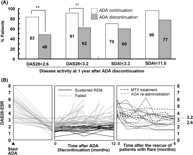
The DAS28-ESR remission rate (83%) in the ADA continuation group was significantly higher (48%) than that in the ADA discontinuation group 1 year after the continuation or discontinuation decision was made (p=0.0056; figure 1A). However, when SDAI was used for evaluation, there was no marked difference in the remission rates (≤3.3) between the ADA continuation and discontinuation groups, as shown by their values of 70% and 60%, respectively (p=0.4502). Similar outcomes were observed in the rates of low disease activity (LDA), that is, there was a significant difference in the evaluation using DAS28 (91% in ADA continuation, 62% in ADA discontinuation, p=0.0122), but there was no significant difference in the evaluation using SDAI (≤11.0) between the groups (96% in ADA continuation, 77% in ADA discontinuation, p=0.5690). Although all the proportions were higher in the ADA continuation group, at least 60% of the ADA discontinuation group showed LDA on DAS28-ESR and SDAI evaluations.
Figure 1.
Clinical outcomes evaluated by DAS28- erythrocyte sedimentation rate (ESR) or simplified disease activity index (SDAI) after ADA discontinuation and effects of ADA readministration to patients with flare. (A) shows the proportion of patients with sustained remission and low disease activity (LDA) evaluated using DAS28-ESR (DAS28) or SDAI. Each rate at 1 year after ADA discontinuation was compared with that in the ADA continuation group (Fisher's exact test). (B) shows the time course of changes in DAS28 including rescues of patients with flare (Left: ADA initiation to discontinuation, Middle: ADA discontinuation to 1 year later, Right: Flare to 6 months following rescue with methotrexate (MTX) or ADA). **p<0.01: ADA discontinuation versus ADA continuation using DAS28. In the comparison using SDAI, no significant difference was observed (p=0.4502 for remission, p=0.5690 for under LDA).
Effects of ADA readministration
During the ADA-free period, approximately 40% patients experienced flare (DAS28-ESR ≥3.2; figure 1B). Although MTX dose was escalated to rescue the failure, it was not effective in most patients (75%); furthermore, reinitiating ADA with or without MTX dose escalation resulted in the reinduction of LDA by 90% within 6 months and by 100% within 9 months. ADA restart due to a relapse was not associated with any harmful effects.
Possibility of becoming ADA-free
Characteristics of patients with sustained remission
When patient backgrounds were compared between those who experienced sustained (n=25) and unsustained (n=27) DAS28-ESR remission for 1 year, a statistically significant difference was observed in four items: (1) RA disease periods (p=0.0488), (2) ADA treatment periods (p=0.0264), (3) ESR value (p=0.0019) and (4) DAS28-ESR score (p=0.001; table 2).
Factors affecting sustaining remission
In the analysis of predictive factors related to sustaining remission for 1 year, only DAS28-ESR had a marked correlation with sustained remission in univariate and multivariate analyses (table 3). Subsequent ROC analysis for high estimation of sustained remission indicated a lower cut-off value for the biologic-free remission of 1.98 than the threshold for DAS28-ESR remission of 2.6. This value was similar to 2.16, which was calculated using the data to estimate sustaining remission for 6 months with the sensitivity 90%, specificity 68.2% and AUC 0.86, indicating that deep remission before discontinuing ADA would be a key in established patients with RA.
ADA continuation versus discontinuation in patients with deep remission
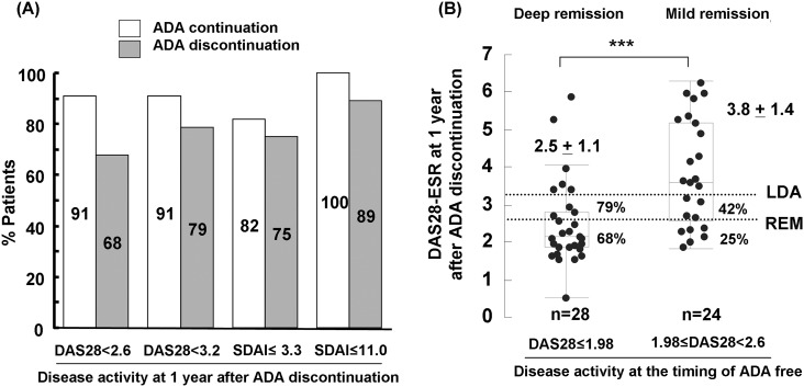
Disease activity in patients with deep remission (DAS28-ESR ≤1.98) was investigated 1 year after ADA discontinuation (figure 2A). In the ADA discontinuation group, 79% and 89% patients had values of DAS28-ERS <3.2 and SDAI ≤11.0, respectively, and their remission rates were approximately 70% in both cases (DAS28-ESR <2.6: 68%, SDAI ≤3.3: 75%). Comparison of the data in patients with deep remission with those of the ADA continuation group revealed no significant difference (p=0.2282–0.7067).
Figure 2.
Clinical outcomes in patients with deep remission and influence of the degree of remission. The percentages of patients in remission or with low disease activity (LDA) at 1 year after fulfilling the ADA-free criteria were investigated in patients with deep remission and compared between the ADA discontinuation and continuation groups, using a cut-off value of DAS28-4 erythrocyte sedimentation rate (ESR) ≤1.98 identified using receiver operating characteristics (ROC) analysis. No significant differences were observed between the groups (p=0.228 for DAS28-ESR<2.6, p=0.649 for DAS28-4ESR<3.2, p=0.707 for simplified disease activity index (SDAI) ≤3.3, p=0.545 for SDAI≤11; Fisher's exact test). (B) shows disease activity at 1 year after ADA discontinuation according to the difference in the degree of remission (deep or mild) when ADA was discontinued. ***p<0.001: deep (DAS28 ≤1.98) versus mild (1.98<DAS28<2.6) remission (Wilcoxon rank sum test).
Comparison between mild and deep remission
The prognosis 1 year after discontinuing ADA was compared between patients with mild (1.98 < DAS28-ESR<2.6) and deep (DAS28-ESR ≤1.98) remission (figure 2B). As shown in figure 2A, approximately 80% patients with deep remission were able to sustain LDA, whereas, only 42% patients with mild remission were able to do so, suggesting that mild remission may be insufficient for ADA discontinuation in established RA.
Influence of ADA discontinuation on structural and functional remission
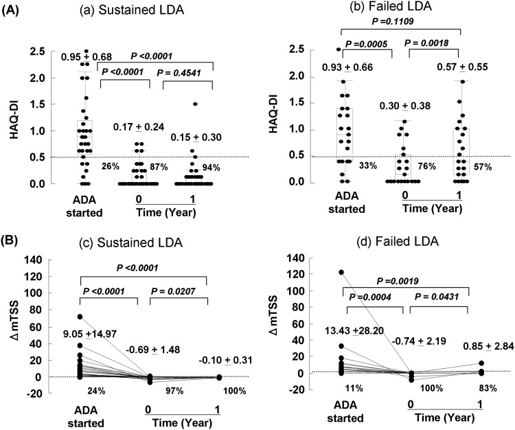
In patients with LDA (DAS28-ESR <3.2) 1 year after ADA discontinuation (n=31), the mean HAQ-DI (0.15) and functional remission (HAQ-DI ≤0.5) rate (94%) were similar to those 1 year earlier (figure 3). The structural remission rate was 100% (mTSS <0.5), demonstrating that maintaining LDA makes it possible to sustain functional and structural remission for at least 1 year after becoming ADA-free. In patients with flare (DAS28-ESR ≥3.2) during the year after ADA discontinuation (n=21), the mean HAQ-DI and mTSS significantly increased from 0.30 to 0.57 (p=0.0018) and −0.74 to 0.85 (p=0.0431), respectively, and the functional and structural remission rates decreased from 76% to 57% and from 100% to 83%, respectively. In the ADA continuation group, 21 patients sustained LDA during the year, but there were only two patients with flare; thus statistical comparison was not performed for the patients with flare. In the patients with sustained LDA, there were no statistically significant differences in HAQ (p=0.1579) and ΔmTSS (p=0.6422) between the ADA continuation and discontinuation groups (see online supplementary figure S1).
Figure 3.
Functional and structural remission in patients with sustained or failed low disease activity (LDA). (A) and (B) show values of health assessment questionnaire-disability index (HAQ-DI) and ΔmTSS in patients with sustained LDA (n=31) at 1 year after ADA discontinuation or failed LDA (n=21) when evaluated by DAS28-4 erythrocyte sedimentation rate (ESR). The percentages show the proportion of patients with sustained functional remission (HAQ<0.5) and structural remission (ΔmTSS<0.5). p Values by Kruskal–Wallis test; mTSS, modified total sharp score.
Discussion
The design of the HONOR study has several characteristics that make it unique and important in the quest for the possibility of biologic-free therapy in established RA by addressing four questions as described in the introduction. The study will follow patients throughout the extended treatment period, and here we evaluated the 1-year data of ADA with the concept of a biologic ‘treatment holiday.’ Of 197 patients who received ADA + MTX/DMARDs, 75 patients (38%) met the ADA-free criteria (maintenance of remission status for 6 months at least) and the majority of the patients who once attained DAS28-ESR remission could maintain stable remission with ADA+MTX/DMARDs under steroid-free conditions. This finding was also supported by the results of a retrospective HARMONY study in Japanese patients treated with ADA.22 Of the 52 patients who agreed to ADA discontinuation, 25 (48%) sustained DAS28-ESR remission for 1 year. Evaluation using SDAI revealed a remission (≤3.3) rate of 60%, which was similar to the percentage of patients with LDA (62%) evaluated using DAS28, despite our understandings that SDAI has more stringent criteria. As shown in table 2, a marked difference in ESR was observed between patients with and without sustained DAS28 remission. It is well known that ESR level is influenced by many factors, such as infection, or other autoimmune diseases. We also calculated the remission rate using the Boolean approach as a reference (Boolean definition: number of swollen and tender joints each ≤1, C-reactive protein (CRP) ≤1 mg/dL and PGA ≤1); the remission rate was 50%, which was 10% lower than the SDAI remission rate (see online supplementary figure S2). The reason for this was that PGA was >1, probably due to damaged HAQ in established RA because tender and swollen joint counts were 0 in the patients (n=5) who did not meet Boolean remission within those who met SDAI remission. Therefore, the remission rate using SDAI (60%) seems to be more accurate than that obtained using DAS28 (48%), considering that all patients who sustained DAS28 <3.2 (62%) showed 100% structural remission 1 year after ADA discontinuation.
Although the evaluation using DAS28-ESR revealed statistically significant better outcomes in the ADA continuation group than the ADA discontinuation group, evaluation using SDAI or assessing prevention of radiographic damage in patients with LDA by DAS28 revealed no difference between the groups. Consequently, it would not be an overstatement to say that patient outcomes 1 year after ADA discontinuation were the same as those in some patients of the ADA continuation group. In fact, there was no statistically significant difference between the two groups regarding patients with deep remission (DAS28-4ESR ≤1.98), which we identified as a factor necessary for successful ADA discontinuation. Meanwhile, 60% patients with mild remission (1.98≤DAS28-4ESR < 2.6) experienced flaring within a year, suggesting that ADA should be continued in such patients even under DAS28 remission. However, there is a risk that some patients discontinue ADA because of no pain or economic burden, both of which are experienced in daily clinical practice. The good news was that ADA readministration to all patients with flare during ADA discontinuation was effective without harmful effects.
This study had some limitations. This was an open-label, non-randomised study with a limited number of subjects who were divided into two groups partly based on patients’ consent, which could have introduced a selection bias. Nonetheless, the study design allowed a comparison between ADA discontinuation approaches (unknown outcomes after ADA discontinuation, expectation of biologic-free disease control in established RA, economical matters, etc) and ADA continuation approaches in routine clinical settings in an ethical manner, with shared decisions between patients and rheumatologists. Confidence in the outcomes would most likely be supported by the results of the RRR and OPTIMA studies that examined biologic-free potential. In the RRR study14 with long-standing patients with RA, DAS28 ≤2.22 was identified by logistic regression and ROC analysis as a necessary condition for a biologic-free remission, and demonstrated that 71.4% patients with deep remission (DAS28 ≤2.22) were able to continue DAS28 <3.2 for 1 year, whereas only 32.6% patients with 2.22 < DAS28 < 3.2 were able to continue. These results suggest that patients in deep remission (DAS28 of approximately 2.0) have a possibility to achieve biologic-free remission. In the OPTIMA study with early RA,13 a multinational, double-blind randomised controlled study, with results similar to those of our study were obtained for comparisons between ADA continuation and discontinuation groups. The remission (86%) and LDA (91%) rates in the ADA continuation group in the OPTIMA study were significantly higher than the remission (66%) and LDA (81%) rates in the ADA discontinuation group when compared using the DAS28-CRP criteria, but there was no statistical difference between ADA continuation and discontinuation (remission: 62% vs 51%, LDA: 92% vs 84%, respectively) groups when SDAI criteria were used, and functional and structural outcomes were comparable between the groups. Thus, despite some limitations, the results of the present study are supported by those of the RRR and OPTIMA studies. Additionally, the results of this study demonstrate the potential of remaining ADA-free in established patients with RA, and provide valuable insights into the paradigms of RA treatment in routine clinical settings, considering safety and economical aspects.
Taken together, these results demonstrate that among the patients who met the ADA-free criteria, 48% were able to sustain DAS28-ESR remission after discontinuing ADA, and 60% were in SDAI remission or showed LDA in DAS28-ESR 1 year after becoming ADA-free while maintaining functional and joint structural remission; furthermore, regarding patients with deep remission, disease activity in the ADA discontinuation group was comparable to that in the ADA continuation group, whereas for patients with flare, readministration of ADA was effective. These data indicate that ADA ‘treatment holiday’ is now feasible in established patients with RA with long-term remission, no steroids and deep remission.
Supplementary Material
Acknowledgments
The author thanks all medical staff at all institutions for providing the data.
Footnotes
Contributors: YT contributed to study design, overall review, making the manuscript, and the others were involved in performance of the study and review of the manuscript. SH, KS, YT participated in its design and coordination. All authors, except FS, enrolled and managed the patients in clinic. SH, SK, SF participated in radiographic evaluation. SH performed the statistical analysis, and FS helped to draft the manuscript and contributed to reviewers’ responses. All authors read and approved the final manuscript.
Funding: The series of studies were supported in part by a Research Grant-In-Aid for Scientific Research by the Ministry of Health, Labour and Welfare of Japan, the Ministry of Education, Culture, Sports, Science and Technology of Japan, and the University of Occupational and Environmental Health, Japan. Although F Sawamura is an AbbVie employee, AbbVie had no role in funding this study or in the data collection or analysis. Other than F Sawamura's contributions to meet the International Committee of Medical Journal Editors (ICMJE) authorship criteria, no other AbbVie employee had input to the content of the publication.
Competing interests: YTanaka, has received consulting fees, speaking fees and/or honoraria from Mitsubishi-Tanabe Pharma, Eisai, Chugai Pharma, Abbott Japan, Astellas Pharma, Daiichi-Sankyo, Abbvie, Janssen Pharma, Pfizer, Takeda Pharma, Astra-Zeneca, Eli Lilly Japan, GlaxoSmithKline, Quintiles, MSD and Asahi-Kasei Pharma and has received research grants from Bristol-Myers, Mitsubishi-Tanabe Pharma, Abbvie, MSD, Chugai Pharma, Astellas Pharma and Daiichi-Sankyo. The other authors declare no conflict of interest.
Ethics approval: Ethics review board of the University of Occupational and Environmental Health, Japan
Provenance and peer review: Not commissioned; externally peer reviewed.
References
- 1.Lee DM, Weinblatt ME. Rheumatoid arthritis. Lancet 2001;358:903–11. [DOI] [PubMed] [Google Scholar]
- 2.Pope RM. Apoptosis as a therapeutic tool in rheumatoid arthritis. Nat Rev Immunol 2002;2:527–35. [DOI] [PubMed] [Google Scholar]
- 3.Choy EH, Panayi GS. Cytokine pathways and joint inflammation in rheumatoid arthritis. N Engl J Med 2001;344:907–16. [DOI] [PubMed] [Google Scholar]
- 4.Aaltonen KJ, Virkki LM, Malmivaara A, et al. Systematic review and meta-analysis of the efficacy and safety of existing TNF blocking agents in treatment of rheumatoid arthritis. PLoS One 2012;7:e30275. [DOI] [PMC free article] [PubMed] [Google Scholar]
- 5.Clark DA. Do anti-TNF-α drugs increase cancer risk in rheumatoid arthritis patients? Inflammopharmacology 2013;21:125–7. [DOI] [PubMed] [Google Scholar]
- 6.Wong AK, Kerkoutian S, Said J, et al. Risk of lymphoma in patients receiving antitumor necrosis factor therapy: a meta-analysis of published randomized controlled studies. Clin Rheumatol 2012;31:631–6. [DOI] [PubMed] [Google Scholar]
- 7.Kaymakcalan Z, Sakorafas P, Bose S, et al. Comparisons of affinities, avidities, and complement activation of adalimumab, infliximab, and etanercept in binding to soluble and membrane tumor necrosis factor. Clin Immunol 2009;131:308–16. [DOI] [PubMed] [Google Scholar]
- 8.Arora T, Padaki R, Liu L, et al. Differences in binding and effector functions between classes of TNF antagonists. Cytokine 2009;45:124–31. [DOI] [PubMed] [Google Scholar]
- 9.Mitoma H, Horiuchi T, Tsukamoto H, et al. Mechanisms for cytotoxic effects of anti-TNF agents on transmembrane TNF-expressing cells: comparison among infliximab, etanercept and adalimumab. Arthritis Rheum 2008;58:1248–57. [DOI] [PubMed] [Google Scholar]
- 10.Quinn MA, Conaghan PG, O'Connor PJ, et al. Very early treatment with inf iximab in addition to methotrexate in early, poor-prognosis rheumatoid arthritis reduces magnetic resonance imaging evidence of synovitis and damage, with sustained benefit after infliximab withdrawal: results from a twelve-month randomized, double-blind, placebo-controlled trial. Arthritis Rheum 2005;52:27–35. [DOI] [PubMed] [Google Scholar]
- 11.Goekoop-Ruiterman YPM, de Vries-Bouwstra JK, Allaart CF, et al. Clinical and radiographic outcomes of four different strategies in patients with early rheumatoid arthritis (the BeSt study): arandomizedcontrolledtrial. Arthritis Rheum 2005;52:3381–90. [DOI] [PubMed] [Google Scholar]
- 12.Detert J, Bastian H, Listing J, et al. Induction therapy with adalimumab plus methotrexate for 24 weeks followed by methotrexate monotherapy up to week 48 versus methotrexate therapy alone for DMARD-naive patients with early rheumatoid arthritis: HIT HARD, an investigator-initiated study. Ann Rheum Dis 2013;72:844–50. [DOI] [PubMed] [Google Scholar]
- 13.Smolen JS, Emery P, Fleischmann R, et al. Adjustment of therapy in rheumatoid arthritis on the basis of achievement of stable low disease activity with adalimumab plus methotrexate or methotrexate alone: the randomized controlled OPTIMA trial. Lancet 2013. Epub ahead of print. doi:pii: S0140-6736(13)61751-1. 10.1016/S0140-6736(13)61751-1. [DOI] [PubMed] [Google Scholar]
- 14.Tanaka Y, Takeuchi T, Mimori T, et al. RRR study investigators. Discontinuation of infliximab after attaining low disease activity in patients with rheumatoid arthritis: RRR (remission induction by Remicade in RA) study. Ann Rheum Dis 2010;69:1286–91. [DOI] [PMC free article] [PubMed] [Google Scholar]
- 15.Arnett FC, Edworthy SM, Bloch DA, et al. The American Rheumatism Association 1987 revised criteria for the classification of rheumatoid arthritis. Arthritis Rheum 1988;31:315–24. [DOI] [PubMed] [Google Scholar]
- 16.Koike R, Takeuchi T, Eguchi K, Miyasaka N; Japan College of Rheumatology. Mod Rheumatol 2007;17:451–8. [DOI] [PubMed] [Google Scholar]
- 17.Japan College of Rheumatology, Official Guidelines for the Use of Anti-TNF Agents for Rheumatoid Arthritis (in Japanese). 2012. http://www.ryumachi-jp.com/info/guideline TNF 120704.html
- 18.Prevoo ML, van 't Hof MA, Kuper HH, et al. Modified disease activity scores that include twenty-eight-joint counts. Development and validation in a prospective Discontinuation of Adalimumab in rheumatoid arthritis (HONOR study): longitudinal study of patients with rheumatoid arthritis. Arthritis Rheum 1995;38:44–8. [DOI] [PubMed] [Google Scholar]
- 19.Tanaka Y. Intensive treatment and treatment holiday of TNF-inhibitors in rheumatoid arthritis. Curr Opin Rheumatol 2012;24:319–26. [DOI] [PubMed] [Google Scholar]
- 20.Fries JF, Spitz P, Kraines RG, et al. Measurement of patient outcome in arthritis. Arthritis Rheum 1980;23:137–45. [DOI] [PubMed] [Google Scholar]
- 21.van der Heijde D. How to read radiographs according to the Sharp/van der Heijde method. J Rheumatol 2000;27:261–3. [PubMed] [Google Scholar]
- 22.Takeuchi T, Tanaka Y, Kaneko Y, et al. Effectiveness and safety of adalimumab in Japanese patients with rheumatoid arthritis: retrospective analyses of data collected during the first year of adalimumab treatment in routine clinical practice (HARMONY study). Mod Rheumatol 2012;22:327–38. [DOI] [PMC free article] [PubMed] [Google Scholar]
Associated Data
This section collects any data citations, data availability statements, or supplementary materials included in this article.


