Abstract
Mutations in the epidermal growth factor receptor (EGFR) are the most common actionable genetic abnormalities yet discovered in lung cancer. However, targeting these mutations with kinase inhibitors is not curative in advanced disease and has yet to demonstrate an impact on potentially curable, early-stage disease, with some data suggesting adverse outcomes. Here, we report that treatment of EGFR-mutated lung cancer cell lines with erlotinib, while showing robust cell death, enriches the ALDH+ stem-like cells through EGFR-dependent activation of Notch3. Additionally, we demonstrate that erlotinib treatment increases the clonogenicity of lung cancer cells in a sphere-forming assay, suggesting increased stem-like cell potential. We demonstrate that inhibition of EGFR kinase activity leads to activation of Notch transcriptional targets in a gamma secretase inhibitor sensitive manner and causes Notch activation. leading to an increase in ALDH high+ cells. We also find a kinase-dependent physical association between the Notch3 and EGFR receptors and tyrosine phosphorylation of Notch3. This could explain the worsened survival observed in some studies of erlotinib treatment at early-stage disease, and suggests that specific dual targeting might overcome this adverse effect.
Keywords: EGFR, Notch3, stem-like cells, erlotinib and tyrosine phosphorylation
Introduction
Modern approaches to cancer therapeutic development are becoming increasingly selective, sometimes targeting single inter-molecular interactions or mutated oncoproteins, allowing for dramatic efficacy in defined subsets of patients with specific genetic abnormalities. This is the case for epidermal growth factor receptor (EGFR) tyrosine kinase inhibitors (TKIs), gefitinib and erlotinib, in the setting of exon 19 deleted or L858R point mutated lung cancer tumors(1, 2). However, responses to TKIs are often short-lived and have failed to result in cures, alone or in combinations, in the metastatic setting. TKIs have also failed to improve survival after curative-intent therapy of early stage disease. For example, one clinical trial, SWOG 0023, randomized stage III patients to gefitinib therapy or placebo after curative-intent chemotherapy and radiation (3). Surprisingly, the study had to be stopped because the overall survival was significantly worse in the arm treated with the kinase inhibitor, unrelated to drug toxicity. In earlier stage tumors, stages I and II, where complete surgical resection results in cures in more than half of patients, addition of gefitinib after surgery might be expected to dramatically improve survival, particularly in patients with EGFR mutated tumors. However, gefitinib failed to increase either PFS or OS, with a strong trend toward worsened overall survival with a hazard ratio of 1.2 in the entire group (4). The group with the largest expected benefit, those with activating EGFR mutations, also trended to doing worse than the unselected group with a hazard ratio of 3.1 for an increased risk of death(4). We hypothesized that the worsened survival in the curative-intent setting is a consequence of the fact that while debulking of tumors underlies the clinical benefit observed for incurable metastatic disease, EGFR inhibition could paradoxically increase clonogens in the tumor decreasing long-term survival in curative-intent therapy, which in turn becomes apparent after the cessation of inhibitor therapy. We set out to test these hypotheses in this study.
Notch receptors are highly conserved, single pass type I transmembrane proteins known to play a role in cell proliferation, cell death and differentiation (5). They have also been linked to multiple human disorders including cancers where different Notch family members are implicated as oncogenes or tumor suppressor genes in different settings (6–8). In lung cancers, Notch was originally implicated in an epithelial tumor by the discovery of a chromosome translocation causing massive overexpression of Notch3 (9) and subsequently Notch receptors and ligands have been found to be overexpressed in a majority of solid tumors, including non-small cell lung cancer (NSCLC) (9, 10). Transgenic mice overexpressing an activated Notch3 in the bronchial epithelium showed perinatal lethality due to arrested differentiation of type II progenitors, and the absence of type I pneumocytes (11).
More recently, multiple studies have shown the Notch pathway to be important in stem cell biology(12–15). Notch has also been shown to play a role in cancer stem cells in many tumor types, including breast, brain, and lung cancer (16–19). In breast ductal carcinoma in situ (DCIS), formation of mammospheres, an indicator of stem-like cells, is decreased upon treatment with Notch inhibitors (18, 20, 21).
In lung cancer, ALDH positivity has been convincingly associated with stem-like cell characteristics and inhibition of Notch3 abrogated the colony and tumor-forming ability of ALDH-positive cells (15, 19). High expression of ALDH1 by immunohistochemistry was also associated with a worsened survival after curative-intent surgical resection(19). This was recently corroborated by studies of ALDH7 in resected lung tumors(22).
In the present study, we sought to determine if Notch signaling could be enhanced by EGFR inhibition resulting in differential effects on the stem-like cell population of human lung cancer cells with mutated or wild type EGFR. We found that while inhibition of EGFR leads to a dramatic reduction of tumor cell numbers, it also leads to a potent activation of the Notch pathway with an increase in the relative abundance of ALDH+ stem-like cells in a Notch3-dependent fashion and an increase in the clonogenicity as determined by spheroid assay. Combined inhibition of EGFR and Notch3 receptors substantially reduces the expansion of stem-like cells. This is the first report showing erlotinib treatment activates Notch in human lung cancer, resulting in an enriched stem-like populations in a Notch3-, and not Notch1-dependent manner. Additionally, this is also the first study to demonstrate kinase-dependent complex formation of these two receptors leading to the tyrosine phosphorylation of Notch3 in human lung cancer cells.
Materials and Methods
Cells and transfections
HCC2429, HCC827, and HCC4006 cells were maintained in RPMI with 10% fetal bovine serum. H358, HCC827 and HCC4006 cells were obtained from ATCC within 6 months of the experiments reported, and were identity-verified by STR analysis and certified as mycoplasma-free. Transfections were performed with Lipofectamine 2000 (Invitrogen) reagent according to the manufacturer’s instructions.
Ligands and inhibitors
EGF was purchased from R & D Systems. Erlotinib was a generous gift from Dr. William Pao at Vanderbilt University. Gamma secretase inhibitor (PF-03084014) was kindly provided by Pfizer Global Research and Development, La Jolla Laboratories (San Diego, CA) and was described previously(23, 24).
Antibodies
Following antibodies were used in this study: EGFR (1005) Santa Cruz Biotechnology, EGFR (Ab12) and EGFR (Ab15) are from Neomarker, Notch1 (5B5), Notch3 (8G5), and Notch3 (D11B8) and EGFR (pY1173) obtained from Cell Signaling Technology. Mouse-anti phosphotyrosine is from BD Transduction Laboratories. β-tubulin antibodies were obtained from Sigma.
Plasmid constructs
The pCDNA-EGFR and pCDNA-EGFR (D816A) and Renilla luciferase constructs were provided by Graham Carpenter (Vanderbilt University). Dr. Thao P. Dang provided pCMV-FLAG-N3DA, and pHES1-luciferase constructs. The TP1-luc reporter construct contains 12 tandem repeats of CSL binding sites upstream of luciferase.
Co-immunoprecipitation, immunoprecipitation and western blotting
Cells were washed twice in ice-cold phosphate buffered saline, harvested and lysed with NP40 buffer (10 mM phosphate buffer, 120 mM NaCl, 2.7 mM KCl, 1% Nonidet P40, 10% glycerol) for co-immunoprecipitation experiments or lysed with RIPA buffer (10 mM phosphate buffer, 120 mM NaCl, 2.7 mM KCl, 1% Nonidet P-40, 0.5% DOC, 0.1% SDS) supplemented with complete mini-EDTA free protease inhibitor mixture (Roche) and phosphatase inhibitor mixture cocktails 2 and 3 (sigma), 2 mM NaF and pervanadate for immunoprecipitation for detection of phosphorylation. Equal amount of lysates were precipitated using appropriate antibodies and protein G magnetic beads, or equal amounts of protein were mixed with SDS sample buffer and separated on SDS-PAGE prior to Western analysis.
Aldefluor assay and Flow cytometry
The aldefluor assay kit (Stem cell Technologies) was used to determine the ALDH+ cells. The assay was performed according to manufacturer’s instructions with modifications. Cells were suspended in aldefluor assay buffer and divided into two groups. One group was pretreated for 10 min with ALDH-specific inhibitor Diethylaminobenzaldehyde (DEAB) before incubation with ALDH enzyme substrate Bodipy-Aminoacetaldehyde (BAA) for 45 minutes at 37° C. Cells were centrifuged and re-suspended in a fresh aldefluor assay buffer to remove the unutilized substrate. Cells were analyzed on a FACSCalibur (BD Biosciences) Flow Cytometer. For the analysis of ALDH+ cells, DEAB treated sample was used as a negative control and ALDH activity in presence of DEAB was considered as a baseline.
Pulmosphere formation assay
To study the stem-like cell phenotype, sphere formation assays were performed as described previously (25) with modifications. HCC827 cells treated with vehicle control or erlotinib were trypsinized and counted using Luna automated cell counter. Cells were seeded in 96-well plates at 1000 cells per well in RPMI supplemented with 10% fetal bovine serum, 35 μg/ml bovine pituitary extract (Life Technologies), N2 supplement (Invitrogen), 20 ng/ml EGF, 20 ng/m (Life Technologies), basic fibroblast growth factor (Roche) and 50% Geltrex LDEV-free, hESC-qualified, reduced growth factor basement membrane matrix (Geltrex) (Life Technologies). Cells containing the semi solid medium were seeded in triplicate. The culture was allowed to solidify at 37°C for 30 min followed by layering of 200 μl of similar growth medium without 50% geltrex and incubated for one to 3 weeks. Pulmosphere number was determined using the GelCount mammalian cell colony counter (Oxford Optronix).
Soft agar assay
To measure in vitro tumorigenicity due to erlotinib treatment treated and untreated H358 cells at a density of 10,000 cells per well in 6-well plate were plated in soft agar, in triplicate. The assay was performed using 0.5% and 0.35% agar in RPMI1640 supplemented with 10% FBS as the base and top layers, respectively. Cells were incubated for 21 days and medium was refreshed twice per week. Colonies were counted using GelCount (Oxford Optronix). The colony efficiency was calculated as proportion of colonies per total number of seeded cells. The data was analyzed using GelCount software.
Results
Pharmacological inhibition of EGFR increases the fraction of ALDH+ cells in lung cancer cell lines
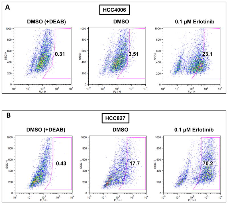
We tested whether there is a relationship between EGFR inhibition and the fraction and number of stem-like cells in NSCLC cell lines. EGFR mutated lung adenocarcinoma cells were treated with DMSO or 0.1μM erlotinib and the medium was changed every day with fresh drug. Equal numbers of cells were subjected to ALDH enzymatic activity assays with the ALDH+ and ALDH− cell populations quantified using flow cytometry. We found that HCC4006 and HCC827 NSCLC cell lines, each with an EGFR activating mutation (EGFR ΔE746–A750) (Fig. 1a and b), showed dramatic increases in the fraction of ALDH+ cells upon treatment with erlotinib compared to DMSO treated cells (Table 1). Interestingly, for HCC4006 cells, which have very low basal ALDH activity, while treatment with erlotinib killed much of the ALDH− population, it appeared to maintain or even increase the total number of ALDH+ cells by a small percentage. However, unlike HCC4006 cells, HCC827 cells appear to have a large fraction of cells with low to moderate baseline ALDH activity (3.5% for HCC4006 vs 17.7% for HCC827). This basal activity may not accurately reflect the stem-like cell population. When the analysis is based on those cells with the highest ALDH activity, by gating on the DMSO treated cells without DEAB, the total number of ALDH+ cells with very high activity increased in the HCC827 cells from 491,200 to 782,100 (Supplementary Fig. 1A and 1B; Supplementary Table 1). This demonstrates that while erlotinib treatment causes a large reduction in the total cell numbers for each cell lines, the total number of ALDH high+ cellsis increased. Supplementary Fig. 1a and b shows that both low positive and high positive fractions increase between 3 and 5 days of erlotinib treatment in HCC827 cells.
Figure 1.
Erlotinib treatment of EGFR mutant lung cancer cells increases the fraction of ALDH+ cells. A, HCC4006 and B, HCC827 cells were treated with DMSO, 0.1 μM of erlotinib for five days and subjected to aldefluor assay to detect the ALDH+ cells. A portion of the cells was pre-incubated with the ALDH inhibitor DEAB (+DEAB) to provide a gate (ALDH negative cells) for flow cytometry.
Table 1.
Treatment with erlotinib enriches ALDH+ cells in EGFR mutant cancer cells. Cells were treated with DMSO or erlotinib for indicated periods for 7 days and subjected to aldefluor assay to determine the ALDH positive cells. Also, after the drug treatment total number of cells was determined and percentage of cell death was counted. Using total number of cells and ALDH+ cells data percentage of ALDH was determined.
| Cell line | Treatment | % Cell death | Total number of live cells | ALDH+ cells | % ALDH+ cells |
|---|---|---|---|---|---|
| HCC4006 | DMSO | 0 | 28,118,750 | 984156 | 3.5 |
| (0.1 μM) Erlotinib | 84.5 | 4,343,750 | 1,003,406 | 23.1 | |
|
| |||||
| HCC827 | DMSO | 0 | 23,466,650 | 4,153,597 | 17.7 |
| (0.1 μM) Erlotinib | 78.5 | 5,050,000 | 3,545,100 | 70.2 | |
We further wanted to explore if this phenomenon was also present in H1650 cells, which are also EGFR mutant but resistant to erlotinib due to loss of PTEN(27). Interestingly, erlotinib treatment also had no effect on the ALDH populations in these cells (Supplementary Fig. 2). Additional studies with A549 and H358 cells carrying wild type EGFR and a K-RAS mutation (28) (Supplementary Fig. 3a and b) also showed increased ALDH activity upon exposure to erlotinib. Unlike H1650, which are completely insensitive to erlotinib, the H358 cell line and A549 cell line are somewhat sensitive to erlotinib (H358 more so than A549) (29) and also show increases in the fraction of ALDH+ cells with erlotinib treatment. These data suggest that this phenomenon is not just restricted to cells with an EGFR activating mutation, but limited to those with some EGFR-signaling dependence, and that EGFR activity is coupled with ALDH activity.
Erlotinib treatment enhances pulmosphere-forming potential in EGFR mutated lung cancer cells
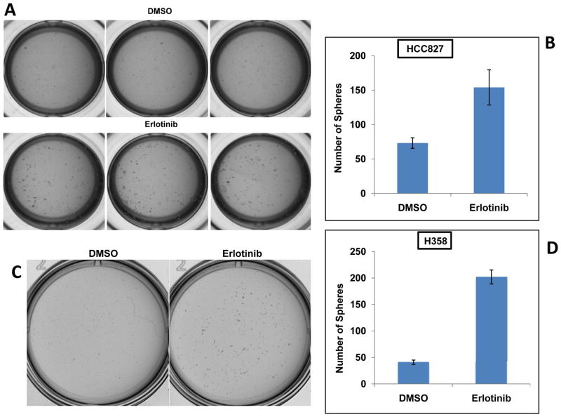
Previous studies have demonstrated that ALDH+ cells possess features similar to cancer stem cells such as increased pulmosphere-forming ability. To determine if the residual cell population after erlotinib treatment is more stem-like we performed the pulmosphere formation assay. HCC827 cells were treated with 0.1 μM erlotinib for five days. Remaining cells were allowed to recover under the regular growth conditions for four days and subjected to sphere formation culture assay. As expected from the ALDH+ data, compared to DMSO, erlotinib treated cells showed an increased number and size of the pulmospheres in semisolid matrix (Fig. 2a and b). We also performed a soft agar colony formation assay, which measures anchorage-independent growth and is an indicator for cell transformation using H358 cells. Cells treated with erlotinib showed significant increase in number of colonies formed compared to DMSO control (Fig. 2c and d). These data clearly suggest that erlotinib treatment increases the clonogenic potential in the surviving population of cells.
Figure 2.
Erlotinib treatment increases the sphere-forming ability in EGFR mutant lung cancer cells. HCC827 cells were treated with DMSO or 0.1 μM of erlotinib for five days and allowed to recover for 4 days prior to subjected to sphere-forming assay in a 96 well plate. A, Spheres were imaged and B, their total number was quantitated (p < 0.008). C, H358 cells were treated with DMSO or erlotinib (1uM) and equal number of cells were subjected to soft agar colony formation assay and D, colony number was quantitated.
EGFR signaling down-regulates Notch-mediated transcriptional activity in a kinase-dependent manner
To assess if the erlotinib-mediated stem-like cell phenotype is due to modulation of transcriptional activity of Notch, we examined the Notch transcriptional activity in the presence of wild type- or kinase inactive-EGFR. HEK293 cells were transiently transfected with EGFR and Notch3-ICD along with Notch inducible CSL-synthetic-(Fig. 3a) or full-length Hes1 promoter-driven luciferase reporters (Fig. 3b). Two days after transfection transcriptional activity was measured using a luciferase assay. The coexpression of EGFR with Notch3-ICD decreased Notch3-ICD-mediated CSL and Hes1 reporter activities in a dose-dependent manner. We further determined if kinase activity is essential for EGFR-mediated negative regulation of Notch3. Notch3-ICD was co-expressed with a kinase-inactive mutant of EGFR and co-expression of this kinase-inactive EGFR did not result in a decrease in Notch-mediated transcriptional activity, demonstrating that EGFR negatively regulates Notch activity through its tyrosine kinase activity. Interestingly, there was a slight increase in reporter activity compared to baseline when EGFR (KD) was transfected Fig. 3A and 3B), which may have been due to interference with endogenous EGFR activity. To make certain that the EGFR transfection was not having any other effect on the cell populations or the reporter in the absence of Notch, EGFR and EGFR (KD) were transfected with reporter alone (Supplementary Figure 4). We did not observe any EGFR mediated Notch reporter activity in the absence of Notch overexpression. Additionally, we tested if pharmacological inhibition of EGFR using erlotinib can stimulate the expression of Notch target genes in lung cancer cells. The qRT-PCR analysis of Hes1 expression showed a significant increase after 24 or 48 hours of treatment with erlotinib in HCC827 cells (Fig. 3c). This demonstrates EGFR kinase activity inhibits Notch signaling, and thus erlotinib treatment relieves this inhibition resulting in Notch transcriptional activation.
Figure 3.
EGFR mediated negative regulation of Notch3 transcriptional activity in a tyrosine kinase-dependent manner. A–B, HEK293FT cells were transiently transfected with either 12X-CSL reporter (A) or full length HES1 reporter (B) with empty vector or N3DA construct and increasing amounts of wild type or kinase inactive EGFR to determine the role of EGFR on transcriptional activity of Notch3-ICD. The data presented are the average of three assays. C, Erlotinib stimulates gene expression of Hes1. HCC827 cells were treated with erlotinib for the indicated time periods and RNA was analyzed by qRT-PCR for the expression of Hes1.
Gamma secretase inhibitor treatment eliminates erlotinib-induced stem-like cells by decreasing Notch activity
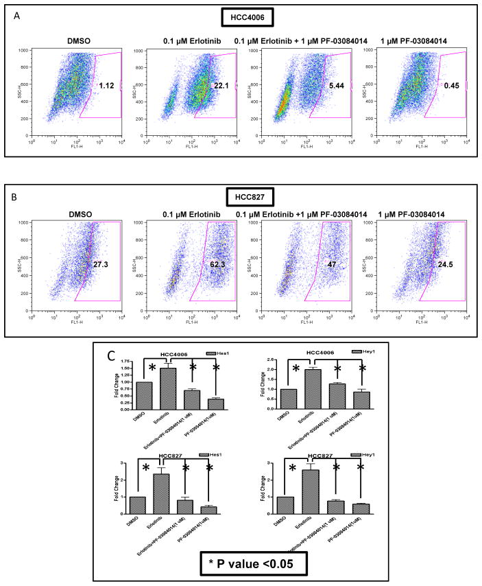
The major Notch pathway inhibitors in clinical testing are gamma secretase inhibitors (GSIs) (30). These compounds block the final activation (S3 cleavage) step in canonical Notch signaling. GSIs inhibit the cleavage of the Notch receptor preventing release of the intracellular domain into the cytoplasm and subsequent translocation to the nucleus, thus abrogating canonical signal activation for all the Notch receptors. Since we find that EGFR inhibition increases ALDH+ cells and also activates Notch transcriptional activity, we sought to determine if the erlotinib-induced increase of ALDH+ cells is inhibited by treatment with a GSI. Concomitant treatment with GSI PF-03084014 and erlotinib reduced the number of ALDH+ cells in both HCC4006 and HCC827 cells observed with erlotinib alone (Fig. 4a and b). Next we sought to identify if erlotinib-stimulated Notch transcriptional activity is sensitive to GSI. Consistent with the GSI sensitivity of erlotinib-induced stem-like cells, both HCC827 and HCC4006 showed increased expression of Hes1 and Hey1 with erlotinib treatment, which was sensitive to PF-0308401 following 3 days of treatment (Fig. 4c). These data support the hypothesis that erlotinib increases ALDH positivity through activation of Notch signaling and suggests a potential clinical strategy for overcoming this effect through combined EGFR and Notch inhibition.
Figure 4.
Inhibition of Notch activation by a gamma secretase inhibitor prevents erlotinib-induced ALDH+ cells. A, HCC4006 and B, HCC827 cells were treated with DMSO (i), 0.1 μM erlotinib (ii), a combination of 0.1 μM erlotinib and 1 μM PF-03084014 (iii), or 1 μM PF-03084014 alone (iv)for 7 days and subjected to aldefluor assay. C, Erlotinib stimulated Notch transcriptional gene targets Hes1 and Hey1 are sensitive to GSI. HCC4006 (top) and HCC827 (bottom) were treated with 0.1 μM of erlotinib, PF-03084014 alone or in combination and RNA was analyzed by qRT-PCR for expression of Notch target genes Hes1 and Hey1.
The erlotinib-induced increase in the ALDH+ fraction in lung cancer cells is dependent on Notch3, but not Notch1
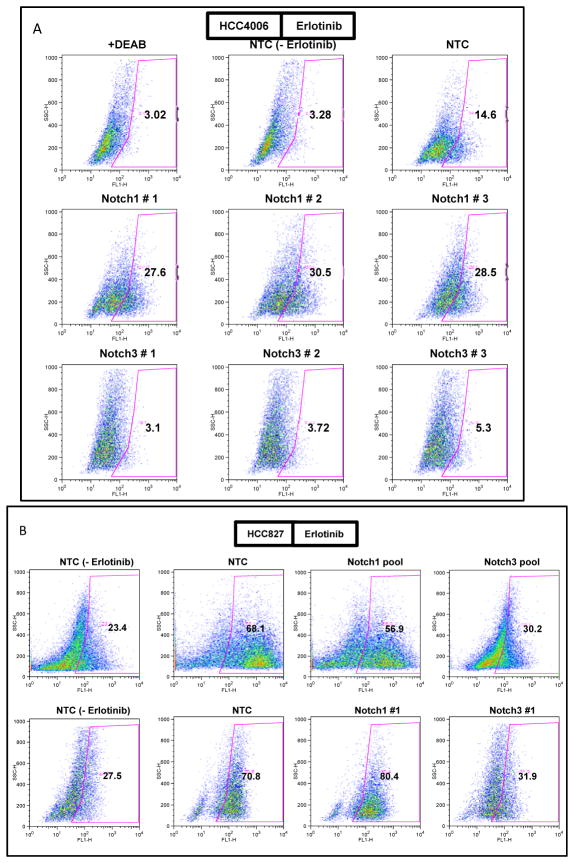
To determine whether erlotinib mediated expansion of ALDH+ cells is mediated by the Notch1 and/or Notch3 receptors, we evaluated the role of Notch1 and 3 in the erlotinib-induced increase of ALDH+ cells using the EGFR-mutated HCC4006 and HCC827 cell lines. Cells were transfected with non-targeting control (NTC) or three separate Notch1 or Notch3 siRNAs and treated with erlotinib. Knockdown for both Notch1 and Notch3 receptors was efficient(Supplementary Fig. 5a and b). As expected, erlotinib treatment stimulated a fivefold and threefold increase in ALDH+ cells in HCC4006 and HCC827, respectively. While knockdown of Notch1 had no effect on erlotinib-stimulated cells, the Notch3 knockdown completely abolished this effect to baseline levels, (Fig. 5a and b) suggesting that erlotinib induces stem-like cells through Notch3 and not Notch1.
Figure 5.
Depletion of Notch3 abolishes erlotinib-induced ALDH+ cells. A, HCC4006 cells were transfected with either non-targeting control (NTC-siRNA) (top) in the absence or presence of erlotinib as indicated, Notch1 siRNAs -1, -2 and -3 (middle), or Notch3 siRNAs -1, -2, and -3 (bottom). Forty-eight hours post-transfection, cells were treated with 0.1μM erlotinib for 3 days and ALDH activity was measured. As a control, NTC siRNA treated cells were also treated with DMSO (top, middle panel). A portion of the cells was pre-incubated with the ALDH inhibitor DEAB (+DEAB) to provide a gate for ALDH-negative cells (top left panel). B, HCC827 cells were transfected with non-targeting control pool (NTC-siRNA), pool of Notch3 or Notch1 siRNAs. Forty-eight hours following transfection, cells were treated with 0.1 μM erlotinib (B) for 3 days and ALDH activity was measured. As a control, NTC siRNA treated cells were also treated with DMSO (top, left panel). A similar experiment was also performed with an individual siRNA (bottom panel).
Notch and EGFR receptors co-precipitate and this interaction is dependent on EGFR kinase activity
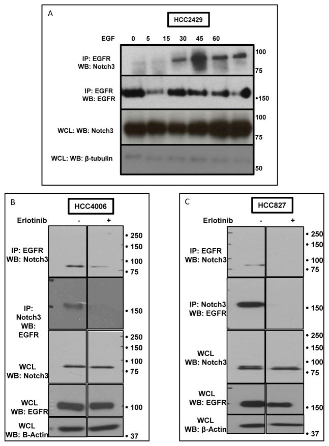
Next, we sought to determine if there is an EGFR activation-dependent functional association between EGFR and Notch receptors in HCC2429 cells, a cell line that expresses wild-type EGFR and both Notch1 and 3 receptors and multiple Notch ligands, and in which the combination of Notch and EGFR-targeted therapies show an increased efficacy(10, 31). Cells were stimulated with EGF for increasing amounts of time and total EGFR was precipitated, and blotted for Notch3. These analyses identified clear EGF-dependent co-precipitation of Notch3 with EGFR (Fig. 6a). In the absence of ligand, little or no association was observed between EGFR and Notch receptors, indicating that the association is ligand (EGF)-dependent. To further validate the interaction between EGFR and Notch3, co-immunoprecipitations were performed using three different antibodies that target distinct epitopes on the EGFR protein. Western blot analyses showed that Notch3 co-immunoprecipitates with all three EGFR antibodies, and this association was further enhanced when stimulated with EGF (Supplementary Fig. 6).
Figure 6.
EGF-mediated association between EGFR and Notch 3 receptors in HCC2429 cells is dependent on EGFR kinase activity. A. HCC2429 cells were stimulated with EGF (25 ng/ml) for the indicated times. Cell lysate was prepared from each time point and immunoprecipitated with EGFR antibody. Western analysis of precipitated proteins was performedand probed with a Notch3 antibody (top) and an EGFR antibody (middle). Whole cell lysate was subjected to western analyses using Notch3, and β-tubulin antibodies to detect the total expression of these proteins. B, HCC4006 C, HCC827cells were treated with DMSO or 0.1 μM erlotinib for 24 hours. Cell lysate was prepared from each treatment and immunoprecipitated with EGFR antibody and blotted for Notch3, or reciprocal immunoprecipitation was performed by precipitating Notch3 and blotting for EGFR. Whole cell lysate was subjected to western analyses using Notch3, EGFR and actin antibodies to detect the total expression of these proteins. IP= Immunoprecipitation, WCL = Whole Cell Lysate.
We further tested if the association between EGFR and Notch receptor is dependent on the kinase activity of EGFR. HCC4006 and HCC827 cells expressing constitutively active EGFR were treated with vehicle control or erlotinib and subjected to co-immunoprecipitation. As expected, control cells showed strong interaction between Notch and EGFR, and erlotinib treatment completely abolished this interaction(Fig. 6b and c). Reciprocal immunoprecipitation revealed the same result (Fig. 6b and c). These experiments demonstrate that EGFR associates with Notch3 receptor in a kinase-dependent fashion.
EGFR mediated tyrosine phosphorylation of Notch receptors
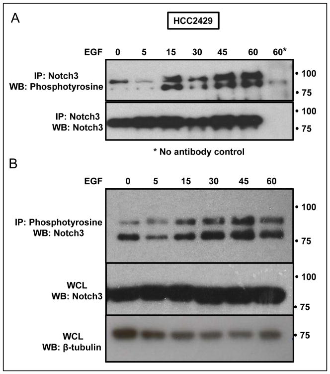
Little is known about post-translational modifications of the Notch receptor and their role in regulating Notch activity(32). The co-immunoprecipitation and functional associations above led us to speculate that Notch3 could be a novel direct or indirect substrate for EGFR mediated tyrosine phosphorylation, which may mediate its role as a negative regulator. To demonstrate tyrosine phosphorylation of the Notch receptor, HCC2429 cells that overexpress Notch3 due to translocation, and wild-type EGFR were stimulated with EGF for increasing durations of exposure. Total protein lysates were immunoprecipitated with a Notch3 antibody and analyzed with either phosphotyrosine or Notch3 antibody (Fig. 7a) or immunoprecipitated with phosphotyrosine and subjected to western analysis with Notch3 (Fig. 7b). These data clearly show that following the addition of EGF, Notch3 is phosphorylated in a time-dependent manner. These data are the first to demonstrate tyrosine phosphorylation of Notch and to identify Notch as a novel direct or indirect substrate for the EGFR tyrosine kinase.
Figure 7.
EGF mediated tyrosine phosphorylation of the Notch3 receptor. A&B, HCC2429 cells were stimulated with EGF (25 ng/ml) for the indicated times to detect the ligand induced tyrosine phosphorylation of Notch3 receptor. A Cell lysates were immunoprecipitated with Notch3 antibody followed by western analysis using a phosphotyrosine antibody. The blot was stripped and re-probed with anti-Notch3 (bottom). B, A reciprocal immunoprecipitation was done with a phosphotyrosine antibody with a subsequent western blot probed for Notch3 (top). Equal amount of cell lysate was analyzed for Notch3 expression (middle) and tubulin (bottom).
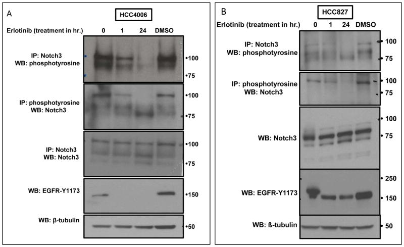
To confirm that the observed tyrosine phosphorylation of Notch3 is dependent on EGFR kinase activity, cells were treated with vehicle or erlotinib for 1 or 24 hours. Treatment with erlotinib reduced the tyrosine phosphorylation of Notch3 in a time-dependent manner (Fig. 8a and b). Furthermore, the phosphorylation was detected reciprocally, demonstrating that Notch3 phosphorylation is due to EGFR kinase activation. A similar pattern was observed for tyrosine-1173 auto phosphorylation of EGFR, further confirming that the Notch3 receptor is tyrosine phosphorylated in an EGFR kinase-dependent fashion.
Figure 8.
EGFR kinase activity is necessary for the tyrosine phosphorylation of the Notch3 receptor. A, HCC4006 and B, HCC827 cells were treated with DMSO or erlotinib (0.1 μM) for indicated periods of time. Cell lysates from each time point were either precipitated with a phosphotyrosine antibody followed by a western blot probed with Notch3 antibody (top), or immunoprecipitated with Notch3 antibody followed by a western blot probed with a phosphotyrosine antibody. Whole cell lysate was also subjected to western analysis to check for of levels Notch3, EGFR-pY1173, and tubulin.
To further address if EGFR mediated Notch3 tyrosine phosphorylation is indeed a key regulatory event that modulates the stem-like phenotype in lung cancer cells we used H1650 cells, which do not show an increase in ALDH activity in response to erlotinib treatment (Supplementary Fig. 2), and also show a continuously active EGFR in the presence of erlotinib (Supplementary Fig. 7). Phosphotyrosine analysis of Notch3 revealed that under basal conditions Notch3 is tyrosine phosphorylated. Treatment with erlotinib did not prevent the Notch3 tyrosine phosphorylation (Supplementary Fig. 7) suggesting that inhibition of EGFR is a key event required for the Notch3 activation.
Discussion
Cellular signaling pathways are complex and interconnected with inhibition of one pathway often resulting in feedback regulation and parallel compensatory activation of other pathways. This causes unexpected results when highly specific inhibitors are used, but understanding the mechanisms and consequences of these events leads to identification of novel signaling cascades and often to targeted solutions. A good example of this is the upregulation of the MEK pathway and the increased incidence of squamous skin cancers with BRAF inhibition in BRAF mutant melanomas, and combinations with MEK inhibitors is not only more effective against the tumor, but also reduces the skin cancer incidence(33). Similarly, inhibition of mTOR results in the feedback activation of AKT signaling(34). These changes are not “adaptive resistance” pathways in the sense that cancers are acquiring secondary drivers to overcome a primary driver, but rather a consequence of normal physiologic counter-regulatory pathways observable even in normal cells, that are made apparent by pathway-specific intervention in cancer.
In this study, we show that erlotinib treatment of EGFR mutated lung cancer cells, while dramatically reducing the growth and number of tumor cells, also directly induces Notch signaling, and that this effect is observed with both mutated, constitutively activated, EGFR and wild-type EGFR. As it is becoming increasingly evident that Notch signaling is important in the maintenance of subsets of tumor cells with a high clonogenic capacity, often referred to as “cancer stem-like cells” or “tumor-initiating cells” (16, 35), this upregulation may have clinical consequences. The involvement of Notch in this cell sub-population is well characterized in brain, breast, and embryonic tumors (18, 36, 37). In lung cancer, ALDH positivity has been found to be a good marker for a tumor cell subset with stem-like cell properties and this subset is dependent on Notch3 for clonogenicity and tumorigenic potential (19). In the present study, we document a reciprocal activation of EGFR and Notch in human lung cancer, and thus a previously unrecognized role for constitutively activated EGFR in repressing stem-like cells in EGFR driven NSCLC cell lines. This may underlie the fact that advanced lung tumors with mutated EGFR have a much better prognosis than those with wild-type EGFR, even when treated with standard, non-kinase inhibitor therapies. Treatment with erlotinib, while resulting in dramatic cell death, also releases this Notch inhibition and paradoxically promotes the stem-like cell population. It is unclear whether erlotinib is sufficient to induce ALDH positivity in an ALDH negative cell, increase ALDH expression in ALDH low cells, or promote proliferation of ALDH high cells.
We find that this phenomenon was reversed with knockdown of Notch3, but not Notch1 suggesting that Notch3 plays a crucial role in regulating stem-like cells, which is in agreement with previous studies (38). Interestingly, Notch1 seems to play a sometimes-opposing role, suppressing the growth of stem-like cells, which also is in line with the earlier work (39), and suggests that pan-Notch inhibition may not be optimal in countering this effect.
Cross-regulation between EGFR and Notch signaling pathways has long been observed in genetic studies and has been shown to be both cooperative and antagonistic depending on the cellular context(40). Similar to what we observe in lung cancer, in Drosophila EGFR opposes Notch signaling in multiple developmental processes, and in fact loss of function mutations in the Notch pathway can compensate for hypomorphic alleles of EGFR (41, 42). In C. elegans vulvar development, it has also been shown that EGFR and LIN-12/Notch have opposing effects on cell fate determination (43). The functional interaction of these pathways has been less clear in mammals, but has recently been shown to be important in neural stem cell number and renewal, where EGFR signaling expands the progenitor cells and depletes the stem cells by ubiquitination mediated down-regulation of Notch1 (44). In skin cancer, antagonistic effects of Notch1 and EGFR have been shown, where inhibition of EGFR leads to increased differentiation of squamous cell carcinoma cells and increased resistance to apoptosis. However, combined inhibition of EGFR and Notch activity significantly induced the Notch mediated apoptosis and differentiation (45). Nevertheless the biochemical basis of the EGFR and Notch interaction has been unclear, and likewise its role in lung cancer biology.
We show here that EGFR directly modulates Notch activity in human lung cancers, and that there is a kinase dependent physical association of the two receptors. EGFR has previously been shown to interact with transcription factors such as STAT3 and regulate gene transcription in the nucleus (25). In this context our findings suggest another mode of EGFR-driven transcriptional regulation mediated through the interaction of EGFR with Notch3. Our studies also demonstrated erlotinib-sensitive tyrosine phosphorylation of Notch3 associated with negative regulation of Notch transcriptional activation. Tyrosine phosphorylation on the Notch-ICD is novel and represents a potential mechanism for the crosstalk of these two pathways.
The discovery of mutations in the EGFR receptor has revolutionized the management of patients with advanced lung cancer and defined a new era of highly targeted therapies selected for tumors with aberrantly activated pathways. Patients whose tumors contain activating mutations of EGFR have dramatic tumor regressions when treated with EGFR-selective TKIs. However, none of these patients are cured and all eventually relapse and die from their disease. It is very clear that these inhibitors must be used continuously for life in most cases, as cessation results in a rapid tumor “flare” (46). In fact, in the setting of curative-intent therapy and defined periods of adjuvant EGFR TKI treatment, while relapse-free survival appears good while on drug, ultimately worsened survival or trends in that direction are seen compared to placebo, suggesting that modulation of EGFR in a selective way may have unforeseen adverse consequences not apparent in the metastatic setting.
The most common cause of acquired resistance in EGFR mutated tumors is the development of a “gatekeeper” mutation, T790M that re-activates the EGFR tyrosine kinase and makes it resistant to erlotinib. The other common mechanism is the development of a bypass signal (such as MET amplification) that circumvents the loss of EGFR signaling. These types of resistance mechanisms cannot explain the adverse outcomes observed with treatment in early stage disease. In this study, we find that erlotinib activates the developmentally important Notch pathway. This provides a potential explanation for the clinical observations in that while tumors bearing these activating mutations may undergo dramatic reduction in tumor bulk after EGFR-targeted therapies, EGFR inhibition may actually promote stem-like cells by activating Notch. The tonic suppression of Notch by constitutively activated EGFR may also explain the good prognosis of EGFR-mutated lung cancers compared to EGFR wild-type tumors when treated with standard chemotherapies, with median survivals of 20 to 30 months in several studies compared to 10 to 12 months (47). Inhibition of the EGFR may thus shrink tumors causing clinical benefit in the metastatic setting, but in fact promote stem-like cell characteristics and clonogenicity of the surviving fraction in the adjuvant setting and this becomes apparent upon stopping the inhibitor. It is also intriguing that tumors with mutant EGFR seem to more often clinically relapse with miliary disease and hundreds or thousands of lesions after therapy with EGFR inhibitors than tumors containing wild-type EGFR (48).
These data thus suggest that caution should be observed in treating patients who undergo curative-intent resections or chemoradiation for tumors bearing activating mutations in EGFR with EGFR inhibitors alone. These patients may actually be harmed by adjuvant unopposed selective inhibition of EGFR that becomes apparent upon stopping the therapy. We suggest that in this setting, selective Notch3 inhibition combined with EGFR TKI therapy should be explored.
Supplementary Material
Acknowledgments
Funded by: NCI CA 90949 to DPC
Footnotes
Conflict of Interest: DPC has done advisory boards for Pfizer. No other relevant conflicts.
References
- 1.Mok TS, Wu YL, Thongprasert S, Yang CH, Chu DT, Saijo N, et al. Gefitinib or carboplatin-paclitaxel in pulmonary adeno carcinoma. N Engl J Med. 2009;361:947–57. doi: 10.1056/NEJMoa0810699. [DOI] [PubMed] [Google Scholar]
- 2.Maemondo M, Inoue A, Kobayashi K, Sugawara S, Oizumi S, Isobe H, et al. Gefitinib or chemotherapy for non-small-cell lung cancer with mutated EGFR. N Engl J Med. 2010;362:2380–8. doi: 10.1056/NEJMoa0909530. [DOI] [PubMed] [Google Scholar]
- 3.Kelly K, Chansky K, Gaspar LE, Albain KS, Jett J, Ung YC, et al. Phase III trial of maintenance gefitinib or placebo after concurrent chemoradiotherapy and docetaxel consolidation in inoperable stage III non-small-cell lung cancer: SWOG S0023. J Clin Oncol. 2008;26:2450–6. doi: 10.1200/JCO.2007.14.4824. [DOI] [PubMed] [Google Scholar]
- 4.Goss GD, O’Callaghan C, Lorimer I, Tsao MS, Masters GA, Jett J, et al. Gefitinib versus placebo in completely resected non-small-cell lung cancer: results of the NCIC CTG BR19 study. J Clin Oncol. 2013;31:3320–6. doi: 10.1200/JCO.2013.51.1816. [DOI] [PMC free article] [PubMed] [Google Scholar]
- 5.Artavanis-Tsakonas S, Rand MD, Lake RJ. Notch signaling: cell fate control and signal integration in development. Science. 1999;284:770–6. doi: 10.1126/science.284.5415.770. [DOI] [PubMed] [Google Scholar]
- 6.Koch U, Radtke F. Notch signaling in solid tumors. Curr Top Dev Biol. 2010;92:411–55. doi: 10.1016/S0070-2153(10)92013-9. [DOI] [PubMed] [Google Scholar]
- 7.Lopez C, Delgado J, Costa D, Villamor N, Navarro A, Cazorla M, et al. Clonal evolution in chronic lymphocytic leukemia: analysis of correlations with IGHV mutational status, NOTCH1 mutations and clinical significance. Genes, chromosomes & cancer. 2013;52:920–7. doi: 10.1002/gcc.22087. [DOI] [PubMed] [Google Scholar]
- 8.Weissmann S, Roller A, Jeromin S, Hernandez M, Abaigar M, Hernandez-Rivas JM, et al. Prognostic impact and landscape of NOTCH1 mutations in chronic lymphocytic leukemia (CLL): a study on 852 patients. Leukemia. 2013;27:2393–6. doi: 10.1038/leu.2013.218. [DOI] [PubMed] [Google Scholar]
- 9.Dang TP, Gazdar AF, Virmani AK, Sepetavec T, Hande KR, Minna JD, et al. Chromosome 19 translocation, overexpression of Notch3, and human lung cancer. J Natl Cancer Inst. 2000;92:1355–7. doi: 10.1093/jnci/92.16.1355. [DOI] [PubMed] [Google Scholar]
- 10.Konishi J, Kawaguchi KS, Vo H, Haruki N, Gonzalez A, Carbone DP, et al. Gamma-secretase inhibitor prevents Notch3 activation and reduces proliferation in human lung cancers. Cancer Res. 2007;67:8051–7. doi: 10.1158/0008-5472.CAN-07-1022. [DOI] [PubMed] [Google Scholar]
- 11.Dang TP, Eichenberger S, Gonzalez A, Olson S, Carbone DP. Constitutive activation of Notch3 inhibits terminal epithelial differentiation in lungs of transgenic mice. Oncogene. 2003;22:1988–97. doi: 10.1038/sj.onc.1206230. [DOI] [PubMed] [Google Scholar]
- 12.Chiba S. Notch signaling in stem cell systems. Stem Cells. 2006;24:2437–47. doi: 10.1634/stemcells.2005-0661. [DOI] [PubMed] [Google Scholar]
- 13.Dreger P, Schnaiter A, Zenz T, Bottcher S, Rossi M, Paschka P, et al. TP53, SF3B1, and NOTCH1 mutations and outcome of allotransplantation for chronic lymphocytic leukemia: six-year follow-up of the GCLLSG CLL3X trial. Blood. 2013;121:3284–8. doi: 10.1182/blood-2012-11-469627. [DOI] [PubMed] [Google Scholar]
- 14.Willander K, Dutta RK, Ungerback J, Gunnarsson R, Juliusson G, Fredrikson M, et al. NOTCH1 mutations influence survival in chronic lymphocytic leukemia patients. BMC cancer. 2013;13:274. doi: 10.1186/1471-2407-13-274. [DOI] [PMC free article] [PubMed] [Google Scholar]
- 15.Hassan KA, Wang L, Korkaya H, Chen G, Maillard I, Beer DG, et al. Notch pathway activity identifies cells with cancer stem cell-like properties and correlates with worse survival in lung adenocarcinoma. Clin Cancer Res. 2013;19:1972–80. doi: 10.1158/1078-0432.CCR-12-0370. [DOI] [PMC free article] [PubMed] [Google Scholar]
- 16.Wang Z, Li Y, Banerjee S, Sarkar FH. Emerging role of Notch in stem cells and cancer. Cancer letters. 2009;279:8–12. doi: 10.1016/j.canlet.2008.09.030. [DOI] [PMC free article] [PubMed] [Google Scholar]
- 17.Bolos V, Blanco M, Medina V, Aparicio G, Diaz-Prado S, Grande E. Notch signalling in cancer stem cells. Clin Transl Oncol. 2009;11:11–9. doi: 10.1007/s12094-009-0305-2. [DOI] [PubMed] [Google Scholar]
- 18.Farnie G, Clarke RB, Spence K, Pinnock N, Brennan K, Anderson NG, et al. Novel cell culture technique for primary ductal carcinoma in situ: role of Notch and epidermal growth factor receptor signaling pathways. Journal of the National Cancer Institute. 2007;99:616–27. doi: 10.1093/jnci/djk133. [DOI] [PubMed] [Google Scholar]
- 19.Sullivan JP, Spinola M, Dodge M, Raso MG, Behrens C, Gao B, et al. Aldehyde dehydrogenase activity selects for lung adenocarcinoma stem cells dependent on notch signaling. Cancer Res. 2010;70:9937–48. doi: 10.1158/0008-5472.CAN-10-0881. [DOI] [PMC free article] [PubMed] [Google Scholar]
- 20.Pannuti A, Foreman K, Rizzo P, Osipo C, Golde T, Osborne B, et al. Targeting Notch to target cancer stem cells. Clinical cancer research: an official journal of the American Association for Cancer Research. 2010;16:3141–52. doi: 10.1158/1078-0432.CCR-09-2823. [DOI] [PMC free article] [PubMed] [Google Scholar]
- 21.Creighton CJ, Li X, Landis M, Dixon JM, Neumeister VM, Sjolund A, et al. Residual breast cancers after conventional therapy display mesenchymal as well as tumor-initiating features. Proceedings of the National Academy of Sciences of the United States of America. 2009;106:13820–5. doi: 10.1073/pnas.0905718106. [DOI] [PMC free article] [PubMed] [Google Scholar]
- 22.Giacalone NJ, Den RB, Eisenberg R, Chen H, Olson SJ, Massion PP, et al. ALDH7A1 expression is associated with recurrence in patients with surgically resected non-small-cell lung carcinoma. Future oncology. 2013;9:737–45. doi: 10.2217/fon.13.19. [DOI] [PMC free article] [PubMed] [Google Scholar]
- 23.Lewis HD, Leveridge M, Strack PR, Haldon CD, O’Neil J, Kim H, et al. Apoptosis in T cell acute lymphoblastic leukemia cells after cell cycle arrest induced by pharmacological inhibition of notch signaling. Chem Biol. 2007;14:209–19. doi: 10.1016/j.chembiol.2006.12.010. [DOI] [PubMed] [Google Scholar]
- 24.Arcaroli JJ, Quackenbush KS, Purkey A, Powell RW, Pitts TM, Bagby S, et al. Tumours with elevated levels of the Notch and Wnt pathways exhibit efficacy to PF-03084014, a gamma-secretase inhibitor, in a preclinical colorectal explant model. British journal of cancer. 2013;109:667–75. doi: 10.1038/bjc.2013.361. [DOI] [PMC free article] [PubMed] [Google Scholar]
- 25.Lo HW, Hsu SC, Ali-Seyed M, Gunduz M, Xia W, Wei Y, et al. Nuclear interaction of EGFR and STAT3 in the activation of the iNOS/NO pathway. Cancer cell. 2005;7:575–89. doi: 10.1016/j.ccr.2005.05.007. [DOI] [PubMed] [Google Scholar]
- 26.Huang Y, Lin L, Shanker A, Malhotra A, Yang L, Dikov MM, et al. Resuscitating cancer immunosurveillance: selective stimulation of DLL1-Notch signaling in T cells rescues T-cell function and inhibits tumor growth. Cancer Res. 2011;71:6122–31. doi: 10.1158/0008-5472.CAN-10-4366. [DOI] [PMC free article] [PubMed] [Google Scholar]
- 27.Sos ML, Koker M, Weir BA, Heynck S, Rabinovsky R, Zander T, et al. PTEN loss contributes to erlotinib resistance in EGFR-mutant lung cancer by activation of Akt and EGFR. Cancer Res. 2009;69:3256–61. doi: 10.1158/0008-5472.CAN-08-4055. [DOI] [PMC free article] [PubMed] [Google Scholar]
- 28.Yoon YK, Kim HP, Han SW, Oh do Y, Im SA, Bang YJ, et al. KRAS mutant lung cancer cells are differentially responsive to MEK inhibitor due to AKT or STAT3 activation: implication for combinatorial approach. Molecular carcinogenesis. 2010;49:353–62. doi: 10.1002/mc.20607. [DOI] [PubMed] [Google Scholar]
- 29.Thomson S, Buck E, Petti F, Griffin G, Brown E, Ramnarine N, et al. Epithelial to mesenchymal transition is a determinant of sensitivity of non-small-cell lung carcinoma cell lines and xenografts to epidermal growth factor receptor inhibition. Cancer Res. 2005;65:9455–62. doi: 10.1158/0008-5472.CAN-05-1058. [DOI] [PubMed] [Google Scholar]
- 30.Gounder MM, Schwartz GK. Moving forward one Notch at a time. J Clin Oncol. 2012;30:2291–3. doi: 10.1200/JCO.2012.42.3277. [DOI] [PubMed] [Google Scholar]
- 31.Konishi J, Yi F, Chen X, Vo H, Carbone DP, Dang TP. Notch3 cooperates with the EGFR pathway to modulate apoptosis through the induction of bim. Oncogene. 2010;29:589–96. doi: 10.1038/onc.2009.366. [DOI] [PMC free article] [PubMed] [Google Scholar]
- 32.Fortini ME. Notch signaling: the core pathway and its posttranslational regulation. Developmental cell. 2009;16:633–47. doi: 10.1016/j.devcel.2009.03.010. [DOI] [PubMed] [Google Scholar]
- 33.Flaherty KT, Infante JR, Daud A, Gonzalez R, Kefford RF, Sosman J, et al. Combined BRAF and MEK inhibition in melanoma with BRAF V600 mutations. N Engl J Med. 2012;367:1694–703. doi: 10.1056/NEJMoa1210093. [DOI] [PMC free article] [PubMed] [Google Scholar]
- 34.O’Reilly KE, Rojo F, She QB, Solit D, Mills GB, Smith D, et al. mTOR inhibition induces upstream receptor tyrosine kinase signaling and activates Akt. Cancer Res. 2006;66:1500–8. doi: 10.1158/0008-5472.CAN-05-2925. [DOI] [PMC free article] [PubMed] [Google Scholar]
- 35.Rasheed ZA, Kowalski J, Smith BD, Matsui W. Concise review: Emerging concepts in clinical targeting of cancer stem cells. Stem Cells. 2011;29:883–7. doi: 10.1002/stem.648. [DOI] [PMC free article] [PubMed] [Google Scholar]
- 36.Farnie G, Clarke RB. Mammary stem cells and breast cancer--role of Notch signalling. Stem cell reviews. 2007;3:169–75. doi: 10.1007/s12015-007-0023-5. [DOI] [PubMed] [Google Scholar]
- 37.Sansone P, Storci G, Tavolari S, Guarnieri T, Giovannini C, Taffurelli M, et al. IL-6 triggers malignant features in mammospheres from human ductal breast carcinoma and normal mammary gland. The Journal of clinical investigation. 2007;117:3988–4002. doi: 10.1172/JCI32533. [DOI] [PMC free article] [PubMed] [Google Scholar]
- 38.Zheng Y, de la Cruz CC, Sayles LC, Alleyne-Chin C, Vaka D, Knaak TD, et al. A rare population of CD24(+)ITGB4(+)Notch(hi) cells drives tumor propagation in NSCLC and requires Notch3 for self-renewal. Cancer cell. 2013;24:59–74. doi: 10.1016/j.ccr.2013.05.021. [DOI] [PMC free article] [PubMed] [Google Scholar]
- 39.Westhoff B, Colaluca IN, D’Ario G, Donzelli M, Tosoni D, Volorio S, et al. Alterations of the Notch pathway in lung cancer. Proc Natl Acad Sci U S A. 2009;106:22293–8. doi: 10.1073/pnas.0907781106. [DOI] [PMC free article] [PubMed] [Google Scholar]
- 40.Sundaram MV. The love-hate relationship between Ras and Notch. Genes & development. 2005;19:1825–39. doi: 10.1101/gad.1330605. [DOI] [PubMed] [Google Scholar]
- 41.zur Lage P, Jarman AP. Antagonism of EGFR and notch signalling in the reiterative recruitment of Drosophila adult chordotonal sense organ precursors. Development. 1999;126:3149–57. doi: 10.1242/dev.126.14.3149. [DOI] [PubMed] [Google Scholar]
- 42.Price JV, Savenye ED, Lum D, Breitkreutz A. Dominant enhancers of Egfr in Drosophila melanogaster: genetic links between the Notch and Egfr signaling pathways. Genetics. 1997;147:1139–53. doi: 10.1093/genetics/147.3.1139. [DOI] [PMC free article] [PubMed] [Google Scholar]
- 43.Yoo AS, Bais C, Greenwald I. Crosstalk between the EGFR and LIN-12/Notch pathways in C. elegans vulval development. Science. 2004;303:663–6. doi: 10.1126/science.1091639. [DOI] [PubMed] [Google Scholar]
- 44.Aguirre A, Rubio ME, Gallo V. Notch and EGFR pathway interaction regulates neural stem cell number and self-renewal. Nature. 2010;467:323–7. doi: 10.1038/nature09347. [DOI] [PMC free article] [PubMed] [Google Scholar]
- 45.Kolev V, Mandinova A, Guinea-Viniegra J, Hu B, Lefort K, Lambertini C, et al. EGFR signalling as a negative regulator of Notch1 gene transcription and function in proliferating keratinocytes and cancer. Nature cell biology. 2008;10:902–11. doi: 10.1038/ncb1750. [DOI] [PMC free article] [PubMed] [Google Scholar]
- 46.Chaft JE, Oxnard GR, Sima CS, Kris MG, Miller VA, Riely GJ. Disease flare after tyrosine kinase inhibitor discontinuation in patients with EGFR-mutant lung cancer and acquired resistance to erlotinib or gefitinib: implications for clinical trial design. Clin Cancer Res. 2011;17:6298–303. doi: 10.1158/1078-0432.CCR-11-1468. [DOI] [PMC free article] [PubMed] [Google Scholar]
- 47.Fukuoka M, Wu YL, Thongprasert S, Sunpaweravong P, Leong SS, Sriuranpong V, et al. Biomarker analyses and final overall survival results from a phase III, randomized, open-label, first-line study of gefitinib versus carboplatin/paclitaxel in clinically selected patients with advanced non-small-cell lung cancer in Asia (IPASS) J Clin Oncol. 2011;29:2866–74. doi: 10.1200/JCO.2010.33.4235. [DOI] [PubMed] [Google Scholar]
- 48.Laack E, Simon R, Regier M, Andritzky B, Tennstedt P, Habermann C, et al. Miliary never-smoking adenocarcinoma of the lung: strong association with epidermal growth factor receptor exon 19 deletion. Journal of thoracic oncology: official publication of the International Association for the Study of Lung Cancer. 2011;6:199–202. doi: 10.1097/JTO.0b013e3181fb7cf1. [DOI] [PubMed] [Google Scholar]
Associated Data
This section collects any data citations, data availability statements, or supplementary materials included in this article.


