Abstract
Background
Novel bone substitutes have challenged the notion of autologous bone grafting as the ‘gold standard’ for the surgical treatment of fracture nonunions. The present study was designed to test the hypothesis that autologous bone grafting is equivalent to other bone grafting modalities in the management of fracture nonunions of the long bones.
Methods
A retrospective review of patients with fracture nonunions included in two prospective databases was performed at two US level 1 trauma centers from January 1, 1998 (center 1) or January 1, 2004 (center 2), respectively, until December 31, 2010 (n = 574). Of these, 182 patients required adjunctive bone grafting and were stratified into the following cohorts: autograft (n = 105), allograft (n = 38), allograft and autograft combined (n = 16), and recombinant human bone morphogenetic protein-2 (rhBMP-2) with or without adjunctive bone grafting (n = 23). The primary outcome parameter was time to union. Secondary outcome parameters consisted of complication rates and the rate of revision procedures and revision bone grafting.
Results
The autograft cohort had a statistically significant shorter time to union (198 ± 172–225 days) compared to allograft (416 ± 290–543 days) and exhibited a trend towards earlier union when compared to allograft/autograft combined (389 ± 159–619 days) or rhBMP-2 (217 ± 158–277 days). Furthermore, the autograft cohort had the lowest rate of surgical revisions (17%) and revision bone grafting (9%), compared to allograft (47% and 32%), allograft/autograft combined (25% and 31%), or rhBMP-2 (27% and 17%). The overall new-onset postoperative infection rate was significantly lower in the autograft group (12.4%), compared to the allograft cohort (26.3%) (P < 0.05).
Conclusion
Autologous bone grafting appears to represent the bone grafting modality of choice with regard to safety and efficiency in the surgical management of long bone fracture nonunions.
Keywords: Fracture nonunion, Autograft, Allograft, Bone morphogenetic protein
Introduction
Fracture nonunions of long bones continue to represent a significant clinical challenge and socioeconomic burden, associated with high complication rates and the potential for poor long-term outcomes [1-5]. Autologous bone grafting has received a negative reputation in the past, mainly due to the high risk of postoperative complications related to the harvesting procedure [6]. However, more recent innovative and minimally invasive harvesting techniques have mitigated the historic issue of donor site morbidity and renewed the interest in autologous bone as a preferred source for bone grafting [7-9].
The introduction of new generation bone substitutes and recombinant molecules with osteoinductive properties has recently challenged the role of autologous bone grafting as the ‘gold standard’ for the surgical treatment of nonunions [10-12]. In light of the immense market potential for bone graft substitutes and related products, estimated to be US$1 billion in the USA alone, the push for new ‘osteobiologicals’ may in large part be industry-driven, rather than based on objective patient safety and quality data [13-15]. In fact, well-publicized concerns have recently been raised regarding the safety of the uncritical application of recombinant bone morphogenetic protein (BMP)-2 for a variety of indications, including its off-label use [14,16-18].
The present study was designed to determine the ‘ideal’ modality of adjunctive bone grafting in the management of long bone fracture nonunions. We hypothesized that the use of autograft is equivalent to other bone grafting options with regard to healing times and complication rates.
Methods
Study design
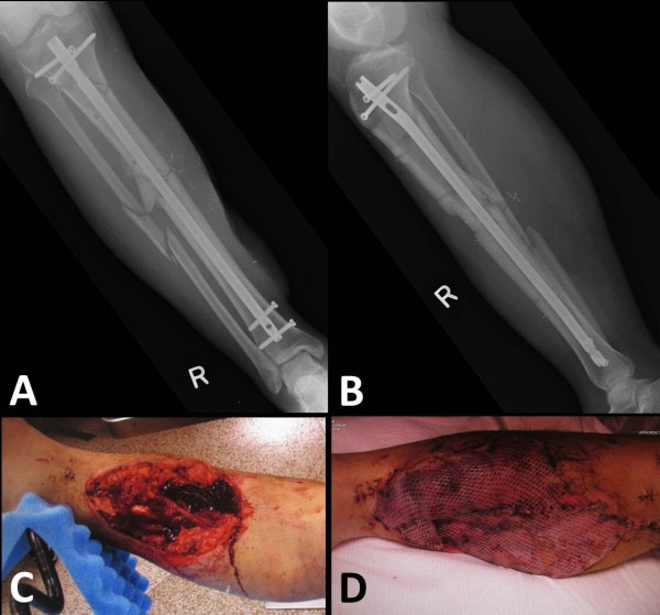
A retrospective cohort study was designed based on two prospective institutional databases from representative level 1 trauma centers in the USA (Denver Health Medical Center, Denver, CO; and Geisinger Medical Center, Danville, PA). Prior to study initiation, approval by the two respective Institutional Review Boards was obtained. The databases included all patients between 18 and 85 years of age, who were admitted to one of the two participating centers for surgical treatment of long bone fracture nonunions between January 1, 1998 and December 31, 2010 (n = 373; Denver Health), and between January 1, 2004 and December 31, 2010 (n = 201; Geisinger), respectively. Patients that did not require adjunctive bone grafting (n = 362) or had a nonunion of the forearm (n = 30) were excluded from the analysis. Patients who were lost to follow-up prior to a minimum of 12 months postoperatively or prior to clinical and radiographic union were excluded. The remaining 182 patients who required adjunctive bone grafting for nonunions of the tibia (n = 98), femur (n = 46), or humerus (n = 38) were included for final analysis. The flowchart in Figure 1 depicts the patient selection process in detail. Figures 2,3,4,5 illustrate a representative case example of a 31-year-old male who sustained an isolated type IIIB open segmental tibia fracture. The patient underwent initial surgical debridement and external fixation, followed by staged conversion to an intramedullary nail. He developed a tibial nonunion, requiring exchange nailing and autologous bone grafting.
Figure 1.
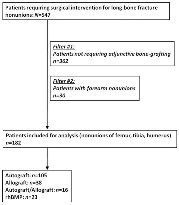
Patient selection flowchart.
Figure 2.
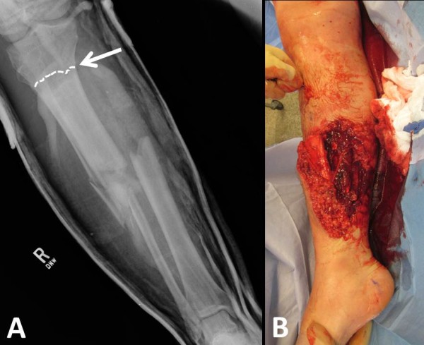
Representative case example. A 31-year-old male pedestrian was hit at high speed by a motor vehicle, sustaining an isolated type IIIB open segmental tibia shaft fracture (A, B) with associated segmental fibula fracture (AO/OTA type 42-C2.2). The arrow in panel A depicts the proximal nondisplaced fracture line of the segmental tibia fracture.
Figure 3.
Identical case as in Figure2. After initial spanning external fixation and surgical debridement, the patient was taken back to the operating room the next day for definitive surgical management by reamed intramedullary interlocking nail fixation (A, B), repeat surgical debridement (C), and soft tissue coverage by free microvascular gracilis flap in the same session (D).
Figure 4.
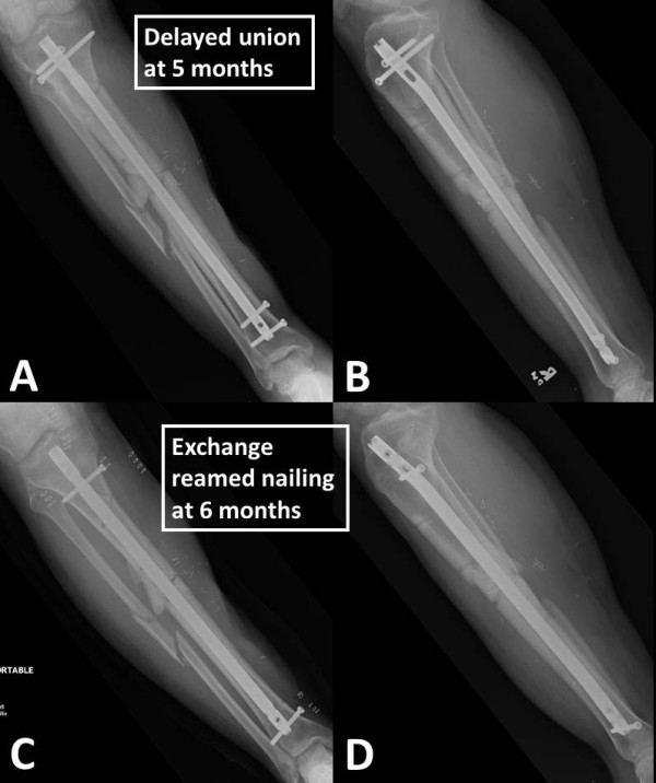
Identical case as in Figures2,3. At 5 months follow-up, the patient showed clinical and radiographic signs of a nonunion of the open tibia fracture (A, B). A surgical revision was performed by exchange reamed nailing at 6 months (C, D).
Figure 5.
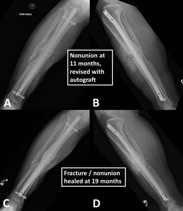
Identical case as in Figures2,3,4. The nonunion persisted at 11 months post injury, and a second revision was performed by decortication and application of a central bone graft (A, B), using a reamer-irrigator-aspirator technique for minimal invasive autograft harvest from the ipsilateral femur. The fracture/nonunion was clinically and radiographically healed at 19 months post injury (C, D), and the patient was asymptomatic with a full functional recovery.
In accordance with the definition by the US Federal Drug Administration (FDA), nonunion was defined as a fractured bone which had not completely healed within 9 months of injury and which had not shown signs of progression towards healing for three consecutive months on serial radiographs [19]. Nonunions were classified according to the Weber and Cech classification system into hypertrophic, normotrophic, or atrophic [19]. Nonunions were treated either by plate osteosynthesis (using conventional plates or locking plate systems), intramedullary nail fixation (including exchange nailing), or by external ring fixator in combination with bone grafting, based on the individual surgeon’s preference.
Patients were stratified into the following cohorts for analysis, based on the bone grafting modality: (1) autograft (n = 105), (2) allograft (n = 38), (3) allograft in combination with autograft (n = 16), and (4) rhBMP-2 with or without adjunctive bone grafting substitute (n = 23). The rhBMP-2 product was mixed with sterile saline and prepared immediately prior to use from a kit containing all necessary components, according to the manufacturer’s instructions (Infuse®, Medtronic, Memphis, TN, USA). In 6 patients, rhBMP-2 was administered without bone grafting adjunct, while in the remaining 17 patients rhBMP-2 was combined with allograft. All patients were followed postoperatively for a minimum of 12 months, or until clinical and radiographic bone healing occurred. ‘Clinical union’ was defined as painless weight bearing and lack of local tenderness over the nonunion site on physical examination [20,21]. ‘Radiographic union’ was defined as bridging bone on a minimum of three cortices on conventional antero-posterior and lateral radiographs. The application of rhBMP-2 for the revision of long bone fracture nonunions was considered an ‘off-label’ indication. Autograft donor sites were as follows: (1) iliac crest (n = 64), (2) femoral reamer-irrigator-aspirator (n = 20), and (3) other (n = 10). The primary outcome parameter was time to union. The secondary outcome parameters included the incidence of new-onset postoperative infection in a previously noninfected nonunion or ongoing infection in infected nonunions, as well as the need of revision surgeries and revision bone grafting.
Statistical analysis
Data were analyzed using SPSS software version 17.0 (SPSS Inc., Chicago, IL, USA). Descriptive statistics were performed to summarize demographic and clinical variables and to evaluate distributional characteristics of continuous variables, using Kruskal-Wallis test, Mann–Whitney U test, and Chi-square test, as appropriate. Data are expressed as means ± standard deviation or means ± 95% confidence interval (95% CI), as deemed appropriate. Statistical significance was defined at P < 0.05.
Results
Patient demographics
A total of 182 patients (102 males and 80 females) met the inclusion criteria (Figure 1). The mean age was 44 ± 13.6 years. Sixty-eight patients were confirmed smokers, and 30 patients had a history of smoking and reported cessation prior to nonunion surgery. The remaining 84 patients reported to have never smoked. Demographic data, stratified by subgroups, are depicted in Table 1. The surgical management of the nonunions consisted of plate osteosynthesis (65%), intramedullary interlocking nail fixation (28%), or application of an external ring fixator (7%). Nonunion characteristics and management strategies in the different cohorts are shown in Table 2.
Table 1.
Patient demographics and fracture characteristics
| Variable | Overall | Autograft | Allograft | Autograft/allograft | rhBMP-2 |
|---|---|---|---|---|---|
| Gender |
|
|
|
|
|
| Male |
102 (56%) |
58 (55%)* |
16 (42%) |
4 (25%) |
7 (30%) |
| Female |
80 (44%) |
47 (45%)* |
22 (58%) |
12 (75%) |
16 (70%) |
| Mean age (years) |
44 (±13.6) |
41 (±12.5)* |
47 (±13) |
46 (±14) |
52 (±15) |
| EtOH |
|
|
|
|
|
| Abuse |
44 (24%) |
22 (21%) |
8 (21%) |
0 |
5 (22%) |
| Occasionally |
45 (25%) |
30 (29%) |
12 (32%) |
6 (37%) |
6 (26%) |
| Never |
97 (51%) |
53 (50%) |
18 (47%) |
10 (63%) |
12 (52%) |
| Tobacco smoking |
|
|
|
|
|
| Current |
68 (37%) |
41 (39%) |
16 (42%) |
4 (25%) |
6 (26%) |
| Former |
30 (17%) |
17 (16%) |
4 (11%) |
4 (25%) |
5 (22%) |
| Never |
84 (46%) |
47 (45%) |
18 (47%) |
8 (50%) |
12 (52%) |
| Illicit drug use |
|
|
|
|
|
| Current |
28 (15%) |
17 (16%) |
9 (24%) |
0** |
1 (4%) |
| Former |
7 (4%) |
6 (6%) |
0 |
0** |
1 (4%) |
| Never |
147 (81%) |
82 (78%) |
29 (76%) |
16 (100%) |
21 (92%) |
| Injury mechanism |
|
|
|
|
|
| MVA/MCA |
83 (46%) |
49 (47%) |
18 (47%) |
8 (50%) |
8 (35%) |
| Fall |
39 (21%) |
20 (19%) |
10 (26%) |
3 (19%) |
6 (26%) |
| GSF |
6 (3%) |
5 (4%) |
1 (3%) |
0 |
0 |
| Other |
33 (18%) |
31 (30%) |
9 (24%) |
5 (31%) |
9 (39%) |
| Fracture location |
|
|
|
|
|
| Tibia |
98 (54%) |
57 (54%) |
21 (55%) |
9 (56%) |
11 (48%) |
| Femur |
46 (25%) |
28 (27%) |
9 (24%) |
4 (25%) |
5 (22%) |
| Humerus |
38 (21%) |
20 (19%) |
8 (21%) |
3 (19%) |
7 (30%) |
| Fracture characteristics |
|
|
|
|
|
| Open |
79 (43%) |
51 (49%) |
17 (45%) |
4 (25%) |
7 (30%) |
| Closed | 103 (57%) | 54 (51%) | 21 (55%) | 12 (75%) | 16 (70%) |
MVA motor vehicle accident, MCA motorcycle accident, GSF gunshot fracture, rhBMP-2 recombinant human bone morphogenetic protein-2.
*(P < 0.05), statistically significant difference among autograft cohort vs. allograft, autograft/allograft, and BMP-2 cohorts.
** (P < 0.05) statistically significant difference between autograft/allograft group and remaining cohorts.
Table 2.
Nonunion characteristics and management strategies
| Variable | Overall (%) | Autograft (%) | Allograft (%) | Allograft/autograft (%) | rhBMP-2 (%) |
|---|---|---|---|---|---|
| Nonunion classification |
|
|
|
|
|
| Atrophic |
55 (30) |
35 (34) |
11 (29) |
4 (25) |
6 (26) |
| Normotrophic |
75 (41) |
44 (42) |
16 (43) |
7 (44) |
9 (39) |
| Hypertrophic |
21 (12) |
13 (12) |
4 (10) |
2 (12) |
2 (9) |
| Infected |
31 (17) |
13 (12) |
7 (18) |
3 (19) |
6 (26) |
| Initial surgical fracture management |
|
|
|
|
|
| Plate |
119 (65) |
79 (75) |
22 (58) |
6 (38) |
13 (57) |
| IMN |
50 (28) |
19 (18) |
13 (34) |
8 (50) |
9 (39) |
| Ring fixator | 13 (7) | 7 (7) | 3 (8) | 2 (12) | 1 (4) |
IMN intramedullary nail, rhBMP-2 recombinant human bone morphogenetic protein-2.
Patients in the autograft group had a significantly shorter average time to union (198 days, 95% CI 172–225 days), when compared to the allograft group (416 days, 95% CI 290–543 days), while no statistically significant differences were found among the autograft group, the autograft/allograft cohort (389 days, 95% CI 159–619 days), and the rhBMP-2 cohort (217 days, 95% CI 158–277 days). Differences in time to healing were significant between the autograft and allograft cohorts (P < 0.001).
Furthermore, the autograft group had a significantly lower incidence of surgical revision rates (17.1%) and need for revision bone grafting (8.6%) compared to the allograft group (47.4% and 31.6%, respectively), the allograft/autograft group (25.0% and 31.3%, respectively), and the rhBMP-2 cohort (26.1% and 17.4%, respectively) (P < 0.01 for autograft vs. all other cohorts). Stratified by subgroups, the incidence of new-onset postoperative infection after nonunion surgery was highest in the allograft group (26.3%), followed by the rhBMP-2 group (17.4%), autograft group (12.4%), and was found to be lowest in the autograft/allograft cohort (6.3%). These data were statistically significant between autograft and allograft cohorts (P < 0.05) and were further stratified by superficial and deep infection. A summary of the outcome data is shown in Table 3.
Table 3.
Outcome parameters
| Variable | Autograft | Allograft | Autograft/allograft | rhBMP-2 |
|---|---|---|---|---|
|
n |
105 |
38 |
16 |
23 |
| Time to union in days (mean ± 95% CI) |
198* (172–225) |
416 (290–543) |
389 (159–619) |
217 (158–277) |
| Surgical revision rate (any unplanned return to the OR) |
18** (17.1%) |
18 (47.4%) |
4 (25.0%) |
6 (26.1%) |
| Revision bone grafting/nonunion rate |
9** (8.6%) |
12 (31.6%) |
5 (31.3%) |
4 (17.4%) |
| New-onset postoperative infection |
13*** (12.4%) |
10 (26.3%) |
1 (6.3%) |
4 (17.4%) |
| Superficial |
5 (4.8%) |
1 (2.6%) |
0 (0%) |
0 (0%) |
| Deep |
8 (7.6%) |
9 (23.7%) |
1 (6.3%) |
4 (17.4%) |
| Persisting infection | 3 (2.9%) | 3 (7.9%) | 1 (6.3%) | 1 (4.3%) |
rhBMP-2 recombinant human bone morphogenetic protein-2.
Data in the time to union row are expressed as mean ± 95% CI.
* (P < 0.001), statistically significant between autograft and allograft cohorts.
** (P < 0.01), statistically significant between autograft and all other cohorts.
*** (P < 0.05), statistically significant between autograft and allograft groups.
Discussion
The present study was designed to analyze the efficacy of distinct adjunctive bone grafting modalities for surgical revisions of long bone fracture nonunions. Our data revealed that in this particular patient population, the use of autograft had a significantly shorter time to union when compared to allograft, the lowest incidence of surgical revision rates and revision bone grafting, and the lowest postoperative infection rates, compared to the allograft alone, allograft/autograft or rhBMP-2 comparative cohorts. There was a statistically nonsignificant trend towards earlier time to union in autograft patients compared to autograft/allograft or rhBMP-2 cohorts. These findings support the notion that autograft remains the gold standard for bone grafting of nonunions [22], despite the emerging availability of a multiplicity of novel osteobiologicals that have recently received considerable attention as bone grafting adjuncts [23]. The indication spectrum of human recombinant rhBMP-2 remains highly controversial, particularly in light of some recent concerns regarding its questionable safety and efficiency profile [14,16-18]. The historic ‘rhBMP-2 Evaluation in Surgery for Tibial Trauma (BESTT)’ multicenter prospective randomized trial on 450 patients with open tibial fractures managed by unreamed interlocking nail fixation revealed that the local application of rhBMP-2 resulted in a significant reduction of nonunion rates [24]. Jones et al. reported the results from a randomized, controlled clinical trial on 30 patients, which revealed that rhBMP-2 in combination with allograft had similar times to union and outcomes compared to autograft in tibial shaft fractures with cortical defects [25]. A more recent prospective, randomized trial failed to demonstrate accelerated healing in open tibia fractures treated by reamed intramedullary nailing and rhBMP-2, confirming the notion that reaming represents a more crucial parameter for successful fracture healing, rather than the adjunctive use of rhBMP-2 [26].
Our present study supports the notion that autograft remains at least as effective as rhBMP-2, with similar time to union and decreased incidence of postoperative complications and requirement for surgical revisions. These findings are challenged by a different recent publications, which compared rhBMP-2 with cancellous allograft and iliac crest autograft in a retrospective study of 89 patients with long bone nonunions [27].The authors failed to find a difference in union rates between the treatment groups and reported an increased incidence of postoperative infections in the autologous iliac crest bone grafting group, compared to the rhBMP-2/allograft cohort [27]. In addition, iliac crest autograft was associated with longer operative procedures and a higher amount of intraoperative blood loss. The authors concluded that rhBMP-2 may provide a viable alternative to autologous iliac bone grafting in the management of long bone nonunions [27].
The lack of long-term safety and efficiency data on the use of BMPs as bone grafting adjuncts, in conjunction with realistic concerns that BMPs may be associated with the long-term induction of bone tumors [28,29], substantiates the ongoing discussion against liberal application of BMPs until long-term data are available [16,30]. Ritting et al. recently published a pediatric case report describing a massive local inflammatory reaction after use of rhBMP-2 for repair of a symptomatic forearm nonunion in a child [31]. Such serious complications question the standard use of rhBMP-2 as routine bone grafting adjuncts in nonunion surgery.
Demineralized bone matrix (DBM) represents the most commonly used bone substitute with osteoinductive properties [32]. With the use of DBM, however, an adjunctive scaffold is needed to add osteoconductive properties [32]. Comparison of combined osteogenic products have demonstrated significant differences in osteogenic potential, depending on processing techniques, carriers, sterilization methods, and storage conditions [33,34]. Such variations result in considerable inconsistency in product safety and efficiency. Bae and colleagues recently reported substantial ‘lot-to-lot’ variability in terms of BMP concentrations and in vivo fusion rates in DBM/BMP products, challenging the reliability of such adjuncts in bone grafting [35,36].
Autograft possesses the combined osteogenic properties of osteoinduction and osteoconduction, thus representing superior mechanical and biologic properties than any allogeneic or synthetic bone substitutes [37]. Traditionally, autograft was harvested from the iliac crest, providing sizeable quantities of autograft and the possibility of a tricortical structural grafting [38]. However, disadvantages of using the iliac crest as a donor site are well established in the literature and include persistent pain, infections, and the induction of iatrogenic pelvic fractures and nonunions [6,39,40].
An emerging alternative to iliac crest harvesting is represented by the Reamer-Irrigator-Aspirator (RIA) system, which allows the harvest of significant amounts of autograft from the intramedullary femoral canal [7,41].
Compared to iliac crest harvest, the RIA produces comparable volumes of bone graft and comparable harvesting times, but with a markedly reduced incidence of postoperative pain and lower rates of postoperative complications [42]. The grafting material obtained by RIA possesses osteogenic properties and, although morselized, appears to provide three-dimensional properties similar to trabecular bone and offers the presence of osteoinductive BMPs and growth factors in the grafting material [43,44].
Our study has several limitations. First, the retrospective nature of the study design is associated with the known pitfalls of any retrospective analysis and thus limits the scientific value of the conclusions. Secondly, our study includes data from two different level 1 trauma centers, adding not only increased surgeon variability, but also resulting in significant differences in the modality of data acquisition between the study sites. On the other hand, this weakness may, at the same time, represent a strength, since the inclusion of two different study sites may adjust for confounding variables specific to a single institution, and thus make the findings more generally applicable. Another drawback of our study is the combined analysis of three different long bones with unequal biomechanical and biological properties with regard to the exposure to axial loading (femur, tibia) vs. rotational forces (humerus), quality of the surrounding soft tissue envelope and blood flow to the fracture site, etc. However, when attempting a subanalysis for individual long bones (humerus vs. femur vs. tibia), the sample sizes and power became too small to make reasonable statistical assumptions without the risk of a type-II error. In line, while differences exist between gender distribution and age between cohorts, subanalyses rendered patient numbers and power too small to extract meaningful statistical data without significant risk for a type-II error. Moreover, the incidence of surgical revision in the autograft group may have been reduced as these patients have received the current gold standard treatment. Finally, there is the possibility of selection bias in the present study. As bone grafting modality was left to the treating surgeon’s discretion, it is possible that patients were treated differently based upon various pre-existing patient factors. As this is a nonrandomized retrospective study, it is difficult to address these factors. In an attempt to shed light on possible differences in demographics and pre-existing social factors, patients in all groups were analyzed for gender and social factors, such as smoking status, intravenous drug use, and alcohol abuse. There were statistically significant differences (P < 0.05) between autograft cohort vs. allograft, autograft/allograft, and BMP-2 cohorts with regards to age and gender. In addition, a statistically significant difference (P < 0.05) was found between the illicit drug use status in the autograft/allograft group and remaining cohorts. All other demographics (smoking and alcohol abuse) failed to demonstrate any statistically significant differences.
Nevertheless, our study provides valuable insights into the efficacy of different bone grafting adjuncts in nonunion surgery of long bones, implying that the use of autograft as a grafting adjunct may continue to represent the best practice until higher quality data are available.
Conclusion
Autograft appears to represent the most efficacious bone grafting adjunct for nonunion surgery of long bones. Time to union, need for revision surgery, and revision bone grafting, as well as new onset or continuing postoperative infection rates were most favorable when autograft was used, compared to allograft, combined autograft/allograft, or rhBMP-2. In light of the growing critical reviews related to the safety and efficacy of rhBMP-2 in other indications, namely in spine surgery, the use of this product should be critically evaluated prior to surgical revision of long bone fracture nonunions.
Competing interests
The authors declare that they have no competing interests.
Authors’ contributions
WRS, DJH, and PFS designed the study. MAF, GP, ER, and KI obtained the data. AEW performed the statistical analysis of the data. MAF, WRS, and PFS wrote the manuscript. CM reviewed the manuscript. All authors read and approved the final manuscript.
Contributor Information
Michael A Flierl, Email: michael.flierl@ucdenver.edu.
Wade R Smith, Email: wadesmith2@gmail.com.
Cyril Mauffrey, Email: cyril.mauffrey@dhha.org.
Kaan Irgit, Email: kaanirgit@yahoo.com.
Allison E Williams, Email: allisonwil@gmail.com.
Erin Ross, Email: philip.stahel@dhha.org.
Gabrielle Peacher, Email: philip.stahel@dhha.org.
David J Hak, Email: david.hak@dhha.org.
Philip F Stahel, Email: philip.stahel@dhha.org.
Acknowledgements
The authors are indebted to Andrea Baron (Denver Health) and Kent Strohecker (Geisinger) for assistance in obtaining institutional review board approval from both participating centers.
References
- Marsh D. Concepts of fracture union, delayed union, and nonunion. Clin Orthop Relat Res. 1998;355:S22–S30. doi: 10.1097/00003086-199810001-00004. [DOI] [PubMed] [Google Scholar]
- Hak DJ. Management of aseptic tibial nonunion. J Am Acad Orthop Surg. 2011;19(9):563–573. doi: 10.5435/00124635-201109000-00007. [DOI] [PubMed] [Google Scholar]
- Tzioupis C, Giannoudis PV. Prevalence of long-bone non-unions. Injury. 2007;38(Suppl 2):S3–S9. doi: 10.1016/s0020-1383(07)80003-9. [DOI] [PubMed] [Google Scholar]
- Lynch JR, Taitsman LA, Barei DP, Nork SE. Femoral nonunion: risk factors and treatment options. J Am Acad Orthop Surg. 2008;16(2):88–97. doi: 10.5435/00124635-200802000-00006. [DOI] [PubMed] [Google Scholar]
- Kanakaris NK, Giannoudis PV. The health economics of the treatment of long-bone non-unions. Injury. 2007;38(Suppl 2):S77–S84. doi: 10.1016/s0020-1383(07)80012-x. [DOI] [PubMed] [Google Scholar]
- Oakley MJ, Smith WR, Morgan SJ, Ziran NM, Ziran BH. Repetitive posterior iliac crest autograft harvest resulting in an unstable pelvic fracture and infected nonunion: case report and review of the literature. Patient Saf Surg. 2007;1(1):6. doi: 10.1186/1754-9493-1-6. [DOI] [PMC free article] [PubMed] [Google Scholar]
- Newman JT, Stahel PF, Smith WR, Resende GV, Hak DJ, Morgan SJ. A new minimally invasive technique for large volume bone graft harvest for treatment of fracture nonunions. Orthopedics. 2008;31(3):257–261. doi: 10.3928/01477447-20080301-29. [DOI] [PubMed] [Google Scholar]
- Dimitriou R, Mataliotakis GI, Angoules AG, Kanakaris NK, Giannoudis PV. Complications following autologous bone graft harvesting from the iliac crest and using the RIA: a systematic review. Injury. 2011;42(Suppl. 2):S3–S15. doi: 10.1016/j.injury.2011.06.015. [DOI] [PubMed] [Google Scholar]
- Herscovici D Jr, Scaduto JM. Use of reamer-irrigator-aspirator technique to obtain autograft for ankle and hindfoot arthrodesis. J Bone Joint Surg Br. 2012;94(1):75–79. doi: 10.1302/0301-620X.94B1.27690. [DOI] [PubMed] [Google Scholar]
- Sen MK, Micalu T. Autologous iliac crest bone graft: should it still be the gold standard for treating nonunions? Injury. 2007;38(Suppl. 1):S75–S80. doi: 10.1016/j.injury.2007.02.012. [DOI] [PubMed] [Google Scholar]
- Kanakaris NK, Paliobeis C, Nlanidakis N, Giannoudis PV. Biological enhancement of tibial diaphyseal aseptic non-unions: the efficacy of autologous bone grafting, BMPs and reaming by-products. Injury. 2007;38(Suppl 2):S65–S75. doi: 10.1016/s0020-1383(07)80011-8. [DOI] [PubMed] [Google Scholar]
- Giannoudis PV, Dinopoulos HT. BMPs: options, indications, and effectiveness. J Orthop Trauma. 2010;24(Suppl 1):S9–S16. doi: 10.1097/BOT.0b013e3181cde5be. [DOI] [PubMed] [Google Scholar]
- Garrison KR, Shemilt I, Donell S, Ryder JJ, Mugford M, Harvey I, Song F, Alt V. Bone morphogenetic protein (BMP) for fracture healing in adults. Cochrane Database Syst Rev. 2010;6 doi: 10.1002/14651858.CD006950.pub2. CD006950. [DOI] [PMC free article] [PubMed] [Google Scholar]
- Burks MV, Nair L. Long-term effects of bone morphogenetic protein-based treatments in humans. J Long Term Eff Med Implants. 2010;20(4):277–293. doi: 10.1615/JLongTermEffMedImplants.v20.i4.30. [DOI] [PubMed] [Google Scholar]
- Argintar E, Edwards S, Delahay J. Bone morphogenetic proteins in orthopaedic trauma surgery. Injury. 2011;42(8):730–734. doi: 10.1016/j.injury.2010.11.016. [DOI] [PubMed] [Google Scholar]
- Carragee EJ, Hurwitz EL, Weiner BK. A critical review of recombinant human bone morphogenetic protein-2 trials in spinal surgery: emerging safety concerns and lessons learned. Spine J. 2011;11(6):471–491. doi: 10.1016/j.spinee.2011.04.023. [DOI] [PubMed] [Google Scholar]
- Carragee EJGAJ, Weiner BK, Rothman DJ, Bono CM. A challenge to integrity in spine publications: years of living dangerously with the promotion of bone growth factors. Spine J. 2011;11(6):463–468. doi: 10.1016/j.spinee.2011.06.001. [DOI] [PubMed] [Google Scholar]
- Mroz TE, Wang JC, Hashimoto R, Norvell DC. Complications related to osteobiologics use in spine surgery: a systematic review. Spine (Phila Pa 1976) 2010;35(9):S86–S104. doi: 10.1097/BRS.0b013e3181d81ef2. [DOI] [PubMed] [Google Scholar]
- Frolke JP, Patka P. Definition and classification of fracture non-unions. Injury. 2007;38(Suppl 2):S19–S22. doi: 10.1016/s0020-1383(07)80005-2. [DOI] [PubMed] [Google Scholar]
- Corrales LA, Morshed S, Bhandari M, Miclau T 3rd. Variability in the assessment of fracture-healing in orthopaedic trauma studies. J Bone Joint Surg Am. 2008;90(9):1862–1868. doi: 10.2106/JBJS.G.01580. [DOI] [PMC free article] [PubMed] [Google Scholar]
- Morshed S, Corrales L, Genant H, Miclau T 3rd. Outcome assessment in clinical trials of fracture-healing. J Bone Joint Surg Am. 2008;90(Suppl 1):62–67. doi: 10.2106/JBJS.G.01556. [DOI] [PubMed] [Google Scholar]
- Giannoudis PV, Dinopoulos HT. Autologous bone graft: when shall we add growth factors? Foot Ankle Clin. 2010;15(4):597–609. doi: 10.1016/j.fcl.2010.09.005. [DOI] [PubMed] [Google Scholar]
- Marsell R, Einhorn TA. Emerging bone healing therapies. J Orthop Trauma. 2010;24(Suppl 1):S4–S8. doi: 10.1097/BOT.0b013e3181ca3fab. [DOI] [PubMed] [Google Scholar]
- Govender S, Csimma C, Genant HK, Valentin-Opran A, Amit Y, Arbel R, Aro H, Atar D, Bishay M, Borner MG, Chiron P, Choong P, Cinats J, Courtenay B, Feibel R, Geulette B, Gravel C, Haas N, Raschke M, Hammacher E, van der Velde D, Hardy P, Holt M, Josten C, Ketterl RL, Lindeque B, Lob G, Mathevon H, McCoy G, Marsh D. et al. Recombinant human bone morphogenetic protein-2 for treatment of open tibial fractures: a prospective, controlled, randomized study of four hundred and fifty patients. J Bone Joint Surg Am. 2002;84-A(12):2123–2134. doi: 10.2106/00004623-200212000-00001. [DOI] [PubMed] [Google Scholar]
- Jones AL, Bucholz RW, Bosse MJ, Mirza SK, Lyon TR, Webb LX, Pollak AN, Golden JD, Valentin-Opran A. Recombinant human BMP-2 and allograft compared with autogenous bone graft for reconstruction of diaphyseal tibial fractures with cortical defects: a randomized, controlled trial. J Bone Joint Surg Am. 2006;88(7):1431–1441. doi: 10.2106/JBJS.E.00381. [DOI] [PubMed] [Google Scholar]
- Aro HT, Govender S, Patel AD, Hernigou P, Perera De Gregorio A, Popescu GI, Golden JD, Christensen J, Valentin A. Recombinant human bone morphogenetic protein-2: a randomized trial in open tibial fractures treated with reamed nail fixation. J Bone Joint Surg Am. 2011;93(9):801–808. doi: 10.2106/JBJS.I.01763. [DOI] [PubMed] [Google Scholar]
- Tressler MA, Richards JE, Sofianos D, Comrie FK, Kregor PJ, Obremskey WT. Bone morphogenetic protein-2 compared to autologous iliac crest bone graft in the treatment of long bone nonunion. Orthopedics. 2011;34(12):e877–e884. doi: 10.3928/01477447-20111021-09. [DOI] [PubMed] [Google Scholar]
- Weiss KR, Cooper GM, Jadlowiec JA, McGough RL 3rd, Huard J. VEGF and BMP expression in mouse osteosarcoma cells. Clin Orthop Relat Res. 2006;450:111–117. doi: 10.1097/01.blo.0000229333.98781.56. [DOI] [PubMed] [Google Scholar]
- Yoshikawa H, Nakase T, Myoui A, Ueda T. Bone morphogenetic proteins in bone tumors. J Orthop Sci. 2004;9(3):334–340. doi: 10.1007/s00776-004-0764-9. [DOI] [PubMed] [Google Scholar]
- Krause F, Younger A, Weber M. Recombinant human BMP-2 and allograft compared with autogenous bone graft for reconstruction of diaphyseal tibial fractures with cortical defects. J Bone Joint Surg Am. 2008;90(5):1168–1169. [PubMed] [Google Scholar]
- Ritting AW, Weber EW, Lee MC. Exaggerated inflammatory response and bony resorption from BMP-2 use in a pediatric forearm nonunion. J Hand Surg [Am] 2012;37(2):316–321. doi: 10.1016/j.jhsa.2011.10.007. [DOI] [PubMed] [Google Scholar]
- Kinney RC, Ziran BH, Hirshorn K, Schlatterer D, Ganey T. Demineralized bone matrix for fracture healing: fact or fiction? J Orthop Trauma. 2010;24(Suppl 1):S52–S55. doi: 10.1097/BOT.0b013e3181d07ffa. [DOI] [PubMed] [Google Scholar]
- Takikawa S, Bauer TW, Kambic H, Togawa D. Comparative evaluation of the osteoinductivity of two formulations of human demineralized bone matrix. J Biomed Mater Res A. 2003;65(1):37–42. doi: 10.1002/jbm.a.10345. [DOI] [PubMed] [Google Scholar]
- Boyce T, Edwards J, Scarborough N. Allograft bone: the influence of processing on safety and performance. Orthop Clin North Am. 1999;30(4):571–581. doi: 10.1016/S0030-5898(05)70110-3. [DOI] [PubMed] [Google Scholar]
- Bae HW, Zhao L, Kanim LE, Wong P, Delamarter RB, Dawson EG. Intervariability and intravariability of bone morphogenetic proteins in commercially available demineralized bone matrix products. Spine (Phila Pa 1976) 2006;31(12):1299–1306. doi: 10.1097/01.brs.0000218581.92992.b7. discussion 1307–1298. [DOI] [PubMed] [Google Scholar]
- Bae H, Zhao L, Zhu D, Kanim LE, Wang JC, Delamarter RB. Variability across ten production lots of a single demineralized bone matrix product. J Bone Joint Surg Am. 2010;92(2):427–435. doi: 10.2106/JBJS.H.01400. [DOI] [PubMed] [Google Scholar]
- Pape HC, Evans A, Kobbe P. Autologous bone graft: properties and techniques. J Orthop Trauma. 2010;24(Suppl 1):S36–S40. doi: 10.1097/BOT.0b013e3181cec4a1. [DOI] [PubMed] [Google Scholar]
- Marino JT, Ziran BH. Use of solid and cancellous autologous bone graft for fractures and nonunions. Orthop Clin North Am. 2010;41(1):15–26. doi: 10.1016/j.ocl.2009.08.003. [DOI] [PubMed] [Google Scholar]
- Ahlmann E, Patzakis M, Roidis N, Shepherd L, Holtom P. Comparison of anterior and posterior iliac crest bone grafts in terms of harvest-site morbidity and functional outcomes. J Bone Joint Surg Am. 2002;84-A(5):716–720. doi: 10.2106/00004623-200205000-00003. [DOI] [PubMed] [Google Scholar]
- DeOrio JK, Farber DC. Morbidity associated with anterior iliac crest bone grafting in foot and ankle surgery. Foot Ankle Int. 2005;26(2):147–151. doi: 10.1177/107110070502600206. [DOI] [PubMed] [Google Scholar]
- Belthur MV, Conway JD, Jindal G, Ranade A, Herzenberg JE. Bone graft harvest using a new intramedullary system. Clin Orthop Relat Res. 2008;466(12):2973–2980. doi: 10.1007/s11999-008-0538-3. [DOI] [PMC free article] [PubMed] [Google Scholar]
- Conway JD. Autograft and nonunions: morbidity with intramedullary bone graft versus iliac crest bone graft. Orthop Clin North Am. 2010;41(1):75–84. doi: 10.1016/j.ocl.2009.07.006. [DOI] [PubMed] [Google Scholar]
- Stannard JP, Sathy AK, Moeinpour F, Stewart RL, Volgas DA. Quantitative analysis of growth factors from a second filter using the Reamer-Irrigator-Aspirator system: description of a novel technique. Orthop Clin North Am. 2010;41(1):95–98. doi: 10.1016/j.ocl.2009.07.002. [DOI] [PubMed] [Google Scholar]
- Hak DJ, Pittman JL. Biological rationale for the intramedullary canal as a source of autograft material. Orthop Clin North Am. 2010;41(1):57–61. doi: 10.1016/j.ocl.2009.07.005. [DOI] [PubMed] [Google Scholar]


