Abstract
The modification of nuclear, mitochondrial, and cytoplasmic proteins by O-linked β-N-acetylglucosamine (O-GlcNAc) is a dynamic and essential post-translational modification of metazoans. Numerous forms of cellular injury lead to elevated levels of O-GlcNAc in both in vivo and in vitro models, and elevation of O-GlcNAc levels before, or immediately after, the induction of cellular injury is protective in models of heat stress, oxidative stress, endoplasmic reticulum (ER) stress, hypoxia, ischemia reperfusion injury, and trauma hemorrhage. Together, these data suggest that O-GlcNAc is a regulator of the cellular stress response. However, the molecular mechanism(s) by which O-GlcNAc regulates protein function leading to enhanced cell survival have not been identified. In order to determine how O-GlcNAc modulates stress tolerance in these models we have used stable isotope labeling with amino acids in cell culture to determine the identity of proteins that undergo O-GlcNAcylation in response to heat shock. Numerous proteins with diverse functions were identified, including NF-90, RuvB-like 1 (Tip49α), RuvB-like 2 (Tip49β), and several COPII vesicle transport proteins. Many of these proteins bind double-stranded DNA-dependent protein kinase (PK), or double-stranded DNA breaks, suggesting a role for O-GlcNAc in regulating DNA damage signaling or repair. Supporting this hypothesis, we have shown that DNA-PK is O-GlcNAc modified in response to numerous forms of cellular stress.
Keywords: O-GlcNAc, Cellular stress, Glycosylation, Signal transduction, Chaperone, Cell stress
Introduction
The addition of monosaccharides of O-linked β-N-acetylglucosamine to Ser and Thr residues (O-GlcNAc) of nuclear, mitochondrial, and cytoplasmic proteins is one of a growing number of post-translational modifications thought to mediate cellular function (Hart et al. 2007). O-GlcNAc regulates protein function in a manner analogous to protein phosphorylation by changing the localization of proteins, regulating protein–protein or protein-DNA interactions, altering the half-life of proteins, and altering the activity of proteins (Hart et al. 2007). Like phosphorylation, O-GlcNAc levels change rapidly and dynamically in response to many signals, including morphogens, the cell cycle, development, extracellular glucose concentrations, and numerous forms of cellular stress (Hart et al. 2007). Notably, O-GlcNAc and phosphorylation may compete for the same amino acids on some key cellular proteins, including the large sub-unit of RNA polymerase II, endothelial nitric oxide synthase, the c-Myc proto-oncogene, estrogen receptor-β, and SV-40 large T-antigen (Chou et al. 1995; Comer and Hart 1996; Tallent et al. 2009). In support of a complex relationship between O-GlcNAc and phosphorylation (Chou et al. 1995; Comer and Hart 1996; Griffith and Schmitz 1995, 1999; Kamemura and Hart 2003; Lefebvre et al. 1999, 2003; Musicki et al. 2005; Tallent et al. 2009; Wang et al. 2008, 2007), elevating O-GlcNAc levels results in decreased phosphorylation at 148 sites (and elevated on 280) (Wang et al. 2008), and at the midbodies of cells over-expressing the O-GlcNAc transferase 68 peptides showed decreased phosphorylation (Wang et al. 2007, 2010a, 2010b).
Growing evidence suggests that O-GlcNAc is a novel regulator of the cellular stress response (Champattanachai et al. 2007; Chatham et al. 2008; Cheung and Hart 2008; Fulop et al. 2007a, b, c; Guinez et al. 2008; Jones et al. 2008; Laczy et al. 2009; Liu et al. 2007, 2006; Ngoh et al. 2009; Ngoh and Jones 2008; Ngoh et al. 2008; Ohn et al. 2008; Sohn et al. 2004; Yang et al. 2006; Zachara and Hart 2004a, b, 2006; Zachara et al. 2004). In response to numerous forms of cellular stress or injury, O-GlcNAc levels are dynamically elevated in both in vitro and in vivo models. Moreover, elevating levels of O-GlcNAc before or after the induction of cellular injury protects both cells and tissues (Champattanachai et al. 2007; Chatham et al. 2008; Cheung and Hart 2008; Fulop et al. 2007a, b, c; Guinez et al. 2008; Jones et al. 2008; Laczy et al. 2009; Liu et al. 2007, 2006; Ngoh et al. 2009; Ngoh and Jones 2008; Ngoh et al. 2008; Ohn et al. 2008; Sohn et al. 2004; Yang et al. 2006; Zachara and Hart 2004a, b, 2006; Zachara et al. 2004). These data support previous studies demonstrating the importance of glucose uptake into cells, and subsequent conversion to UDP-GlcNAc (the donor sugar for O-GlcNAc) through the hexosamine biosynthetic pathways in diverse stress models (Heilig et al. 2003; Hoorens et al. 1996; Loberg et al. 2002; Malhotra and Brosius 1999; Moley and Mueckler 2000; Pang et al. 2002), as well as data showing that glucosamine also promotes survival in some models of tissue/cell injury (Champattanachai et al. 2007, 2008; Chatham et al. 2008; Not et al. 2007; Yang et al. 2006). Taken together, these data suggest that O-GlcNAc is a key post-translational modification employed by cells and whole animals to rapidly respond to, and survive, stress.
Numerous pathways appear to be modulated by O-GlcNAc in a manner that would promote cell survival, including (1) HSP expression (Zachara et al. 2004); (2) protein solubility (Cheung and Hart 2008; Lim and Chang 2006); (3) cytosolic Ca2+ influx (Liu et al. 2007, 2006; Nagy et al. 2006);(4) calpain activity (Liu et al. 2007); (5) p38 MAP kinase phosphorylation (Fulop et al. 2007b); (6) circulating IL-6 and TNF-α levels (Liu et al. 2006; Yang et al. 2006; Zou et al. 2007); and (7) maintenance of mitochondrial membrane potential, which is possibly dependent on VDAC (Jones et al. 2008). However, it is unclear at a molecular level how O-GlcNAc regulates these pathways and what additional proteins/pathways are regulated by stress-induced O-GlcNAcylation.
To gain a greater understanding of the pathways and mechanisms by which the addition of O-GlcNAc to nuclear, cytoplasmic, and mitochondrial proteins alters cellular pathways, we have identified proteins that are dynamically O-GlcNAc modified in response to heat stress. Interestingly, these data not only highlight expected roles for O-GlcNAc in regulating transcription and nuclear transport, but also possible roles for O-GlcNAc in regulating unexpected processes such as vesicle transport, microRNA processing, DNA damage repair, and protein arginine methylation.
Methods
Reagents and antibodies
The following antibodies were used in this study: CTD110.6, AL28 (Anti-OGT), anti-Actin (SIGMA–Aldrich; A-5060), anti-αTubulin (SIGMA–Aldrich; T-5168), anti-NF-90 (BD Transduction laboratories; D39920), anti-NF-90 (Gift), anti-NF45 (Gift), anti-Sec24 (Gift), anti-p125i (Gift), Anti-Carm1 (Upstate; 07-080), Anti-WNK1 (Santa Cruz Biotechnology; SC-28897), Anti-WNK1 (Cell Signaling Technology; 4979), anti-Tip49α (Gift), anti-Tip49α (Santa Cruz), anti-Tip49β (Gift), anti-Tip49β (Abcam), Anti-DNA-PK (Calbiochem; PC127), Anti-DNA-PK (Calbiochem; NA57), Anti-HSP 70 (Stressgen Bioreagents; SPA-810), Anti-HSC70 (Santa Cruz Biotechnology; SC-7298), mAb414 (Nuclear Pore Proteins; gift), Anti-SOD1 (Santa Cruz Biotechnology; SC-11407), and Anti-SOD2 (Santa Cruz Biotechnology; SC-30080). PUGNAc was from Toronto Research Biochemicals; Doxorubicin and Bleocin were from Merck. All other chemicals were of the highest grade.
Cell culture and treatments
Typically, Cos-7 cells were plated at 5 × 10⁵ cells per 100 mm plate in DMEM (1 g/l glucose), 10% FBS and Pen/Strep and maintained in a humidified incubator at 37°C with 5% CO2. 36 h post-plating media was replaced, and 48 h post-plating cell stress treatments were initiated. Cells were heat-stressed at 45°C for 1 h, and recovered at 37°C for the indicated length of time (typically 1 h). Unless otherwise noted, Cos-7 cells were treated as follows: sodium chloride (100 mM, 6 h), PUGNAc (50 μM, 8 h), Doxorubicin (2 μM, 4 or 8 h), H2O2 (500 μM, 6 h), bleocin (2.5 μg/ml, 6 h), and Tunicamycin (25 μg/ml, 18 h).
Stable isotope labeling with amino acids in cell culture
SILAC labeling
Cos-7 cells (ATCC) were passaged six times in DMEM (4.5 g/l glucose), 10% v/v FBS and Pen/Strep, supplemented with arginine (light), 13C6 l-arginine (medium), or 13C615N4 l-arginine (heavy) as previously reported (Harsha et al. 2008). Cells (1 × 10⁶) were seeded in 150 mM (Corning) dishes 48 h prior to treatments. PUGNAc was applied at 50 μM for 12 h prior to harvesting. Cells were heat stressed at 45°C for 1 h, and recovered at 37°C for 1 h before harvesting, as previously reported (Ibarrola et al. 2003; Ong et al. 2002; Wang et al. 2007).
Immunoprecipitations
Cells were washed with ice-cold Phosphate-Buffered Saline pH 7.4 (PBS; 137 mM NaCl, 2.7 mM KCl, 10 mM Na2HPO4, 2 mM KH2PO4, pH7.4) and removed from plates by scraping. Cell pellets were stored at −70°C until extraction. Total nuclear and cytoplasmic extracts were made as previously reported. Equal protein (11 mg) from each sample (control, heat shocked and PUGNAc) was combined (total protein 33 mg). O-GlcNAc-modified proteins were purified using 1.5 mg of anti-O-GlcNAc antibody, CTD110.6, coupled to CNBR-activated agarose.
Protein extracts were incubated for 12 h at 4°C with mixing. Unbound proteins were re-precipitated with 1.5 mg of anti-O-GlcNAc antibody for an additional 8 h. Proteins bound to CTD110.6 were washed with 15 column volumes of wash buffer (50 mM Tris pH8.0, 200 mM NaCl, 0.5 mM EDTA), followed by 15 column volumes of 100 mM galactose in wash buffer. Proteins were eluted from CTD110.6 using five column volumes of 1 M GlcNAc in wash buffer. Elution's from the first and second CTD110.6 immunoprecipitation were combined and then precipitated with 10 volumes of ice-cold methanol. Proteins were resuspended in SDS–PAGE sample buffer and separated on a 16-cm 7.5% discontinuous SDS–PAGE gel before being stained with coommassie G250.
Mass spectrometry
Protein bands were excised and digested with trypsin as described previously (Amanchy et al. 2005). Briefly, proteins were reduced and alkylated in gel, before being trypsinized overnight at 37°C. Tryptic peptides were extracted from the gel and concentrated in a vacufuge to approximately 10 μl.
Peptides were analyzed by reversed-phase liquid chromatography tandem mass spectrometry (LC–MS/MS). Using an 1100 Series CapLC system (Agilent Technologies, Palo Alto, CA) peptides were desalted on a home-built trap column (12 μm C18 ODS-A (YMC Co, Kyoto, Japan)) connected to an analytical column (5 μm Vydac C18 resin, Nest Group, Southboro, MA). Peptides were loaded and desalted at 4 μl/min: 95% mobile phase A (0.4% v/v acetic acid and 0.005% v/v heptafluorobutyric acid, v/v) and 5% mobile phase B (90% v/v acetonitrile, 0.4% v/v acetic acid, and 0.005% v/v heptafluorobutyric acid, v/v). Hereafter peptides were eluted by changing the solvent composition to 40% mobile phase A/60% mobile phase B, in 34 min at 300 nl/min. Eluted peptides were analyzed using a hybrid quadrupole time-of-flight mass spectrometer (Micromass Q-TOF US-API, Manchester, UK) equipped with an nanoES source (Proxeon A/S, Odense, Denmark). MS/MS data were queried against NCBInr Primates using Mascot 2.2 (MatrixScience, London, UK). The following variable modifications were allowed: Acetyl (N-term), Glu- > pyro-Glu (N-term E), 13C6-Arg, 13C615N4-Arg, O-GlcNAc (ST), and Oxidation of methionines. All cysteines were treated as being carbamidomethylated. Trypsin, allowing for three missed cleavages, was used as enzyme specificity. Mass accuracies of 1.15 Da for precursor peptide ions and 0.15 Da for fragment ions were used. A decoy search approach was taken and a peptide-based false discovery rate of 2% was calculated. Calculation of stable isotope labeling with amino acids in cell culture (SILAC) ratios was conducted by manual inspection of each peptide.
Protein extractions and immunoprecipitations
Cells were washed with ice-cold PBS, harvested by scraping, and stored at −70°C until extraction. Total nuclear and cytoplasmic extracts were made as previously reported. Extracts were diluted 1:1 with 1% v/v NP-40 in TBS (10 mM Tris–HCl pH 7.5, 150 mM NaCl). Typically, 0.5 mg of total nucleocytoplasmic extract was incubated with 1 μg of antibody for 18 h at 4°C with mixing. Extracts were incubated for a further 2 h with protein A/G. Antibody/protein A/G complexes were washed with buffer 50 mM Tris pH 7.5, 175 mM NaCl, 0.5% v/v NP-40. Proteins were released by incubation in SDS–PAGE buffer at 100°C for 5 min. Proteins were separated by SDS–PAGE on Criterion Tris–HCl gels (BIORAD) and blotted to nitrocellulose. Typically, 20% of the precipitate (or the IP from 100 μg) was loaded on gels. Blots were blocked with 3% w/v milk in TBST (10 mM Tris–HCl pH 7.5, 150 mM NaCl, 0.05% v/v Tween-20).
Results
Numerous proteins are dynamically O-GlcNAc modified in response to stress
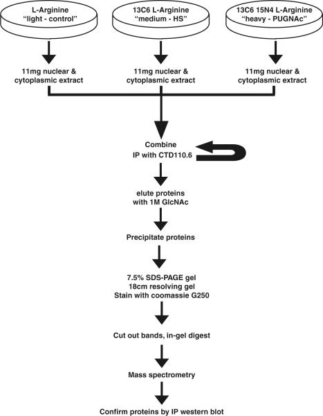
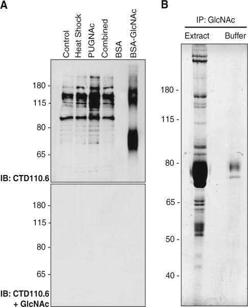
In order to identify proteins that are O-GlcNAc modified in response to stress, we used the SILAC method as outlined in Fig. 1 (Ibarrola et al. 2003; Ong et al. 2002; Wang et al. 2007). O-GlcNAc-modified proteins from isotope labeled populations were enriched by an immunoprecipitatio step, before identification and relative quantitation by mass spectrometry. Cos-7 cells were adapted and grown in media containing normal l-arginine, 13C6 l-arginine (medium), or 13C6, 15N4 l-arginine arginine (designated light, medium and heavy, respectively). Cos-7 cells were treated with heat stress 45°C (1 h, 45°C) and recovered for 1 h (medium) or PUGNAc (50 μM, 12 h; heavy). Cells grown in normal l-arginine were not treated and served as a control. Consistent with our previous data, cells subjected to a heat stress displayed elevated levels of O-GlcNAc, as did proteins isolated from cells treated with PUGNAc (an inhibitor of the O-GlcNAcase which removes O-GlcNAc). Equal protein from each treatment was combined and precipitated twice with CTD110.6, an O-GlcNAc specific antibody. To further enhance the specificity of the immunoprecipitation, the resin was washed in buffer containing 100 mM galactose, and we only analyzed proteins that were competitively eluted with 1 M GlcNAc in TBS. Proteins eluted with GlcNAc were separated by SDS–PAGE, stained with G250 coomassie (Fig. 2b), and analyzed further by mass spectrometry.
Fig. 1.
SILAC strategy used in this study
Fig. 2.
Samples used in the SILAC study. a 20 μg of isotope labeled extract (control, heat-stressed, PUGNAc-treated), or the combined extract, were separated by SDS–PAGE and O-GlcNAc levels were detected with CTD110.6. As a control, CTD110.6 was competed away with 100 mM GlcNAc. b Proteins eluted from the CTD110.6 immunoprecipitate were separated by SDS–PAGE and detected with G250 colloidal coomassie
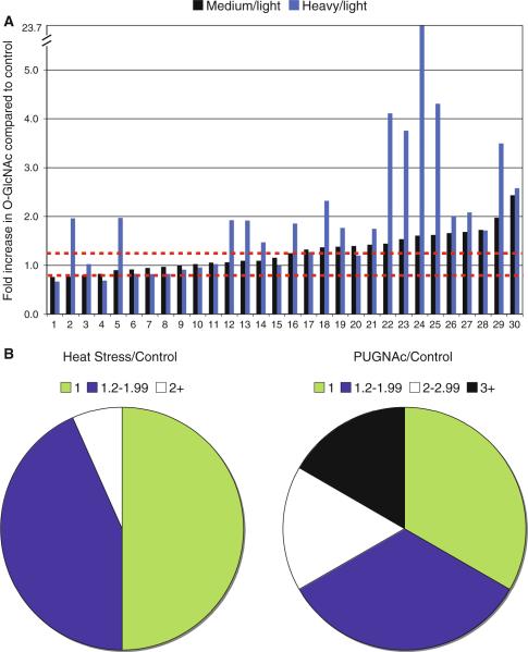
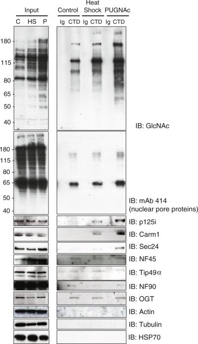
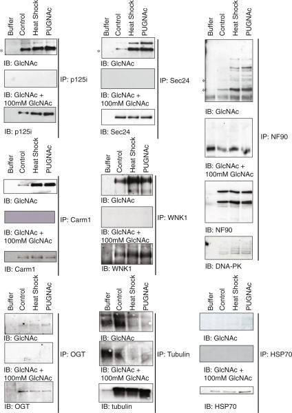
Using this stringent approach, 51 proteins were identified (Supplemental Table 1, Supplemental Table 2) of which 30 proteins contained peptides labeled by heavy isotopes. 21 proteins were statically O-GlcNAcylated, and 15 were dynamically O-GlcNAcylated in response to heat stress (Fig. 3; Table 1). Notably, several peptides appeared to be modified by O-GlcNAc, although we unable to assign which Ser/Thr residue was modified (Table 2; Supplemental Table 2). To confirm these data, and to ensure that identified proteins were pulled out specifically, we immunoprecipitated non-labeled extract (Control, Heat Stressed, and PUGNAc) with either a non-specific IgM or CTD110.6 and blotted for proteins of interest. As shown in Fig. 4, proteins identified and quantified in the screen appear to have been precipitated specifically.
Fig. 3.
Numerous proteins showed elevated levels of O-GlcNAc in response to either heat stress or PUGNAc treatment. a The fold increase in response to heat stress (black bars) or PUGNAc treatment (blue bars) are indicated. The red dashed line represents a 25% increase or decrease, which represents the error in the experiment. Proteins are numbered as in Table 1. b The distribution of proteins with a 1.2−1.99 fold or 2+ fold increase in response to heat stress are shown (left panel). Whereas proteins with a 1.2−1.99 fold, 2−2.99 fold, or 3+ fold increase in response to PUGNAc treatment (right panel) are indicated
Table 1.
Proteins identified in the MS screen
| # | kDa | Protein name (alternative names) | Heat shock | PUGNAc | Confirmed by IP/WB | Modified in Tr-H* | Previously ID'd as O-GlcNAcylated |
|---|---|---|---|---|---|---|---|
| 1 | 100 | Importin subunit beta-1 (Karyopherin subunit beta-1, Nuclear factor p97, Pore targeting complex 97 kDa subunit, Importin-90) | 0.8 | 0.7 | |||
| 2 | 42 | RAE1 (mRNA export factor, mRNA-associated protein mrnp 41, Rae1 protein homolog) | 0.8 | 2.0 | ✔ | ||
| 3 | 120 | OGT (O-linked GlcNAc transferase isoform 1) | 0.8 | 1.0 | ✔ | ✔ | ✔ |
| 4 | 100 | Transportin 1 (Importin beta-2, Karyopherin beta-2, M9 region interaction protein) | 0.8 | 0.7 | |||
| 5 | 95 | NUP98 (Nucleoporin 98 kDa) | 0.9 | 2.0 | ✔ | ✔ | |
| 6 | 150 | Nice-4 (Ubiquitin associated protein 2) | 0.9 | 0.8 | |||
| 7 | 68 | HSP70-8 (Heat shock 70 kDa protein 8) | 0.9 | 0.8 | ✔ | ||
| 8 | 55 | elF1α (Eukaryotic translation elongation factor 1 alpha 1) | 1.0 | 0.8 | |||
| 9 | 60 | Tubulin, alpha 1b | 1.0 | 0.9 | ✔ | ||
| 10 | 95 | NUP88 (Nucleoporin 88 kDa) | 1.0 | 1.0 | ✔ | ✔ | ✔ |
| 11 | 63 | NUP 62 (Nucleoporin 62 kDa) | 1.1 | 1.0 | ✔ | ||
| 12 | 140 | HCF1 (Host cell factor C1) | 1.1 | 1.9 | |||
| 13 | 120 | VP-16 (Host cell factor C1—VP16-accessory protein) | 1.1 | 1.9 | |||
| 14 | 60 | NUP54 (Nucleoporin 54 kDa) | 1.1 | 1.5 | ✔ | ✔ | ✔ |
| 15 | 240 | NUP214 (Nucleoporin 214 kDa) | 1.2 | 1.0 | ✔ | ✔ | ✔ |
| 16 | 46 | Carm-1 (Coactivator-associated arginine methyltransferase 1, Protein arginine N-methyltransferase 4) | 1.2 | 1.9 | ✔ | ||
| 17 | 55 | Tubulin, beta 5 | 1.3 | 1.3 | |||
| 18 | 150 | SEC31-like 1 isoform 1 | 1.4 | 2.3 | ✔ | ✔ | |
| 19 | 150 | SEC24b (SEC24 family, member B) | 1.4 | 1.8 | ✔ | ✔ | |
| 20 | 190 | WNK1 (WNK lysine deficient protein kinase 1, Erythrocyte 65 kDa protein) | 1.4 | 1.2 | ✔ | ✔ | ✔ |
| 21 | 150 | Nice-4 (Ubiquitin associated protein 2-like) | 1.4 | 1.7 | |||
| 22 | 140 | Zinc Finger RNA binding protein (hZFR; M-phase phosphoprotein homolog; ZFR protein; Zinc finger RNA-binding protein) | 1.4 | 4.1 | ✔ | ||
| 23 | 140 | p125i (Sec23-interacting protein p125) | 1.5 | 3.8 | ✔ | ✔ | |
| 24 | 130 | SEC24c (SEC24-related protein C) | 1.6 | 23.7 | ✔ | ✔ | ✔ |
| 25 | 46 | NF45 (Interleukin enhancer binding factor 2) | 1.6 | 4.3 | ✔ | ||
| 26 | 55 | Tip49α (RuvB-like; Nuclear matrix protein 238, 54 kDa erythrocyte cytosolic protein, TIP60-associated protein 54-alpha, INO80 complex subunit H | 1.7 | 2.0 | ✔ | ||
| 27 | 190 | NUP153 (Nucleoporin 153 kDa) | 1.7 | 2.1 | ✔ | ||
| 28 | 190 | HB×Ag transactivated protein 2 (BAT212) | 1.7 | 1.7 | |||
| 29 | 240 | Similar to gene trap ankyrin repeat | 2.0 | 3.5 | |||
| 30 | 55 | Tip49β (RuvB-like 2, Reptin 48 kDa, Repressing pontin 5, 51 kDa erythrocyte cytosolic protein, TIP60-associated protein 54-beta, INO80 complex subunit J | 2.4 | 2.6 | ✔ |
Proteins identified as differentially O-GlcNAcylated in a model of trauma hemorrhage (Teo et al. 2010)
Table 2.
O-GlcNAc modified peptides identified in this study
| Protein | Peptide |
|---|---|
| Zinc finger RNA binding protein | AGYSQGATQYTQAQQTR |
| NUP214 | LGELLFPSSLAGETLGSFSGLR |
| RAE1 | KGGRTLQLPER |
| NUP214 | ASSTSLTSTQPTK |
| Unassigned | KVIITIQYQK |
| Triosephosphate isomerase 1 | MNGRKQSLGELIGTLNAAK |
| Sec24B | SSPVVSTVLSGSSSTR |
| HCF1 | VMSVVQTKPVQTSAVTGQASTGPVTQIIQTK |
| Unassigned | ASQGVLRLLVQDR |
| Unassigned | MSVAFPSARSR |
Fig. 4.
Confirmation of proteins identified by the SILAC screen. Control, heat-stressed, or PUGNAc-treated cells were immunoprecipitated with either CTD110.6 or control IgM covalently coupled to cyanogen bromide activated Sepharose. Cell extract and immunoprecipitates were separated by SDS–PAGE and nuclear pore proteins (mAb414), NF-90, NF45, Carm1, Sec24, p125i, Tip49α, OGT, Actin, Tubulin, and HSP70 were detected by immunoblot
Based on the quantitative results we divided the O-GlcNAc-modified proteins into three groups: (1) O-GlcNAcylated in response to heat stress; (2) O-GlcNAcylated but not in response to heat stress; and (3) not O-GlcNAcylated (Fig. 3). Proteins falling into group three do not appear to be O-GlcNAc modified as the SILAC ratio is unchanged in either the heat-stressed or PUGNAc-treated group. Moreover, these proteins were not immuno-precipitated by CTD110.6 (Fig. 3) and O-GlcNAc was not detected on these proteins by IP/Western Blot (Fig. 4). We conclude that these proteins were isolated as they either interact with an O-GlcNAc-modified protein, or alternatively are present as they are highly expressed proteins in the cell and are a contaminant. Changes in the levels of O-GlcNAc on a protein could results from: (1) the O-GlcNAcylation status changing with stress; (2) The O-GlcNAcylation status of an interacting proteins changing with stress; (3) The expression of a protein changed with stress; or (4) the expression of an interacting glycoprotein changing with stress. To confirm that the proteins identified in this screen were O-GlcNAcylated in response to stress (option 1), rather that changes in expression or protein–protein interactions, a subset of the proteins were immunoprecipitated from non-labeled extract (Control, Heat Stressed, and PUGNAc) with the appropriate antibody and CTD110.6 was used to detect O-GlcNAc levels (Fig. 5; Table 1). Immunoprecipitations were also performed with either rabbit or mouse non-specific immunoglobulin (data not shown) and the signals shown in Fig. 5 appear to be specific.
Fig. 5.
Numerous proteins identified by the SILAC screen are O-GlcNAcylated dynamically in response to heat stress. Individual proteins were immunoprecipitated from control, heat-stressed, or PUGNAc-treated cells and the levels of protein, or O-GlcNAc were detected by immunoblot. An immuno-precipitation containing extraction buffer (B) was performed with the antibody of interest to confirm that bands detected by CTD110.6 did not arise from the primary antibody. As a control, CTD110.6 was competed away with 100 mM GlcNAc. In cases, such as NF-90, were numerous O-GlcNAcylated proteins are present the molecular weight of the protein of interest is indicated by an asterisk
Fifteen proteins isolated in the SILAC screen do not appear to be O-GlcNAcylated in response to heat stress; however, PUGNAc resulted in an increase in the SILAC ratio on five of these proteins suggesting that OGT, RAE1, NUP98, VP16, Sec23A and NUP54 are O-GlcNAcylated. These data suggest that in spite of the apparent global increase in O-GlcNAc levels in response to heat stress, not every O-GlcNAcylated protein is a target of OGT during heat stress. The remaining proteins appear to fall into two groups: Group 1 includes proteins such as Carm1, SEC24b, p125i, WNK1, and a subset of nuclear pore proteins. Here we observed significant changes in the SILAC ratio for both heat-stressed and PUGNAc-treated cells suggesting that the samples were O-GlcNAcylated and that the glycosylation state responds to heat stress. Much of this data was confirmed by IP/Western blot. Group 2 includes proteins such as NF45 and a sub-set of nuclear pore proteins which exhibited changed SILAC ratios, but data from IP/Western blot showed little difference in O-GlcNAcylation in response to heat stress. Together these data suggest that these proteins are likely associated with O-GlcNAcylated proteins. To investigate the O-GlcNAcylation of NF45, we immunoprecipitated NF45 and its binding partner NF90. While NF90 appears O-GlcNAc modified, and associated with numerous O-GlcNAcylated proteins, NF45 does not appear to be O-GlcNAc modified (Supplemental Fig. 1). Interestingly, during our analysis we isolated numerous peptides from the protein NICE-4. These peptides fell into two groups, those in which there was no change in the SILAC ratio, and those where we observed a consistent increase in the SILAC ratio (Heat stress = 1.4 and PUGNAc = 1.7). These data suggest that there are two isoforms of NICE-4, only one of which is O-GlcNAc modified in response to stress. Alternatively, these data suggest that there may be two populations of NICE-4, only one of which is O-GlcNAc modified in response to stress. For a number of proteins, such as Tip49α/β we were unable to determine the O-GlcNAcylation status due to their molecular weight (co-migrate with heavy chain), whereas for others we were unable to obtain an antibody that immunoprecipitated.
Turnover of O-GlcNAc on proteins appears different
As a control, one population of cells was treated with PUGNAc (an inhibitor of the O-GlcNAcase) and as expected this resulted in an increase in O-GlcNAc on many of the proteins isolated in this screen (Fig. 2a). Interestingly, the elevation in O-GlcNAc on different proteins varied (Fig. 3; Table 1), and this was supported by immuno-precipitation data (Figs. 4, 5). For some proteins PUGNAc only resulted in a small (<twofold) increase (Tubulin-β, WNK1, NUP54, and Zinc finger RNA binding protein, Fig. 3), whereas large increases in O-GlcNAcylation were observed for proteins such as Sec24c (24-fold). Possible interpretations of this data are that for proteins with a low increase in O-GlcNAc in response to PUGNAc treatment, (1) the protein is heavily populated by O-GlcNAc and thus inhibiting O-GlcNAc has little effect; or (2) the turnover of O-GlcNAc relative to the turnover of the protein is low. For proteins where we observed a robust increase in the level of O-GlcNAc in response to PUGNAc, interpretations are: (1) the protein is barely O-GlcNAcylated in basal conditions, and the fold increase is artificially inflated; or (2) that O-GlcNAc is cycling quickly on these proteins, and as such the addition of PUGNAc has a dramatic effect. Proteins involved in COPII vesicle transport fall into this group showing a dramatic increase in the levels of O-GlcNAc post-heat stress or PUGNAc treatment (Fig. 5).
DNA dependent protein kinase is a target of the O-GlcNAc transferase
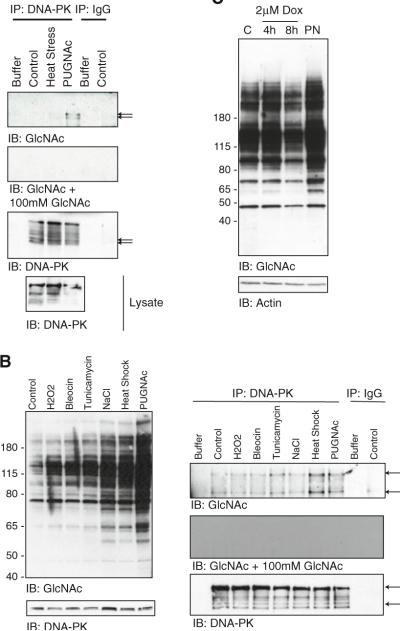
Many of the proteins identified in this screen participate in the DNA damage response or are phosphorylated during DNA damage (Table 3). As O-GlcNAc levels are known to increase in response to UV irradiation (Zachara et al. 2004), we investigated a role for O-GlcNAc in mediating DNA damage. Previously, NF90 has been reported to bind DNA-PK and while we were able to confirm this association (Fig. 5), it was not reproducibly affected by either heat stress or PUGNAc treatment (Ting et al. 1998). Interestingly, there was a high-molecular-weight “O-GlcNAc-responsive” band in the NF90 immunoprecipitation. To determine if this band was O-GlcNAcylated DNA-PK, we immunoprecipitated DNA-PK from control, heat-stressed and PUGNAc-treated Cos-7 cells and observed O-GlcNAc responsive bands that were competed away by free GlcNAc (Fig. 6a). DNA-PK appears as a number of immunoreactive bands, the source of which is unknown. Notably, O-GlcNAcylation of DNA-PK also increased in response to ER stress (Tunicamycin; Fig. 6b). We did not observe increases in DNA-PK O-GlcNAcylation in response to oxidative stress, osmotic stress, or DNA damage induced by Bleocin (also known as Bleomycin which induces double stranded DNA breaks). However, as we only immunoprecipitated DNA-PK from one time point and dose, we cannot conclude that DNA-PK is not O-GlcNAcylated in response to these forms of stress. We assume that due to the very large molecular weight of DNA-PK (460 kDa), this protein was not resolved well in the SDS–PAGE gel and as such was not identified in the SILAC screen. Unfortunately, both Bleocin and UV irradiation result in cellular damage other than DNA breaks. To determine if O-GlcNAc levels respond specifically to DNA damage, Cos-7 cells were treated with Doxorubucin which induces double stranded DNA breaks through inhibition of Topoisomerase II. In Doxorubicin treated Cos-7 cells we observed both increased and decreased levels in O-GlcNAc (Fig. 6c). Interestingly, we and others have shown, that O-GlcNAc levels can be elevated in response to stress followed by an decline (Jones et al. 2008). Together these data suggest that O-GlcNAc levels change in response to DNA damage and that O-GlcNAc may regulate the repair of DNA damage through modification of DNA-PK.
Table 3.
Functions of proteins O-GlcNAcylated in response to heat-stress
| kDa | Protein name | O-GlcNAcylated in response to stress | Complexes with O-GlcNAcylated proteins | Function | Stress-associated functions |
|---|---|---|---|---|---|
| 60 | NUP54 | – | ? | Nuclear transport | O-GlcNAcylation in response to oxidative stress is associated with inhibition of nuclear import and export (Crampton et al. 2009; Kodiha et al. 2009) |
| 240 | NUP214 | – | ✔ | Nuclear transport | O-GlcNAcylation in response to oxidative stress is associated with inhibition of nuclear import and export (Crampton et al. 2009; Kodiha et al. 2009) |
| 46 | CARM1 | ✔ | – | Methylates histone residues | CARM1 is involved in DNA damage-induced activation of GADD45 (Wysocka et al. 2006); co-activator of P53 (Scoumanne and Chen 2008); enhances tolerance to oxidative stress (Bauer et al. 2010); interacts with OGT (Cheung et al. 2008) |
| 150 | SEC31 | ? | ? | ER to Golgi Vesicle transport; binds Sec13 and p250—a phospholipase | ER stress reduces ER export (Amodio et al. 2009) |
| 150 | SEC24 B | ✔ | – | ER to Golgi Vesicle transport; binds Sec24 and p125i | Enhances survival under oxidative stress in Arabidopsis (Belles-Boix et al. 2000) |
| 190 | WNK1 | ✔ | – | Protein kinase | Involved in renal transport and hypertension (Wilson et al. 2001; Xu et al. 2005); regulation of TRPV4 (Fu et al. 2006), protects C. Elegans from hypertonic conditions (Choe and Strange 2007), regulated by hyperosmotic stress (Zagorska et al. 2007), regulates SPAK and OSR1(Vitari et al. 2005); Activates ERK5 (Xu et al. 2004); WNK3 regulates the interaction of caspase 3 activation (Verissimo et al. 2006) |
| 150 | NICE4 | ? | ? | ? | Phosphorylated upon DNA damage, probably by ATM or ATR |
| 140 | Zinc finger RNA-binding protein | ? | ? | Involved in postimplantation and gastrulation stages of development. Involved in the nucleocytoplasmic shuttling of STAU2—a protein that transports and targets mRNA | Enhances p53 transcript levels during stress |
| 140 | p125i | ✔ | – | ER to Golgi Vesicle transport; phospholipase binds sec23 | ER stress reduces ER export (Amodio et al. 2009) |
| 130 | SEC24-C | ✔ | – | ER to Golgi Vesicle transport; binds Sec24 and p125i | Enhances survival under oxidative stress in Arabidopsis (Belles-Boix et al. 2000) |
| 46 | NF45 | – | ✔ | Can regulate protein arginine N-methyltransferase 1 activity. May regulate transcription of the IL2 gene during T-cell activation. Can promote the formation of stable DNA-dependent protein kinase holoenzyme complexes on DNA. Negatively regulates miroRNA processing | Negatively regulates miroRNA processing (Sakamoto et al. 2009), micro rna expression changes in response to stress (Leung et al. 2006; Marsit et al. 2006; Reyes et al.; Sunkar; Xu et al. 2003); binds PKR (Langland et al. 1999), binds Ku80 and Ku70 (Shi et al. 2007); interacts with DNA-PK (Ting et al. 1998) |
| 55 | TIP49α | ? | ✔ | Chromatin remodeling. ATP-binding proteins that belong to the AAA + (ATPase associated with diverse cellular activities) family of ATPases | DAN Damage repair & Apoptosis (Jha and Dutta 2009) |
| 190 | NUP153 | ✔ | – | Nuclear transport | O-GlcNAcylation in response to oxidative stress is associated with inhibition of nuclear import and export (Crampton et al. 2009; Kodiha et al. 2009) |
| 240 | Similar to gene trap ankyrin repeat | ? | ? | This gene encodes a protein with ankyrin repeats, which are associated with protein–protein interactions; also binds RNA | May be involved in host-virus interactions (Yeo and Chow 2007) |
| 55 | Tip49β | ? | ? | See Pontin (Above) | See Pontin (Above) |
| Proteins Identified as O-GlcNAcylated in response to stress, but not identified in the SILAC screen | |||||
| 90 & 110 | NF-90 | ✔ | ✔ | Can regulate protein arginine N-methyltransferase 1 activity. May regulate transcription of the IL2 gene during T-cell activation. Can promote the formation of stable DNA-dependent protein kinase holoenzyme complexes on DNA. Negatively regulates miroRNA processing | Negatively regulates miroRNA processing (Sakamoto et al. 2009), micro rna expression changes in response to stress (Leung et al. 2006; Marsit et al. 2006; Reyes et al. 2010; Sunkar 2010; Xu et al. 2003); binds PKR (Langland et al. 1999), binds Ku80 and Ku70 (Shi et al. 2007); interacts with DNA-PK (Ting et al. 1998) |
| 460 | DNA -PK | ✔ | ✔ | Protein Kinase | Serine/threonine-protein kinase that acts as a molecular sensor for DNA damage. Involved in DNA nonhomologous end joining (NHEJ) required for double-strand break (DSB) repair and V(D)J recombination |
√ Indicates that definitive evidence for this option was obtained in the study
? Indicates that suggestive evidence for this option was obtained in the study
- Indicates that no evidence for this option, or alternatives, was obtained in the study
Fig. 6.
DNA-PK appears to be O-GlcNAc modified. a DNA-PK was immunoprecipitated from control, heat-stressed, or PUGNAc-treated Cos-7 cells. O-GlcNAc was detected by immunoblot. b DNA-PK immunoprecipitated from cells treated sodium chloride (100 mM, 6 h), PUGNAc (50 μM, 8 h), H2O2 (500 μM 6 h), bleocin (2.5 μg/ml, 6 h), and Tunicamycin (25 μg/ml, 18 h) was separated by SDS–PAGE and O-GlcNAc and DNA-PK were detected by immunoblot. As a control, O-GlcNAc was competed away with 100 mM GlcNAc. c Total cell extracts from Cos-7 cells treated with Doxorubicin (2 μM, 4 or 8 h), was separated by SDS–PAGE, and O-GlcNAc and Actin levels were detected by immunoblot
Discussion
Recently, the modification of nuclear, cytoplasmic, and mitochondrial proteins by O-GlcNAc has emerged as a regulator of the cellular stress response (Champattanachai et al. 2007; Chatham et al. 2008; Cheung and Hart 2008; Fulop et al. 2007a, b, c; Guinez et al. 2008; Jones et al. 2008; Laczy et al. 2009; Liu et al. 2007, 2006; Ngoh et al. 2009; Ngoh and Jones 2008; Ngoh et al. 2008; Ohn et al. 2008; Sohn et al. 2004; Yang et al. 2006; Zachara and Hart 2004a, b, 2006; Zachara et al. 2004). In response to diverse forms of cellular injury, O-GlcNAc levels are dynamically upregulated on myriad of proteins. Moreover, increasing the O-GlcNAc modification before or after cellular injury appears to regulate the ability of cells to survive a lethal insult. Given that O-GlcNAc appears to protect cells in models of heat stress, hypoxia, oxidative stress, ischemia reperfusion injury, and trauma hemorrhage, it would appear that O-GlcNAc is an integral component of the cellular stress response (Champattanachai et al. 2007; Chatham et al. 2008; Cheung and Hart 2008; Fulop et al. 2007a, b, c; Guinez et al. 2008; Jones et al. 2008; Laczy et al. 2009; Liu et al. 2007, 2006; Ngoh et al. 2009; Ngoh and Jones 2008; Ngoh et al. 2008; Ohn et al. 2008; Sohn et al. 2004; Yang et al. 2006; Zachara and Hart 2004a, b, 2006; Zachara et al. 2004). In support of this hypothesis, we and others have shown that O-GlcNAc can regulate the expression of heat shock proteins—the sentinels of the stress response (Lim and Chang 2006; Singleton and Wischmeyer 2008; Sohn et al. 2004; Zachara et al. 2004). In order to determine how O-GlcNAc mediates stress-tolerance at a molecular level, we have identified proteins that are dynamically O-GlcNAc-modified in response to heat stress.
Using SILAC technology we have identified 15 proteins that are dynamically O-GlcNAc modified (Tables 1, 3; Fig. 3), and have validated these data by immunoprecipitation and western blot (Figs. 4, 5). These proteins fall into diverse groups, including proteins involved in DNA damage, nuclear transport, transcriptional regulation, vesicle transport, and microRNA biosynthesis (Table 3). Interestingly, not every O-GlcNAc-modified protein appeared to be a target of the O-GlcNAc transferase during heat stress (Fig. 3). Recently, an independent study identified proteins that were O-GlcNAc modified in response to trauma hemorrhage. The overlap between O-GlcNAc-modified proteins identified in each study is indicated in Table 1. While some proteins are modified in response to both forms of cellular injury, other proteins only appear O-GlcNAcylated in response to one form of stress. While further research needs to be performed, these data suggest that different profiles of proteins are O-GlcNAcylated in response to different injuries. Thus some O-GlcNAcylated proteins may play a role in the central stress response others may play more specific roles.
Two of the proteins identified in this SILAC screen were Tip49α and Tip48β, which are components of many nuclear complexes involved in chromatin remodeling (Jha and Dutta 2009). Interestingly, Tip49β (also known as reptin) is one component of the Polycomb group complex PRC1. Polycomb group proteins repress transcription though a number of mechanisms, one of which is methylation of histones. Notably, the polycomb group protein, Supersexcombs has recently been shown to encode OGT. O-GlcNAcylation appears important for the activity of the PRC1 complex, of which Polyhomeotic (PH) appears to be O-GlcNAc modified (Gambetta et al. 2009; Schwartz and Pirrotta 2009; Sinclair et al. 2009). Tip49α and Tip48β are also important for the DNA damage response at multiple steps: (1) In association with Tip60, Tip49α appears important for efficient autophosphorylation and acetylation of the kinase ATM which not only phosphorylates H2AX but also promotes the induction of cell cycle checkpoints; (2) termination of the DNA damage signal through dephosphorylation of H2AX requires Tip49α; and (3) remodeling of chromatin around DNA damage to promote repair also requires Tip49α/β (Jha and Dutta 2009). Our data has highlighted a possible role for O-GlcNAc in regulating the DNA damage response. In support of O-GlcNAc playing a role in stabilizing DNA after stress, we have shown that O-GlcNAc levels respond to DNA damaging agents such as Bleocin and Doxorubicin. Moreover, DNA-PK is O-GlcNAc modified in response to various forms of cellular stress.
We isolated several proteins that are associated with protein methylation (though not in a PcG dependent manner): Carm1 and NF90. Carm1 methylates arginine residues and this has been implicated in regulating both protein function and chromatin structure though modification of histones (Barr et al. 2009; Chen et al. 2002; Cheng et al. 2007; Cheung et al. 2008; El Messaoudi et al. 2006; Scoumanne and Chen 2008; Wysocka et al. 2006). The homolog of NF-90, ILF3, is thought to associate with CARM1 and regulate its methyl transferase activity, although the biological consequence of this interaction has not been defined (Tang et al. 2000). Together, these data suggest that O-GlcNAc may regulate transcription post stress through regulating both the methylation and acetylation status of histones.
Monosaccharides of O-linked β-N-acetylglucosamine may alter transcriptional profiles in a manner independent of chromatin remodeling. Recently, two of the proteins isolated from this study have been implicated in the biogenesis of microRNAs: similar to gene trap ankyrin repeat and NF90. While no role for O-GlcNAc in regulation microRNA profiles has been defined, it has been shown that various stressors including heat stress and ischemia reperfusion injury results in profound changes in the expression of micro RNA (Cheng et al. 2009; Kulshreshtha et al. 2007a, b; Leung et al. 2006; Marsit et al. 2006; Phillips et al. 2007; Ren et al. 2009; Tang et al. 2009; Xu et al. 2009, 2003; Yin et al. 2009, 2008). Moreover, deletion of Argonaut a key protein in the biogenesis of microRNAs makes cells more susceptible to various forms of stress (Cheng et al. 2009; Tang et al. 2009; van Rooij et al. 2007; Xu et al. 2003). This study suggests that a complex network of proteins are modified and regulated by O-GlcNAc in response to stress, and this study should provide insight into the molecular basis of O-GlcNAc mediated stress tolerance.
Supplementary Material
Acknowledgments
The authors acknowledge the generous gift of antibodies by Dr. Peter N. Kao (Anti-NF-90 and NF-45), Dr. Jean-Pierre Paccaud (Anti-Sec24), Dr. Katsuko Tani (Anti-p125i), and Dr. Taka-aki Tamura (anti-Tip49α, antiTip49β). The author's work is supported by NIH grants HD R37-13563, CA R01-42486, DK-R01-61671, DK-R21/33-71280 (GWH), the National Heart, Lung, and Blood Institute, National Institutes of Health, contract No. N01-HV-28180 (GWH and AP), an NIH roadmap grant for Technology Centers of Networks and Pathways (U54RR020839), and an A*Star Research Grant to Johns Hopkins Singapore (NEZ). Under a licensing agreement between Covance Research Products and The Johns Hopkins University, Dr. Hart receives a share of royalties received by the university on sales of the CTD 110.6 antibody. The terms of this arrangement are being managed by The Johns Hopkins University in accordance with its conflict of interest policies.
Abbreviations
- Carm1 CK II
Casein kinase II
- DNA-PK
DNA-dependent protein kinase
- DMSO
Dimethylsulfoxide
- DON
6-diazo-5-oxonorleucine
- GAPDH
Glyceraldehyde-3-phosphate dehydrogenase
- FBS
Fetal bovine serum
- HSP
Heat shock protein
- MEFs
Mouse embryonic fibroblasts
- O-GlcNAc
Monosaccharides of O-linked β-N-acetylglucosamine
- O-GlcNAcase
O-GlcNAc hexosaminidase (EC 3.2.1.52)
- OGT: UDP-GlcNAc
Polypeptide O-β-N-acetylglucosaminyltransferase (EC 2.4.1.94)
- PBS
Phosphate-buffered saline
- PUGNAc
O-(2-acetamido-2-deoxy-d-glucopyranosylidene) amino-N-phenylcarbamate
- TBS
Tris-buffered saline
Footnotes
Electronic supplementary material The online version of this article (doi:10.1007/s00726-010-0695-z) contains supplementary material, which is available to authorized users.
References
- Amanchy R, Kalume DE, Pandey A. Stable isotope labeling with amino acids in cell culture (SILAC) for studying dynamics of protein abundance and posttranslational modifications. Sci STKE 2005. 2005:12. doi: 10.1126/stke.2672005pl2. [DOI] [PubMed] [Google Scholar]
- Amodio G, Renna M, Paladino S, Venturi C, Tacchetti C, Moltedo O, Franceschelli S, Mallardo M, Bonatti S, Remondelli P. Endoplasmic reticulum stress reduces the export from the ER and alters the architecture of post-ER compartments. Int J Biochem Cell Biol. 2009;41:2511–2521. doi: 10.1016/j.biocel.2009.08.006. [DOI] [PubMed] [Google Scholar]
- Barr FD, Krohmer LJ, Hamilton JW, Sheldon LA. Disruption of histone modification and CARM1 recruitment by arsenic represses transcription at glucocorticoid receptor-regulated promoters. PLoS One. 2009;4:e6766. doi: 10.1371/journal.pone.0006766. [DOI] [PMC free article] [PubMed] [Google Scholar]
- Bauer I, Graessle S, Loidl P, Hohenstein K, Brosch G. Novel insights into the functional role of three protein arginine methyltransferases in Aspergillus nidulans. Fungal Genet Biol. 2010 doi: 10.1016/j.fgb.2010.03.006. [DOI] [PubMed] [Google Scholar]
- Bauer I, Graessle S, Loidl P, Hohenstein K, Brosch G. Novel insights into the functional role of three protein arginine methyltransferases in Aspergillus nidulans. Fungal Genet Biol. 2010 doi: 10.1016/j.fgb.2010.03.006. [DOI] [PubMed] [Google Scholar]
- Belles-Boix E, Babiychuk E, Montagu MV, Inze D, Kushnir S. CEF, a sec24 homologue of Arabidopsis thaliana, enhances the survival of yeast under oxidative stress conditions. J Exp Bot. 2000;51:1761–1762. doi: 10.1093/jexbot/51.351.1761. [DOI] [PubMed] [Google Scholar]
- Champattanachai V, Marchase RB, Chatham JC. Glucosamine protects neonatal cardiomyocytes from ischemia-reperfusion injury via increased protein-associated O-GlcNAc. Am J Physiol Cell Physiol. 2007;292:C178–C187. doi: 10.1152/ajpcell.00162.2006. [DOI] [PubMed] [Google Scholar]
- Champattanachai V, Marchase RB, Chatham JC. Glucosamine protects neonatal cardiomyocytes from ischemia-reperfusion injury via increased protein O-GlcNAc and increased mitochondrial Bcl-2. Am J Physiol Cell Physiol. 2008;294:C1509–C1520. doi: 10.1152/ajpcell.00456.2007. [DOI] [PMC free article] [PubMed] [Google Scholar]
- Chatham JC, Not LG, Fulop N, Marchase RB. Hexosamine biosynthesis and protein O-glycosylation: the first line of defense against stress, ischemia, and trauma. Shock. 2008;29:431–440. doi: 10.1097/shk.0b013e3181598bad. [DOI] [PubMed] [Google Scholar]
- Chen SL, Loffler KA, Chen D, Stallcup MR, Muscat GE. The coactivator-associated arginine methyltransferase is necessary for muscle differentiation: CARM1 coactivates myocyte enhancer factor-2. J Biol Chem. 2002;277:4324–4333. doi: 10.1074/jbc.M109835200. [DOI] [PubMed] [Google Scholar]
- Cheng D, Cote J, Shaaban S, Bedford MT. The arginine methyltransferase CARM1 regulates the coupling of transcription and mRNA processing. Mol Cell. 2007;25:71–83. doi: 10.1016/j.molcel.2006.11.019. [DOI] [PubMed] [Google Scholar]
- Cheng Y, Liu X, Zhang S, Lin Y, Yang J, Zhang C. MicroRNA-21 protects against the H(2)O(2)-induced injury on cardiac myocytes via its target gene PDCD4. J Mol Cell Cardiol. 2009;47:5–14. doi: 10.1016/j.yjmcc.2009.01.008. [DOI] [PMC free article] [PubMed] [Google Scholar]
- Cheung WD, Hart GW. AMP-activated protein kinase and p38 MAPK activate O-GlcNAcylation of neuronal proteins during glucose deprivation. J Biol Chem. 2008;283:13009–13020. doi: 10.1074/jbc.M801222200. [DOI] [PMC free article] [PubMed] [Google Scholar]
- Cheung WD, Sakabe K, Housley MP, Dias WB, Hart GW. O-linked beta-N-acetylglucosaminyltransferase substrate specificity is regulated by myosin phosphatase targeting and other interacting proteins. J Biol Chem. 2008;283:33935–33941. doi: 10.1074/jbc.M806199200. [DOI] [PMC free article] [PubMed] [Google Scholar]
- Choe KP, Strange K. Evolutionarily conserved WNK and Ste20 kinases are essential for acute volume recovery and survival after hypertonic shrinkage in Caenorhabditis elegans. Am J Physiol Cell Physiol. 2007;293:C915–C927. doi: 10.1152/ajpcell.00126.2007. [DOI] [PubMed] [Google Scholar]
- Chou TY, Hart GW, Dang CV. c-Myc is glycosylated at threonine 58, a known phosphorylation site and a mutational hot spot in lymphomas. J Biol Chem. 1995;270:18961–18965. doi: 10.1074/jbc.270.32.18961. [DOI] [PubMed] [Google Scholar]
- Comer FI, Hart GW. Investigating the role of O-GlcNAc on RNA polymerase II. FASEB J. 1996;10(6):A1110–A1119. [Google Scholar]
- Crampton N, Kodiha M, Shrivastava S, Umar R, Stochaj U. Oxidative stress inhibits nuclear protein export by multiple mechanisms that target FG nucleoporins and Crm1. Mol Biol Cell. 2009;20:5106–5116. doi: 10.1091/mbc.E09-05-0397. [DOI] [PMC free article] [PubMed] [Google Scholar]
- El Messaoudi S, Fabbrizio E, Rodriguez C, Chuchana P, Fauquier L, Cheng D, Theillet C, Vandel L, Bedford MT, Sardet C. Coactivator-associated arginine methyltransferase 1 (CARM1) is a positive regulator of the Cyclin E1 gene. Proc Natl Acad Sci USA. 2006;103:13351–13356. doi: 10.1073/pnas.0605692103. [DOI] [PMC free article] [PubMed] [Google Scholar]
- Fu Y, Subramanya A, Rozansky D, Cohen DM. WNK kinases influence TRPV4 channel function and localization. Am J Physiol Renal Physiol. 2006;290:F1305–F1314. doi: 10.1152/ajprenal.00391.2005. [DOI] [PubMed] [Google Scholar]
- Fulop N, Marchase RB, Chatham JC. Role of protein O-linked N-acetyl-glucosamine in mediating cell function and survival in the cardiovascular system. Cardiovasc Res. 2007a;73:288–297. doi: 10.1016/j.cardiores.2006.07.018. [DOI] [PMC free article] [PubMed] [Google Scholar]
- Fulop N, Zhang Z, Marchase RB, Chatham JC. Glucosamine cardioprotection in perfused rat heart associated with increased O-Linked N-acetylglucosamine protein modification and altered p38 activation. Am J Physiol Heart Circ Physiol. 2007b doi: 10.1152/ajpheart.01091.2006. [DOI] [PMC free article] [PubMed] [Google Scholar]
- Fulop N, Zhang Z, Marchase RB, Chatham JC. Glucosamine cardioprotection in perfused rat hearts associated with increased O-linked N-acetylglucosamine protein modification and altered p38 activation. Am J Physiol Heart Circ Physiol. 2007c;292:H2227–H2236. doi: 10.1152/ajpheart.01091.2006. [DOI] [PMC free article] [PubMed] [Google Scholar]
- Gambetta MC, Oktaba K, Muller J. Essential role of the glycosyltransferase sxc/Ogt in polycomb repression. Science. 2009;325:93–96. doi: 10.1126/science.1169727. [DOI] [PubMed] [Google Scholar]
- Griffith LS, Schmitz B. O-linked N-acetylglucosamine is upregulated in Alzheimer brains. Biochem Biophys Res Commun. 1995;213:424–431. doi: 10.1006/bbrc.1995.2149. [DOI] [PubMed] [Google Scholar]
- Griffith LS, Schmitz B. O-linked N-acetylglucosamine levels in cerebellar neurons respond reciprocally to pertubations of phosphorylation. Eur J Biochem. 1999;262:824–831. doi: 10.1046/j.1432-1327.1999.00439.x. [DOI] [PubMed] [Google Scholar]
- Guinez C, Mir AM, Dehennaut V, Cacan R, Harduin-Lepers A, Michalski JC, Lefebvre T. Protein ubiquitination is modulated by O-GlcNAc glycosylation. FASEB J. 2008;22:2901–2911. doi: 10.1096/fj.07-102509. [DOI] [PubMed] [Google Scholar]
- Harsha HC, Molina H, Pandey A. Quantitative proteomics using stable isotope labeling with amino acids in cell culture. Nat Protoc. 2008;3:505–516. doi: 10.1038/nprot.2008.2. [DOI] [PubMed] [Google Scholar]
- Hart GW, Housley MP, Slawson C. Cycling of O-linked beta-N-acetylglucosamine on nucleocytoplasmic proteins. Nature. 2007;446:1017–1022. doi: 10.1038/nature05815. [DOI] [PubMed] [Google Scholar]
- Heilig C, Brosius F, Siu B, Concepcion L, Mortensen R, Heilig K, Zhu M, Weldon R, Wu G, Conner D. Implications of glucose transporter protein type 1 (GLUT1)-haplodeficiency in embryonic stem cells for their survival in response to hypoxic stress. Am J Pathol. 2003;163:1873–1885. doi: 10.1016/S0002-9440(10)63546-8. [DOI] [PMC free article] [PubMed] [Google Scholar]
- Hoorens A, Van de Casteele M, Klöppel G, Pipeleers D. Glucose promotes survival of rat pancreatic beta cells by activating synthesis of proteins which suppress a constitutive apoptotic program. JClin Invest. 1996;98:1568–1574. doi: 10.1172/JCI118950. [DOI] [PMC free article] [PubMed] [Google Scholar]
- Ibarrola N, Kalume DE, Gronborg M, Iwahori A, Pandey A. A proteomic approach for quantitation of phosphorylation using stable isotope labeling in cell culture. Anal Chem. 2003;75:6043–6049. doi: 10.1021/ac034931f. [DOI] [PubMed] [Google Scholar]
- Jha S, Dutta A. RVB1/RVB2: running rings around molecular biology. Mol Cell. 2009;34:521–533. doi: 10.1016/j.molcel.2009.05.016. [DOI] [PMC free article] [PubMed] [Google Scholar]
- Jones SP, Zachara NE, Ngoh GA, Hill BG, Teshima Y, Bhatnagar A, Hart GW, Marban E. Cardioprotection by N-acetylglucosamine linkage to cellular proteins. Circulation. 2008;117:1172–1182. doi: 10.1161/CIRCULATIONAHA.107.730515. [DOI] [PubMed] [Google Scholar]
- Kamemura K, Hart GW. Dynamic interplay between O-glycosylation and O-phosphorylation of nucleocytoplasmic proteins: a new paradigm for metabolic control of signal transduction and transcription. Prog Nucleic Acid Res Mol Biol. 2003;73:107–136. doi: 10.1016/s0079-6603(03)01004-3. [DOI] [PubMed] [Google Scholar]
- Kodiha M, Tran D, Morogan A, Qian C, Stochaj U. Dissecting the signaling events that impact classical nuclear import and target nuclear transport factors. PLoS One. 2009;4:e8420. doi: 10.1371/journal.pone.0008420. [DOI] [PMC free article] [PubMed] [Google Scholar]
- Kulshreshtha R, Ferracin M, Negrini M, Calin GA, Davuluri RV, Ivan M. Regulation of microRNA expression: the hypoxic component. Cell Cycle. 2007a;6:1426–1431. [PubMed] [Google Scholar]
- Kulshreshtha R, Ferracin M, Wojcik SE, Garzon R, Alder H, Agosto-Perez FJ, Davuluri R, Liu CG, Croce CM, Negrini M, et al. A microRNA signature of hypoxia. Mol Cell Biol. 2007b;27:1859–1867. doi: 10.1128/MCB.01395-06. [DOI] [PMC free article] [PubMed] [Google Scholar]
- Laczy B, Hill BG, Wang K, Paterson AJ, White CR, Xing D, Chen YF, Darley-Usmar V, Oparil S, Chatham JC. Protein O-GlcNAcylation: a new signaling paradigm for the cardiovascular system. Am J Physiol Heart Circ Physiol. 2009;296:H13–H28. doi: 10.1152/ajpheart.01056.2008. [DOI] [PMC free article] [PubMed] [Google Scholar]
- Langland JO, Kao PN, Jacobs BL. Nuclear factor-90 of activated T-cells: a double-stranded RNA-binding protein and substrate for the double-stranded RNA-dependent protein kinase, PKR. Biochemistry. 1999;38:6361–6368. doi: 10.1021/bi982410u. [DOI] [PubMed] [Google Scholar]
- Lefebvre T, Alonso C, Mahboub S, Dupire MJ, Zanetta JP, Caillet-Boudin ML, Michalski JC. Effect of okadaic acid on O-linked N-acetylglucosamine levels in a neuroblastoma cell line. Biochim Biophys Acta. 1999;1472:71–81. doi: 10.1016/s0304-4165(99)00105-1. [DOI] [PubMed] [Google Scholar]
- Lefebvre T, Ferreira S, Dupont-Wallois L, Bussiere T, Dupire MJ, Delacourte A, Michalski JC, Caillet-Boudin ML. Evidence of a balance between phosphorylation and O-GlcNAc glycosylation of Tau proteins—a role in nuclear localization. Biochim Biophys Acta. 2003;1619:167–176. doi: 10.1016/s0304-4165(02)00477-4. [DOI] [PubMed] [Google Scholar]
- Leung AK, Calabrese JM, Sharp PA. Quantitative analysis of Argonaute protein reveals microRNA-dependent localization to stress granules. Proc Natl Acad Sci USA. 2006;103:18125–18130. doi: 10.1073/pnas.0608845103. [DOI] [PMC free article] [PubMed] [Google Scholar]
- Lim KH, Chang HI. O-linked N-acetylglucosamine suppresses thermal aggregation of Sp1. FEBS Lett. 2006;580:4645–4652. doi: 10.1016/j.febslet.2006.07.040. [DOI] [PubMed] [Google Scholar]
- Liu J, Pang Y, Chang T, Bounelis P, Chatham JC, Marchase RB. Increased hexosamine biosynthesis and protein OGlcNAc levels associated with myocardial protection against calcium paradox and ischemia. J Mol Cell Cardiol. 2006;40:303–312. doi: 10.1016/j.yjmcc.2005.11.003. [DOI] [PubMed] [Google Scholar]
- Liu J, Marchase RB, Chatham JC. Increased O-GlcNAc levels during reperfusion lead to improved functional recovery and reduced calpain proteolysis. Am J Physiol Heart Circ Physiol. 2007;293:H1391–H1399. doi: 10.1152/ajpheart.00285.2007. [DOI] [PMC free article] [PubMed] [Google Scholar]
- Loberg RD, Vesely E, Brosius FC., III Enhanced glycogen synthase kinase-3beta activity mediates hypoxia-induced apoptosis of vascular smooth muscle cells and is prevented by glucose transport and metabolism. J Biol Chem. 2002;277:41667–41673. doi: 10.1074/jbc.M206405200. [DOI] [PubMed] [Google Scholar]
- Malhotra R, Brosius FC., 3rd Glucose uptake and glycolysis reduce hypoxia-induced apoptosis in cultured neonatal rat cardiac myocytes. J Biol Chem. 1999;274:12567–12575. doi: 10.1074/jbc.274.18.12567. [DOI] [PubMed] [Google Scholar]
- Marsit CJ, Eddy K, Kelsey KT. MicroRNA responses to cellular stress. Cancer Res. 2006;66:10843–10848. doi: 10.1158/0008-5472.CAN-06-1894. [DOI] [PubMed] [Google Scholar]
- Moley KH, Mueckler MM. Glucose transport and apoptosis. Apoptosis. 2000;5:99–105. doi: 10.1023/a:1009697908332. [DOI] [PubMed] [Google Scholar]
- Musicki B, Kramer MF, Becker RE, Burnett AL. Inactivation of phosphorylated endothelial nitric oxide synthase (Ser-1177) by O-GlcNAc in diabetes-associated erectile dysfunction. Proc Natl Acad Sci USA. 2005;102:11870–11875. doi: 10.1073/pnas.0502488102. [DOI] [PMC free article] [PubMed] [Google Scholar]
- Nagy T, Champattanachai V, Marchase RB, Chatham JC. Glucosamine inhibits angiotensin II-induced cytoplasmic Ca2 + elevation in neonatal cardiomyocytes via protein-associated O-linked N-acetylglucosamine. Am J Physiol Cell Physiol. 2006;290:C57–C65. doi: 10.1152/ajpcell.00263.2005. [DOI] [PubMed] [Google Scholar]
- Ngoh GA, Jones SP. New insights into metabolic signaling and cell survival: the role of beta-O-linkage of N-acetylglucosamine. J Pharmacol Exp Ther. 2008;327:602–609. doi: 10.1124/jpet.108.143263. [DOI] [PMC free article] [PubMed] [Google Scholar]
- Ngoh GA, Watson LJ, Facundo HT, Dillmann W, Jones SP. Non-canonical glycosyltransferase modulates post-hypoxic cardiac myocyte death and mitochondrial permeability transition. J Mol Cell Cardiol. 2008;45:313–325. doi: 10.1016/j.yjmcc.2008.04.009. [DOI] [PMC free article] [PubMed] [Google Scholar]
- Ngoh GA, Facundo HT, Hamid T, Dillmann W, Zachara NE, Jones SP. Unique hexosaminidase reduces metabolic survival signal and sensitizes cardiac myocytes to hypoxia/reoxygenation injury. Circ Res. 2009;104:41–49. doi: 10.1161/CIRCRESAHA.108.189431. [DOI] [PMC free article] [PubMed] [Google Scholar]
- Not LG, Marchase RB, Fulop N, Brocks CA, Chatham JC. Glucosamine administration improves survival rate after severe hemorrhagic shock combined with trauma in rats. Shock. 2007 doi: 10.1097/shk.0b013e3180487ebb. [DOI] [PubMed] [Google Scholar]
- Ohn T, Kedersha N, Hickman T, Tisdale S, Anderson P. A functional RNAi screen links O-GlcNAc modification of ribosomal proteins to stress granule and processing body assembly. Nat Cell Biol. 2008;10:1224–1231. doi: 10.1038/ncb1783. [DOI] [PMC free article] [PubMed] [Google Scholar]
- Ong SE, Blagoev B, Kratchmarova I, Kristensen DB, Steen H, Pandey A, Mann M. Stable isotope labeling by amino acids in cell culture, SILAC, as a simple and accurate approach to expression proteomics. Mol Cell Proteomics. 2002;1:376–386. doi: 10.1074/mcp.m200025-mcp200. [DOI] [PubMed] [Google Scholar]
- Pang Y, Hunton DL, Bounelis P, Marchase RB. Hyperglycemia inhibits capacitative calcium entry and hypertrophy in neonatal cardiomyocytes. Diabetes. 2002;51:3461–3467. doi: 10.2337/diabetes.51.12.3461. [DOI] [PubMed] [Google Scholar]
- Phillips JR, Dalmay T, Bartels D. The role of small RNAs in abiotic stress. FEBS Lett. 2007;581:3592–3597. doi: 10.1016/j.febslet.2007.04.007. [DOI] [PubMed] [Google Scholar]
- Ren XP, Wu J, Wang X, Sartor MA, Qian J, Jones K, Nicolaou P, Pritchard TJ, Fan GC. MicroRNA-320 is involved in the regulation of cardiac ischemia/reperfusion injury by targeting heat-shock protein 20. Circulation. 2009;119:2357–2366. doi: 10.1161/CIRCULATIONAHA.108.814145. [DOI] [PMC free article] [PubMed] [Google Scholar]
- Reyes JL, Arenas-Huertero C, Sunkar R. Cloning of stress-responsive microRNAs and other small RNAs from plants. Methods Mol Biol. 2010;639:239–251. doi: 10.1007/978-1-60761-702-0_14. [DOI] [PubMed] [Google Scholar]
- Sakamoto S, Aoki K, Higuchi T, Todaka H, Morisawa K, Tamaki N, Hatano E, Fukushima A, Taniguchi T, Agata Y. The NF90-NF45 complex functions as a negative regulator in the microRNA processing pathway. Mol Cell Biol. 2009;29:3754–3769. doi: 10.1128/MCB.01836-08. [DOI] [PMC free article] [PubMed] [Google Scholar]
- Schwartz YB, Pirrotta V. A little bit of sugar makes polycomb better. J Mol Cell Biol. 2009;1:11–12. doi: 10.1093/jmcb/mjp008. [DOI] [PubMed] [Google Scholar]
- Scoumanne A, Chen X. Protein methylation: a new mechanism of p53 tumor suppressor regulation. Histol Histopathol. 2008;23:1143–1149. doi: 10.14670/hh-23.1143. [DOI] [PMC free article] [PubMed] [Google Scholar]
- Shi L, Qiu D, Zhao G, Corthesy B, Lees-Miller S, Reeves WH, Kao PN. Dynamic binding of Ku80, Ku70 and NF90 to the IL-2 promoter in vivo in activated T-cells. Nucleic Acids Res. 2007;35:2302–2310. doi: 10.1093/nar/gkm117. [DOI] [PMC free article] [PubMed] [Google Scholar]
- Sinclair DA, Syrzycka M, Macauley MS, Rastgardani T, Komljenovic I, Vocadlo DJ, Brock HW, Honda BM. Drosophila OGlcNAc transferase (OGT) is encoded by the Polycomb group (PcG) gene, super sex combs (sxc) Proc Natl Acad Sci USA. 2009;106:13427–13432. doi: 10.1073/pnas.0904638106. [DOI] [PMC free article] [PubMed] [Google Scholar]
- Singleton KD, Wischmeyer PE. Glutamine induces heat shock protein expression via O-glycosylation and phosphorylation of HSF-1 and Sp1. JPEN J Parenter Enteral Nutr. 2008;32:371–376. doi: 10.1177/0148607108320661. [DOI] [PubMed] [Google Scholar]
- Sohn KC, Lee KY, Park JE, Do SI. OGT functions as a catalytic chaperone under heat stress response: a unique defense role of OGT in hyperthermia. Biochem Biophys Res Commun. 2004;322:1045–1051. doi: 10.1016/j.bbrc.2004.08.023. [DOI] [PubMed] [Google Scholar]
- Sunkar R. MicroRNAs with macro effects on plant stress responses. Semin Cell Dev Biol. 2010 doi: 10.1016/j.semcdb.2010.04.001. [DOI] [PubMed] [Google Scholar]
- Tallent MK, Varghis N, Skorobogatko Y, Hernandez-Cuebas L, Whelan K, Vocadlo DJ, Vosseller K. In vivo modulation of O-GlcNAc levels regulates hippocampal synaptic plasticity through interplay with phosphorylation. J Biol Chem. 2009;284:174–181. doi: 10.1074/jbc.M807431200. [DOI] [PubMed] [Google Scholar]
- Tang J, Kao PN, Herschman HR. Protein-arginine methyltransferase I, the predominant protein-arginine methyltransferase in cells, interacts with and is regulated by interleukin enhancer-binding factor 3. J Biol Chem. 2000;275:19866–19876. doi: 10.1074/jbc.M000023200. [DOI] [PubMed] [Google Scholar]
- Tang Y, Zheng J, Sun Y, Wu Z, Liu Z, Huang G. MicroRNA-1 regulates cardiomyocyte apoptosis by targeting Bcl-2. Int Heart J. 2009;50:377–387. doi: 10.1536/ihj.50.377. [DOI] [PubMed] [Google Scholar]
- Teo CF, Ingale S, Wolfert MA, Elsayed GA, Not LG, Chatham JC, Wells L, Boons GJ. Glycopeptide-specific monoclonal antibodies suggest new roles for O-GlcNAc. Nat Chem Biol. 2010;6:338–343. doi: 10.1038/nchembio.338. [DOI] [PMC free article] [PubMed] [Google Scholar]
- Ting NS, Kao PN, Chan DW, Lintott LG, Lees-Miller SP. DNA-dependent protein kinase interacts with antigen receptor response element binding proteins NF90 and NF45. J Biol Chem. 1998;273:2136–2145. doi: 10.1074/jbc.273.4.2136. [DOI] [PubMed] [Google Scholar]
- van Rooij E, Sutherland LB, Qi X, Richardson JA, Hill J, Olson EN. Control of stress-dependent cardiac growth and gene expression by a microRNA. Science. 2007;316:575–579. doi: 10.1126/science.1139089. [DOI] [PubMed] [Google Scholar]
- Verissimo F, Silva E, Morris JD, Pepperkok R, Jordan P. Protein kinase WNK3 increases cell survival in a caspase-3-dependent pathway. Oncogene. 2006;25:4172–4182. doi: 10.1038/sj.onc.1209449. [DOI] [PubMed] [Google Scholar]
- Vitari AC, Deak M, Morrice NA, Alessi DR. The WNK1 and WNK4 protein kinases that are mutated in Gordon's hypertension syndrome phosphorylate and activate SPAK and OSR1 protein kinases. Biochem J. 2005;391:17–24. doi: 10.1042/BJ20051180. [DOI] [PMC free article] [PubMed] [Google Scholar]
- Wang Z, Pandey A, Hart GW. Dynamic interplay between O-linked N-acetylglucosaminylation and glycogen synthase kinase-3-dependent phosphorylation. Mol Cell Proteomics. 2007;6:1365–1379. doi: 10.1074/mcp.M600453-MCP200. [DOI] [PubMed] [Google Scholar]
- Wang Z, Gucek M, Hart GW. Cross-talk between GlcNAcylation and phosphorylation: site-specific phosphorylation dynamics in response to globally elevated O-GlcNAc. Proc Natl Acad Sci USA. 2008;105:13793–13798. doi: 10.1073/pnas.0806216105. [DOI] [PMC free article] [PubMed] [Google Scholar]
- Wang Z, Udeshi ND, O'Malley M, Shabanowitz J, Hunt DF, Hart GW. Enrichment and site mapping of O-linked N-acetylglucosamine by a combination of chemical/enzymatic tagging, photochemical cleavage, and electron transfer dissociation mass spectrometry. Mol Cell Proteomics. 2010a;9:153–160. doi: 10.1074/mcp.M900268-MCP200. [DOI] [PMC free article] [PubMed] [Google Scholar]
- Wang Z, Udeshi ND, Slawson C, Compton PD, Sakabe K, Cheung WD, Shabanowitz J, Hunt DF, Hart GW. Extensive crosstalk between O-GlcNAcylation and phosphorylation regulates cytokinesis. Sci Signal. 2010b;3:ra2. doi: 10.1126/scisignal.2000526. [DOI] [PMC free article] [PubMed] [Google Scholar]
- Wilson FH, Disse-Nicodeme S, Choate KA, Ishikawa K, Nelson-Williams C, Desitter I, Gunel M, Milford DV, Lipkin GW, Achard JM, et al. Human hypertension caused by mutations in WNK kinases. Science. 2001;293:1107–1112. doi: 10.1126/science.1062844. [DOI] [PubMed] [Google Scholar]
- Wysocka J, Allis CD, Coonrod S. Histone arginine methylation and its dynamic regulation. Front Biosci. 2006;11:344–355. doi: 10.2741/1802. [DOI] [PubMed] [Google Scholar]
- Xu P, Vernooy SY, Guo M, Hay BA. The Drosophila microRNA Mir-14 suppresses cell death and is required for normal fat metabolism. Curr Biol. 2003;13:790–795. doi: 10.1016/s0960-9822(03)00250-1. [DOI] [PubMed] [Google Scholar]
- Xu BE, Stippec S, Lenertz L, Lee BH, Zhang W, Lee YK, Cobb MH. WNK1 activates ERK5 by an MEKK2/3-dependent mechanism. J Biol Chem. 2004;279:7826–7831. doi: 10.1074/jbc.M313465200. [DOI] [PubMed] [Google Scholar]
- Xu BE, Lee BH, Min X, Lenertz L, Heise CJ, Stippec S, Goldsmith EJ, Cobb MH. WNK1: analysis of protein kinase structure, downstream targets, and potential roles in hypertension. Cell Res. 2005;15:6–10. doi: 10.1038/sj.cr.7290256. [DOI] [PubMed] [Google Scholar]
- Xu CF, Yu CH, Li YM. Regulation of hepatic microRNA expression in response to ischemic preconditioning following ischemia/reperfusion injury in mice. OMICS. 2009;13:513–520. doi: 10.1089/omi.2009.0035. [DOI] [PubMed] [Google Scholar]
- Yang S, Zou LY, Bounelis P, Chaudry I, Chatham JC, Marchase RB. Glucosamine administration during resuscitation improves organ function after trauma hemorrhage. Shock. 2006;25:600–607. doi: 10.1097/01.shk.0000209563.07693.db. [DOI] [PubMed] [Google Scholar]
- Yeo WM, Chow VT. The VP1 structural protein of enterovirus 71 interacts with human ornithine decarboxylase and gene trap ankyrin repeat. Microb Pathog. 2007;42:129–137. doi: 10.1016/j.micpath.2006.12.002. [DOI] [PubMed] [Google Scholar]
- Yin C, Wang X, Kukreja RC. Endogenous microRNAs induced by heat-shock reduce myocardial infarction following ischemia-reperfusion in mice. FEBS Lett. 2008;582:4137–4142. doi: 10.1016/j.febslet.2008.11.014. [DOI] [PMC free article] [PubMed] [Google Scholar]
- Yin C, Salloum FN, Kukreja RC. A novel role of microRNA in late preconditioning: upregulation of endothelial nitric oxide synthase and heat shock protein 70. Circ Res. 2009;104:572–575. doi: 10.1161/CIRCRESAHA.108.193250. [DOI] [PMC free article] [PubMed] [Google Scholar]
- Zachara NE, Hart GW. O-GlcNAc a sensor of cellular state: the role of nucleocytoplasmic glycosylation in modulating cellular function in response to nutrition and stress. Biochim Biophys Acta. 2004a;1673:13–28. doi: 10.1016/j.bbagen.2004.03.016. [DOI] [PubMed] [Google Scholar]
- Zachara NE, Hart GW. O-GlcNAc a sensor of cellular state: the role of nucleocytoplasmic glycosylation in modulating cellular functions in response to nutrition and stress. Biochim Biophys Acta Gen Subj. 2004b doi: 10.1016/j.bbagen.2004.03.016. in press. [DOI] [PubMed] [Google Scholar]
- Zachara NE, Hart GW. Cell signaling, the essential role of O-GlcNAc! Biochim Biophys Acta. 2006;1761:599–617. doi: 10.1016/j.bbalip.2006.04.007. [DOI] [PubMed] [Google Scholar]
- Zachara NE, O'Donnell N, Cheung WD, Mercer JJ, Marth JD, Hart GW. Dynamic O-GlcNAc modification of nucleocytoplasmic proteins in response to stress. A survival response of mammalian cells. J Biol Chem. 2004;279:30133–30142. doi: 10.1074/jbc.M403773200. [DOI] [PubMed] [Google Scholar]
- Zagorska A, Pozo-Guisado E, Boudeau J, Vitari AC, Rafiqi FH, Thastrup J, Deak M, Campbell DG, Morrice NA, Prescott AR, et al. Regulation of activity and localization of the WNK1 protein kinase by hyperosmotic stress. J Cell Biol. 2007;176:89–100. doi: 10.1083/jcb.200605093. [DOI] [PMC free article] [PubMed] [Google Scholar]
- Zou L, Yang S, Hu S, Chaudry IH, Marchase RB, Chatham JC. The protective effects of PUGNAc on cardiac function after trauma-hemorrhage are mediated via increased protein O-GlcNAc levels. Shock. 2007;27:402–408. doi: 10.1097/01.shk.0000245031.31859.29. [DOI] [PubMed] [Google Scholar]
Associated Data
This section collects any data citations, data availability statements, or supplementary materials included in this article.


