Abstract
Background/Aims
Helicobacter pylori causes numerous extragastric manifestations, including coronary heart disease. The coronary artery calcification (CAC) score, measured using computed tomography (CT) has been used as a screening test for coronary atherosclerosis. This study investigated the association between H. pylori seropositivity and CAC scores in a screening population.
Methods
Patients who underwent a health checkup between October 2003 and July 2007 and who did not have a history of ischemic heart disease were enrolled in the study. Subjects were screened with a multidetector CT scan to determine the CAC score and for anti-H. pylori antibody immunoglobulin G; traditional risks for coronary heart disease were evaluated using a structured questionnaire, anthropometric measurements, and laboratory tests.
Results
Of the 2,029 subjects enrolled (1,295 males), 1,214 (59.8%) subjects were H. pylori positive and 815 were H. pylori negative. There were no significant differences in the baseline characteristics of the seropositive and seronegative patients. When the CAC presence or absence scores were considered, multivariate analysis revealed that H. pylori seropositivity was statistically associated with the presence of CAC and that this association was stronger in the mild CAC score category.
Conclusions
H. pylori seropositive patients are at a higher risk for coronary atherosclerosis regardless of traditional cardiovascular risk factors. This association is particularly applicable for early coronary atherosclerosis.
Keywords: Helicobacter pylori, Seropositivity, Coronary atherosclerosis, Screening
INTRODUCTION
Chronic infection with the bacteria Helicobacter pylori can cause many gastric diseases, including peptic ulcers and chronic gastritis, which can extend to gastric cancers.1-3 Moreover, it has been repeatedly reported that H. pylori infections are associated with several extragastric conditions, such as cardiovascular diseases, pulmonary diseases, neurological diseases and hematological disorders.4-6 In regards to cardiovascular or cerebrovascular diseases, H. pylori infections have been shown to be associated with stroke and coronary heart disease as a result of the atherosclerotic changes in blood vessels that arise from a chronic inflammatory response. However, many of these studies have focused on the relationship between overt diseases and H. pylori infection.6-9
In the case of coronary heart disease, the quiescent gap between asymptomatic atherosclerotic change and symptomatic coronary heart disease should be screened for more aggressively, and practitioners should recommend life style modifications to patients to reduce the likelihood of further morbidity.10 Coronary artery calcium (CAC) levels reported by computed tomography (CT) scans are widely accepted as reproducible and reliable markers for coronary heart disease risk;11,12 furthermore, CAC score is especially useful for the detection of early stage coronary atherosclerosis. According to their CAC score, a subclinical patient can be recommended life style changes and/or medication for the effective prevention of coronary heart disease.13,14
The evaluation of H. pylori infection is important for the promotion of the health of the general population, as well as for the prevention of coronary heart disease. Measuring serum immunoglobulin G antibody to H. pylori is most commonly used for population-based studies, and its accuracy has been sufficiently established through many studies across a diverse range of ethnic groups and countries, including South Korea.15-17 Even though CAC score and H. pylori serology are acceptable tests under screening conditions, to the best of our knowledge, there have not been any studies that have revealed the relationship between H. pylori infection and coronary atherosclerosis using these tests. Therefore, we investigated the association between H. pylori seropositivity and CAC score in the screening population.
MATERIALS AND METHODS
1. Subjects and ethics
We carried out a retrospective, cross-sectional case-control study. Between October 2003 and July 2007, 49,147 subjects (26,588 males, 22,559 females) visited the Seoul National University Hospital Healthcare System Gangnam Center for a routine health check-up. All of the data acquired from the check-ups were collected through an electronic medical record and stored in a data warehouse named HEALTHWATCH® version 1.0 (Seoul National University Hospital Healthcare System Gangnam Center, Seoul, Korea). We retrieved the data for and enrolled 2,029 subjects who had previously undergone their health check-ups; which included CAC scores recorded with multidetector CT, and serological tests for anti-H. pylori antibody immunoglobulin G, both tests had been performed on the same day (between October 2003 and July 2007). We excluded the subjects who had a history of coronary heart disease or acute chest pain.18
This study protocol was approved by the Institutional Review of Board (IRB) of Seoul National University Hospital (IRB No. H-1010-031-335).
2. Check-up programs in detail
1) A structured questionnaire for the analysis of pertinent health and life style factors
The questionnaire consisted of demographic information, past and current medical history, socioeconomic status, and a systematized review of symptoms including acute chest pain, questions on diet, smoking habits (current smokers; defined as having smoked greater than or equal to 100 cigarettes in their lifetime and who smoked every day or on some days around the time of examination)19 and alcohol consumption (≥140 g/wk or ≥20 g/day).20
2) Anthropometric measurements and evaluation for conventional cardiovascular risks
All subjects underwent physical measurements by trained personnel with standardized instruments. Body mass index (BMI) was calculated from weight and height and then categorized according to the modified World Health Organization (WHO) criteria from the Asia-Pacific guidelines.21,22 Waist circumference was measured within the WHO recommended area; i.e., the midpoint between the lower border of the rib cage and the iliac crest.22 On the same day, after 12 hour fasting, we also measured blood pressure and performed laboratory tests for levels of total cholesterol, triglyceride, high density lipoprotein (HDL) cholesterol, fasting serum glucose, glycosylated hemoglobin (hemoglobin A1c, HbA1c), C-reactive protein (CRP), and uric acid.
Subjects were classified with metabolic syndrome by the presence of three or more of the following criteria: 1) abdominal obesity (waist circumference >90 cm in males and >80 cm in females, as defined in the Regional Office of the Western Pacific Region of World Health Organization (WPRO) waist circumference criteria [National Cholesterol Education Program Adult Treatment Panel III]); 2) blood pressure (BP) ≥130/85 mm Hg; 3) fasting serum glucose ≥100 mg/dL; 4) serum triglyceride ≥150 mg/dL; 5) HDL-cholesterol <40 mg/dL in males or <50 mg/dL in females.21
3) Serological test for anti-H. pylori antibody immunoglobulin G
Anti-H. pylori antibody immunoglobulin G (anti-H. pylori Ab Ig G) was measured using an enzyme linked immunosorbent assay kit (Radim Diagnostics, Rome, Italy) and an automatic analyzer, Alisei® (Seac, Pomezia, Italy). H. pylori seropositivity was determined according to the manufacturer's instructions.
4) Coronary artery calcium scores recorded using multidetector CT
Scanning of the coronary artery was performed using a 16-row multi-slice CT scanner (Sensation 16; Siemens Medical Systems, Erlangen, Germany). Following a topogram of the chest, a calcium score scan was performed using a retrospective method with a tube voltage of 120 kV, and 110 effective mAs with a 200 mm field of view.23 Data were reconstructed to a 3-mm slice thickness at a -400 ms acquisition window. Calcium score analysis was performed onsite on a dedicated workstation using the analysis software Wizard VB10B® (Somaris/5 VB10B-W, SynGo; Siemens). Quantitative CAC scores were calculated according to the method described by Agatston et al.24
3. Statistical analysis
The continuous variables in this study were expressed as means with standard deviations (SD). In between-group comparisons, continuous variables were analyzed by Student's t-test and categorical variables by chi-square test. Logistic regression analysis was performed to evaluate the statistical significances of conventional cardiovascular risk factors and probable variables on CAC score. Each variable was given as an odds ratio (OR) with a 95% confidence interval (CI). A two tailed p-value of less than 0.05 was considered statistically significant. Statistical analysis was conducted using the SPSS version 12.0 (SPSS Inc., Chicago, IL, USA).
RESULTS
1. Clinical characteristics and CAC score with regards to H. pylori seropositivity
A total of 2,029 subjects (1,295 males, 734 females, age 60.4±7.9) were enrolled in this study. They were divided into two groups according to H. pylori-seropositivity; classified as either HP-positive (anti-H. pylori Ab Ig G positive) or HP-negative (anti-H. pylori Ab Ig G negative). There were 1,214 (59.8%) HP-positive and 815 (40.2%) HP-negative subjects, respectively.
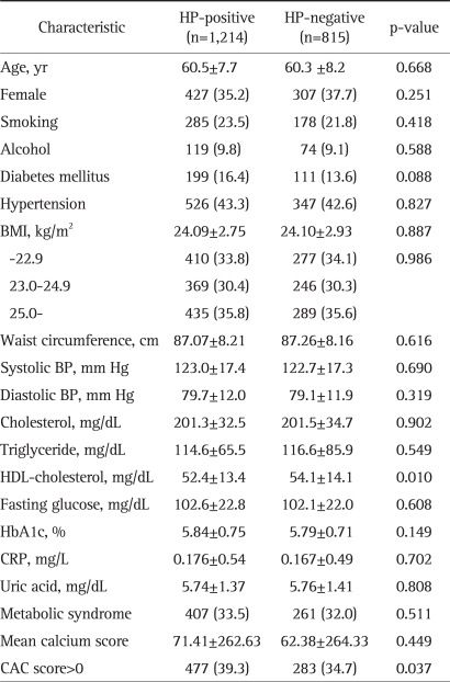
The clinical characteristics of each H. pylori seropositivity group are presented in Table 1. Baseline characteristics were not significantly different between HP-positive and HP-negative subjects. Moreover, there were no significant differences between the groups in the prevalence of metabolic syndrome, in most of the laboratory tests (except HDL-cholesterol level), and in mean CAC scores. The HP-positive group had a significantly lower mean HDL-cholesterol level (p=0.010), and a significantly higher number of subjects with a CAC score >0 (p=0.037). Diabetes was more prevalent in the HP-positive group, but the difference was not statistically significant (p=0.088).
Table 1.
Comparison of Clinical Characteristics according to H. pylori Seropositivity
Data are presented as the mean±SD or number (%).
HP, H. pylori; HP-positive, H. pylori seropositivity; HP-negative, H. pylori seronegativity; BMI, body mass index; BP, blood pressure; HbA1c, glycosylated hemoglobin, hemoglobin A1c; HDL, high density lipoprotein; CRP, C-reactive protein; CAC, coronary artery calcium.
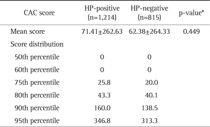
Mean CAC score was not significantly different between the HP-positive and HP-negative groups (p=0.449). The percentile distribution of CAC score according to H. pylori seropositivity is presented in Table 2. The difference of their means and SD was not statistically apparent in these studies since the distribution was highly skewed to upper score and its range was from zero to 5,060.
Table 2.
Distribution of Coronary Artery Calcium Scores according to H. pylori Seropositivity
Data are presented as the mean±SD.
CAC, coronary artery calcium; HP, H. pylori; HP-positive, H. pylori seropositivity; HP-negative, H. pylori seronegativity.
*p-value is from Student's t-test.
2. Clinical characteristics according to presence of CAC score (CAC score >0)
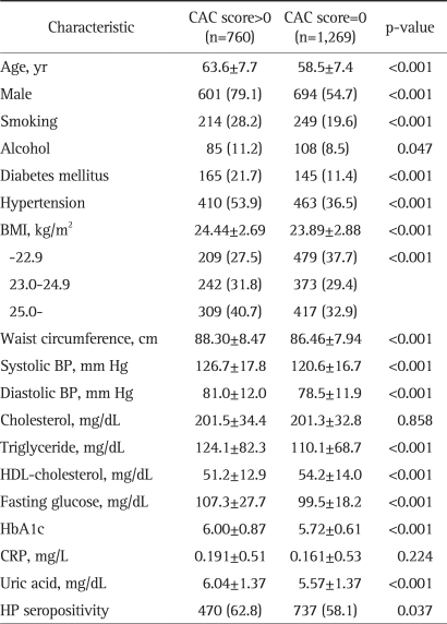
The subjects were divided into two groups according to their CAC scores: the absence of a CAC score group (CAC score=0; Absence-CAC) and the presence of a CAC score group (CAC score >0; Presence-CAC). The clinical characteristics of each group are displayed in Table 3. There were 1,269 (62.5%) subjects in the Absence-CAC and 760 (37.5%) in the Presence-CAC. The presence of a CAC score was significantly associated with multiple baseline characteristics such as age (p<0.001), sex (p<0.001), smoking (p<0.001), alcohol (p=0.047), diabetes (p<0.001), hypertension (p<0.001), BMI (p<0.001), waist circumference (p<0.001), systolic BP (p<0.001), and diastolic BP (p<0.001). From the laboratory parameters, the presence of a CAC score was also significantly related to serum triglyceride (p<0.001), HDL-cholesterol (p<0.001), fasting glucose (p<0.001), HbA1c (p<0.001), uric acid (p<0.001), and H. pylori seropositivity (p=0.037). Serum total cholesterol (p=0.858) and CRP (p=0.224) were not significantly different between the Absence-CAC and Presence-CAC groups.
Table 3.
Comparison of Clinical Characteristics according to the Score for Presence of Coronary Artery Calcium
Date are presented as the mean±SD or number (%).
CAC, coronary artery calcium; BMI, body mass index; BP, blood pressure; HbA1c, glycosylated hemoglobin, hemoglobin A1c; CRP, C-reactive protein; HP, H. pylori.
3. Univariate and multivariate analyses of the relationship between coronary risk factors and the presence of a CAC score
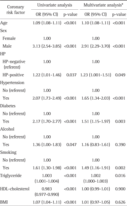
We conducted a logistic regression analysis to test for possible independent associations between the detected variables and the presence of a CAC score; the results are presented in Table 4. In the multivariate model, we included the variables that were statistically significant in the unadjusted univariate analysis, which were: age, sex, BMI, smoking, alcohol, diabetes, hypertension, serum triglyceride, HDL-cholesterol, and H. pylori seropositivity. Among these variables, several were independently associated with the presence of a CAC score in the multivariate logistic regression analysis. The significant variables (p-value, odds ratio with 95% confidence interval) were age (p<0.001; OR, 1.10; 95% CI, 1.08 to 1.11), sex (p<0.001; OR, 2.91; 95% CI, 2.29 to 3.70), H. pylori seropositivity (p=0.049; OR, 1.23; 95% CI, 1.001 to 1.51), hypertension (p<0.001; OR, 1.65; 95% CI, 1.34 to 2.03), diabetes (p=0.003; OR, 1.51; 95% CI, 1.15 to 1.97), smoking (p=0.002; OR, 1.49; 95% CI, 1.16 to 1.91), and serum triglyceride level (p=0.016; OR, 1.002; 95% CI, 1.000 to 1.003).
Table 4.
Univariate and Multivariate Analyses of Coronary Risk Factors and Coronary Artery Calcium Score
OR, odds ratio; CI, confidence interval; CAC, coronary artery calcium; HP, H. pylori; HP-positive, H. pylori seropositivity; HP-negative, H. pylori seronegativity; HDL, high density lipoprotein; BMI, body mass index.
*Multivariate model for age, sex, BMI, smoking, alcohol, diabetes, hypertension, triglyceride, HDL-cholesterol.
4. Analyses of H. pylori seropositivity against the severity categories of CAC score
Multivariate analysis conducted on the categorization of Absence-CAC (CAC score=0) and Presence-CAC (CAC score >0) verified an independent association between H. pylori seropositivity and the presence of a CAC score (p=0.049; OR 1.23; 95% CI 1.001 to 1.51) (Table 4 and detailed in Table 5).
Table 5.
Univariate and Multivariate Analyses of H. pylori Seropositivity and Coronary Artery Calcium Score
OR, odds ratio; CI, confidence interval; CAC, coronary artery calcium; HP, H. pylori; HP-positive, H. pylori seropositivity; HP-negative, H. pylori seronegativity.
*Multivariate model for age, sex, body mass index (BMI), smoking, alcohol, diabetes, hypertension, triglyceride, high density lipoprotein (HDL)-cholesterol.
To evaluate the association between H. pylori seropositivity and CAC score, CAC scores were stratified into four categories according to their severities.12-14 The severity categories were absence (CAC score=0), mild (0<CAC score≤10), moderate (10<CAC score≤100), and severe (100<CAC score). The results of the univariate and multivariate analyses of H. pylori seropositivity against the CAC severity categories are shown in Table 6. H. pylori seropositivity was strongly associated with the mild CAC category (0<CAC score≤10) (p=0.004; OR, 1.82; 95% CI, 1.22 to 2.73). However, this association was no longer discernible in the progressed CAC score categories: moderate (10<CAC score≤100) or severe (100<CAC score).
Table 6.
Univariate and Multivariate Analyses of H. pylori Seropositivity according to Coronary Artery Calcium Score Severity Categories
OR, odds ratio; CI, confidence interval; CAC, coronary artery calcium; HP, H. pylori; HP-positive, H. pylori seropositivity; HP-negative, H. pylori seronegativity; CACS, CAC score.
*Multivariate model for age, sex, body mass index (BMI), smoking, alcohol, diabetes, hypertension, triglyceride, high density lipoprotein (HDL)-cholesterol.
DISCUSSION
To the best of our knowledge, this is the first study clarifying the association between H. pylori seropositivity and CAC scores in a general asymptomatic population. Seropositivity for H. pylori antibody G within the study population was significantly related to the presence of CAC; especially for the mild CAC category, i.e., to early coronary atherosclerosis in asymptomatic subjects. Since chronic infections are known to be a predisposing factor for the development of coronary heart disease, there has been a continuous effort to investigate the relationship between H. pylori infection and coronary heart disease.7,9,25-27 Most of the studies have been conducted on diseased subjects and only revealed a weak association, which became further confused by multivariate analysis. Furthermore, those studies conducted on young healthy subjects did not find any association between H. pylori infection and coronary vessel dysfunction.28,29 This might have originated from too many confounding variables related to the overt disease and the inappropriate selection of screening methods during the subclinical stage. CAC scores regard the overall burden of coronary atherosclerosis in patients, rather than the diagnosis of specific diseased vessels. Therefore, categorized CAC scores are currently being used as a guideline for suggesting life style changes, for recommendations regarding aspirin consumption and for the monitoring of the health of the general population.13,14 It was not surprising that, H. pylori seropositivity was not significantly associated with the manifestation of moderate to severe CAC, since coronary atherosclerosis is a multi-factorial disease. However, the strong relationship between H. pylori infection and a mild CAC score suggests that H. pylori infection can play a role in early atherosclerosis, independently, as well as the many traditional risk factors.
H. pylori infection can cause vascular disease directly, as a pathogen of vessel walls,30,31 or indirectly, through the inflammatory process.32 Regarding inflammation and atherosclerosis as a result of H. pylori infection, acute vascular events, such as plaque disruption and endothelial desquamation, occur in advanced atherosclerotic vessels as a result of the actions of various inflammatory markers. In most of the previous studies conducted, symptomatic subjects have presented with elevated levels of inflammatory markers.26,33,34 In the present study, we could not discern the relationship between H. pylori infection, and acute vascular events or CRP; therefore, our results neither support nor oppose the hypothesis that H. pylori can cause early stage atherosclerosis through the inflammatory pathway. The significantly lower mean HDL-cholesterol in the HP-positive group, compared to the negative group, suggests that endothelial dysfunction might have a relationship with H. pylori infection. Indeed, some studies have found lipid profile changes and endothelial dysfunction in H. pylori infected subjects;25,35 furthermore, in these studies, the lipid profile improved after eradication of H. pylori infection.36,37
Our study has several advantages over previous clinical studies. First, this study was carried out in an asymptomatic general population. Usually, studies conducted on symptomatic study populations can blur the association between H. pylori and coronary heart disease. The subjects enrolled in this study were asymptomatic and subclinical, and, therefore, it was possible to elucidate upon the association between early coronary atherosclerosis and H. pylori infection. Second, we selected an appropriate screening tool for investigating early coronary atherosclerosis. CAC score, measured using CT, is a well validated tool for recording the multiple surrogate markers of coronary heart disease among an asymptomatic Korean population.38 Third, our health check-up program simultaneously recorded the traditional risk factors of coronary heart disease, as well as other potential risk factors. Therefore, we could test for covariation among the traditional risk factors as well as those not yet reported.
There were also limitations to this study. First, there is a gap between H. pylori seropositivity and the real ongoing infection. Although there have always been debates regarding the gold standard tests for H. pylori infection, the serological test for H. pylori is the test most commonly adopted in a mass investigation.15-17 Second, since this was a cross-sectional study, we could not elucidate upon the mechanism by which H. pylori infection causes coronary heart disease. To discover this mechanism, we need to perform a prospective cohort model study. In addition, some cross sectional studies are being designed to investigate early atherosclerosis in organs other than coronary vessels.
In conclusion, our study revealed an association between H. pylori seropositivity and the presence of a CAC score in the subclinical circumstances, and suggested that H. pylori infection could be related to the early stages of coronary atherosclerosis rather than advanced coronary atherosclerosis.
Footnotes
No potential conflict of interest relevant to this article was reported.
References
- 1.Parsonnet J, Friedman GD, Vandersteen DP, et al. Helicobacter pylori infection and the risk of gastric carcinoma. N Engl J Med. 1991;325:1127–1131. doi: 10.1056/NEJM199110173251603. [DOI] [PubMed] [Google Scholar]
- 2.Sipponen P, Hyvärinen H. Role of Helicobacter pylori in the pathogenesis of gastritis, peptic ulcer and gastric cancer. Scand J Gastroenterol Suppl. 1993;196:3–6. doi: 10.3109/00365529309098333. [DOI] [PubMed] [Google Scholar]
- 3.Kuipers EJ, Uyterlinde AM, Peña AS, et al. Long-term sequelae of Helicobacter pylori gastritis. Lancet. 1995;345:1525–1528. doi: 10.1016/s0140-6736(95)91084-0. [DOI] [PubMed] [Google Scholar]
- 4.Moyaert H, Franceschi F, Roccarina D, Ducatelle R, Haesebrouck F, Gasbarrini A. Extragastric manifestations of Helicobacter pylori infection: other Helicobacters. Helicobacter. 2008;13(Suppl 1):47–57. doi: 10.1111/j.1523-5378.2008.00634.x. [DOI] [PubMed] [Google Scholar]
- 5.Kanbay M, Kanbay A, Boyacioglu S. Helicobacter pylori infection as a possible risk factor for respiratory system disease: a review of the literature. Respir Med. 2007;101:203–209. doi: 10.1016/j.rmed.2006.04.022. [DOI] [PubMed] [Google Scholar]
- 6.Heuschmann PU, Neureiter D, Gesslein M, et al. Association between infection with Helicobacter pylori and Chlamydia pneumoniae and risk of ischemic stroke subtypes: results from a population-based case-control study. Stroke. 2001;32:2253–2258. doi: 10.1161/hs1001.097096. [DOI] [PubMed] [Google Scholar]
- 7.Mendall MA, Goggin PM, Molineaux N, et al. Relation of Helicobacter pylori infection and coronary heart disease. Br Heart J. 1994;71:437–439. doi: 10.1136/hrt.71.5.437. [DOI] [PMC free article] [PubMed] [Google Scholar]
- 8.Sawayama Y, Ariyama I, Hamada M, et al. Association between chronic Helicobacter pylori infection and acute ischemic stroke: Fukuoka Harasanshin Atherosclerosis Trial (FHAT) Atherosclerosis. 2005;178:303–309. doi: 10.1016/j.atherosclerosis.2004.08.025. [DOI] [PubMed] [Google Scholar]
- 9.Dai DF, Lin JW, Kao JH, et al. The effects of metabolic syndrome versus infectious burden on inflammation, severity of coronary atherosclerosis, and major adverse cardiovascular events. J Clin Endocrinol Metab. 2007;92:2532–2537. doi: 10.1210/jc.2006-2428. [DOI] [PubMed] [Google Scholar]
- 10.Ford ES, Ajani UA, Croft JB, et al. Explaining the decrease in U.S. deaths from coronary disease, 1980-2000. N Engl J Med. 2007;356:2388–2398. doi: 10.1056/NEJMsa053935. [DOI] [PubMed] [Google Scholar]
- 11.Rumberger JA, Simons DB, Fitzpatrick LA, Sheedy PF, Schwartz RS. Coronary artery calcium area by electron-beam computed tomography and coronary atherosclerotic plaque area: a histopathologic correlative study. Circulation. 1995;92:2157–2162. doi: 10.1161/01.cir.92.8.2157. [DOI] [PubMed] [Google Scholar]
- 12.Budoff MJ, Achenbach S, Blumenthal RS, et al. Assessment of coronary artery disease by cardiac computed tomography: a scientific statement from the American Heart Association Committee on Cardiovascular Imaging and Intervention, Council on Cardiovascular Radiology and Intervention, and Committee on Cardiac Imaging, Council on Clinical Cardiology. Circulation. 2006;114:1761–1791. doi: 10.1161/CIRCULATIONAHA.106.178458. [DOI] [PubMed] [Google Scholar]
- 13.Rumberger JA, Brundage BH, Rader DJ, Kondos G. Electron beam computed tomographic coronary calcium scanning: a review and guidelines for use in asymptomatic persons. Mayo Clin Proc. 1999;74:243–252. doi: 10.4065/74.3.243. [DOI] [PubMed] [Google Scholar]
- 14.Halliburton SS, Stillman AE, White RD. Noninvasive quantification of coronary artery calcification: methods and prognostic value. Cleve Clin J Med. 2002;69(Suppl 3):S6–S11. doi: 10.3949/ccjm.69.suppl_3.s6. [DOI] [PubMed] [Google Scholar]
- 15.Feldman RA, Evans SJ. Accuracy of diagnostic methods used for epidemiological studies of Helicobacter pylori. Aliment Pharmacol Ther. 1995;9(Suppl 2):21–31. [PubMed] [Google Scholar]
- 16.Roberts AP, Childs SM, Rubin G, de Wit NJ. Tests for Helicobacter pylori infection: a critical appraisal from primary care. Fam Pract. 2000;17(Suppl 2):S12–S20. doi: 10.1093/fampra/17.suppl_2.s12. [DOI] [PubMed] [Google Scholar]
- 17.Kim JH, Kim HY, Kim NY, et al. Seroepidemiological study of Helicobacter pylori infection in asymptomatic people in South Korea. J Gastroenterol Hepatol. 2001;16:969–975. doi: 10.1046/j.1440-1746.2001.02568.x. [DOI] [PubMed] [Google Scholar]
- 18.Yoon YE, Chang SA, Choi SI, et al. The absence of coronary artery calcification does not rule out the presence of significant coronary artery disease in Asian patients with acute chest pain. Int J Cardiovasc Imaging. doi: 10.1007/s10554-011-9819-0. Epub 2011 Feb 24. DOI: 10.1007/s10554-011-9819-0. [DOI] [PubMed] [Google Scholar]
- 19.Centers for Disease Control and Prevention (CDC) Cigarette smoking among adults--United States, 1994. MMWR Morb Mortal Wkly Rep. 1996;45:588–590. [PubMed] [Google Scholar]
- 20.Rehm J, Monteiro M, Room R, et al. Steps towards constructing a global comparative risk analysis for alcohol consumption: determining indicators and empirical weights for patterns of drinking, deciding about theoretical minimum, and dealing with different consequences. Eur Addict Res. 2001;7:138–147. doi: 10.1159/000050731. [DOI] [PubMed] [Google Scholar]
- 21.Meigs JB. The metabolic syndrome (insulin resistance syndrome or syndrome X) [Internet] Waltham: UpToDate, Inc.; 2010. [cited 2010 Sep 1]. Available from: http://www.uptodateonline.com. [Google Scholar]
- 22.World Health Organization. Obesity: prevention and managing, the global epidemic. Geneva: World Health Organization; 2000. [Google Scholar]
- 23.Choi EK, Choi SI, Rivera JJ, et al. Coronary computed tomography angiography as a screening tool for the detection of occult coronary artery disease in asymptomatic individuals. J Am Coll Cardiol. 2008;52:357–365. doi: 10.1016/j.jacc.2008.02.086. [DOI] [PubMed] [Google Scholar]
- 24.Agatston AS, Janowitz WR, Hildner FJ, Zusmer NR, Viamonte M, Jr, Detrano R. Quantification of coronary artery calcium using ultrafast computed tomography. J Am Coll Cardiol. 1990;15:827–832. doi: 10.1016/0735-1097(90)90282-t. [DOI] [PubMed] [Google Scholar]
- 25.Danesh J, Collins R, Peto R. Chronic infections and coronary heart disease: is there a link? Lancet. 1997;350:430–436. doi: 10.1016/S0140-6736(97)03079-1. [DOI] [PubMed] [Google Scholar]
- 26.Nikolopoulou A, Tousoulis D, Antoniades C, et al. Common community infections and the risk for coronary artery disease and acute myocardial infarction: evidence for chronic over-expression of tumor necrosis factor alpha and vascular cells adhesion molecule-1. Int J Cardiol. 2008;130:246–250. doi: 10.1016/j.ijcard.2007.08.052. [DOI] [PubMed] [Google Scholar]
- 27.Pasceri V, Patti G, Cammarota G, Pristipino C, Richichi G, Di Sciascio G. Virulent strains of Helicobacter pylori and vascular diseases: a meta-analysis. Am Heart J. 2006;151:1215–1222. doi: 10.1016/j.ahj.2005.06.041. [DOI] [PubMed] [Google Scholar]
- 28.Prasad A, Zhu J, Halcox JP, Waclawiw MA, Epstein SE, Quyyumi AA. Predisposition to atherosclerosis by infections: role of endothelial dysfunction. Circulation. 2002;106:184–190. doi: 10.1161/01.cir.0000021125.83697.21. [DOI] [PubMed] [Google Scholar]
- 29.Khairy P, Rinfret S, Tardif JC, et al. Absence of association between infectious agents and endothelial function in healthy young men. Circulation. 2003;107:1966–1971. doi: 10.1161/01.CIR.0000064895.89033.97. [DOI] [PubMed] [Google Scholar]
- 30.Ameriso SF, Fridman EA, Leiguarda RC, Sevlever GE. Detection of Helicobacter pylori in human carotid atherosclerotic plaques. Stroke. 2001;32:385–391. doi: 10.1161/01.str.32.2.385. [DOI] [PubMed] [Google Scholar]
- 31.Iriz E, Cirak MY, Engin ED, et al. Detection of Helicobacter pylori DNA in aortic and left internal mammary artery biopsies. Tex Heart Inst J. 2008;35:130–135. [PMC free article] [PubMed] [Google Scholar]
- 32.Libby P. Inflammation in atherosclerosis. Nature. 2002;420:868–874. doi: 10.1038/nature01323. [DOI] [PubMed] [Google Scholar]
- 33.Bunch TJ, Day JD, Anderson JL, et al. Frequency of Helicobacter pylori seropositivity and C-reactive protein increase in atrial fibrillation in patients undergoing coronary angiography. Am J Cardiol. 2008;101:848–851. doi: 10.1016/j.amjcard.2007.09.118. [DOI] [PubMed] [Google Scholar]
- 34.Rizzo M, Corrado E, Coppola G, Muratori I, Novo S. Prediction of cerebrovascular and cardiovascular events in patients with subclinical carotid atherosclerosis: the role of C-reactive protein. J Investig Med. 2008;56:32–40. doi: 10.2310/jim.0b013e31816204ab. [DOI] [PubMed] [Google Scholar]
- 35.Chimienti G, Russo F, Lamanuzzi BL, et al. Helicobacter pylori is associated with modified lipid profile: impact on Lipoprotein(a) Clin Biochem. 2003;36:359–365. doi: 10.1016/s0009-9120(03)00063-8. [DOI] [PubMed] [Google Scholar]
- 36.Scharnagl H, Kist M, Grawitz AB, Koenig W, Wieland H, März W. Effect of Helicobacter pylori eradication on high-density lipoprotein cholesterol. Am J Cardiol. 2004;93:219–220. doi: 10.1016/j.amjcard.2003.09.045. [DOI] [PubMed] [Google Scholar]
- 37.Ando T, Minami M, Ishiguro K, et al. Changes in biochemical parameters related to atherosclerosis after Helicobacter pylori eradication. Aliment Pharmacol Ther. 2006;24(Suppl 4):58–64. [Google Scholar]
- 38.Kim D, Choi SY, Choi EK, et al. Distribution of coronary artery calcification in an asymptomatic Korean population: association with risk factors of cardiovascular disease and metabolic syndrome. Korean Circ J. 2008;38:29–35. [Google Scholar]


