Abstract
The cell envelope is a crucial determinant of virulence and drug resistance in Mycobacterium tuberculosis. Several features of pathogenesis and immunomodulation of host responses are attributable to the structural diversity in cell wall lipids, particularly in the mycolic acids. Structural modification of the α-mycolic acid by introduction of cyclopropane rings as catalyzed by the methyltransferase, PcaA, is essential for a lethal, persistent infection and the cording phenotype in M. tuberculosis. Here, we demonstrate the presence of cyclopropanated cell wall mycolates in the nonpathogenic strain Mycobacterium smegmatis and identify MSMEG_1351 as a gene encoding a PcaA homologue. Interestingly, α-mycolic acid cyclopropanation was inducible in cultures grown at 25 °C. The growth temperature modulation of the cyclopropanating activity was determined by high resolution magic angle spinning NMR analyses on whole cells. In parallel, quantitative reverse transcription-PCR analysis showed that MSMEG_1351 gene expression is up-regulated at 25 °C compared with 37 °C. An MSMEG_1351 knock-out strain of M. smegmatis, generated by recombineering, exhibited a deficiency in cyclopropanation of α-mycolates. The functional equivalence of PcaA and MSMEG_1351 was established by cross-complementation in the MSMEG_1351 knock-out mutant and also in a ΔpcaA strain of Mycobacterium bovis BCG. Overexpression of MSMEG_1351 restored the wild-type mycolic acid profile and the cording phenotype in BCG. Although the biological significance of mycolic acid cyclopropanation in nonpathogenic mycobacteria remains unclear, it likely represents a mechanism of adaptation of cell wall structure and composition to cope with environmental factors.
Keywords: Bacterial Metabolism, Fatty Acid, Lipid Structure, Membrane Lipids, S-Adenosylmethionine (AdoMet), Mycobacterium smegmatis, PcaA, Cyclopropane Synthase, Mycolic Acid, Temperature
Introduction
Mycolic acids are major components of the hydrophobic cell wall of Corynebacterineae (1–4). These α-branched β-hydroxylated long chain fatty acids are covalently linked to the arabinogalactan layer that is attached to the peptidoglycan backbone and are also present as extractable lipids conjugated with sugars such as trehalose, forming trehalose dimycolate (4). Mycolic acids from mycobacteria possess a long main carbon chain called meromycolate, which in its nascent form is interrupted by double bonds at two specific sites called the proximal (closer to the β-hydroxy) and the distal positions. Chemical modifications of these double bonds give rise to different types of mycolic acids, depending on the nature of the chemical groups introduced. Mycobacterium tuberculosis carries the following three types of mycolic acids: α-subtypes, containing two cis-cyclopropane rings, and two oxygenated subtypes, harboring methoxy and keto functions on the distal position, with a cis- or trans-cyclopropane ring on the proximal site (1, 3). These chemical modifications are performed by a group of S-adenosylmethionine (AdoMet)3-dependent methyltransferases (3). These enzymes share extensive sequence similarity, but genetic studies have revealed the distinct functional characteristics of each in the biosynthesis of mycolic acids. For example, in M. tuberculosis, PcaA is required for cis-cyclopropanation at the proximal site of α-mycolates (5), whereas MmaA2 converts double bonds to cis-cyclopropane rings at the distal and proximal positions of α- and oxygenated mycolates, respectively (6), and CmaA2 introduces a trans-cyclopropane ring in oxygenated mycolates (7). MmaA4 (Hma) is required for the production of both keto and methoxy mycolates (8) and MmaA3 for the methoxy species (9). Some of these enzymes have been shown to be important for virulence (5, 8, 10, 11) and to be inhibited by (12) or required for the activation of the antitubercular drug thiacetazone (13).
The fast growing nonpathogenic Mycobacterium smegmatis species is characterized by a mycolic acid profile consisting mainly of cis/cis and cis/trans diethylenic α-mycolates, the mono-ethylenic shorter α′ subtype, as well as the epoxy mycolates (supplemental Fig. S1) (3, 14, 15). Therefore, because of the apparent lack of cyclopropane rings, M. smegmatis was used in early experiments as a surrogate strain to identify and investigate the role of M. tuberculosis mycolic acid methyltransferases. For instance, the overexpression of M. tuberculosis cmaA1 leads to the conversion of the distal cis double bond into a cis-cyclopropane ring in the α-mycolate of M. smegmatis (15). A similar strategy was used to characterize other M. tuberculosis methyltransferases, including CmaA2 (16) and MmaA2 (17). It is noteworthy that the heterologous overexpression studies yielded different results from those obtained in null mutants of the methyltransferase genes in M. tuberculosis, thus leading to conflicting interpretations of the substrate specificities of these enzymes.
Mining the M. smegmatis genome revealed the presence of at least seven predicted methyltransferases, from which only one, MSMEG_0913, has recently been characterized and shown to specifically add a methyl branch to the proximal site of α- and epoxy-mycolates (18). The presence of several related genes encoding potential AdoMet-dependent methyltransferases in this species suggests that M. smegmatis has the ability to cyclopropanate its mycolic acids, although this chemical modification may not be apparent under the standard conditions used to grow M. smegmatis in the laboratory. In contrast to M. tuberculosis, which replicates at 37 °C in the human host, M. smegmatis is a saprophytic species that dwells in the soil, where it is likely exposed to rapidly changing environmental conditions (19, 20). Therefore, we postulated that M. smegmatis utilizes this panel of methyltransferases differentially in response to environmental changes. It is well established that bacteria are able to chemically modify their phospholipids in response to environmental changes, usually by altering the acyl chains of their membrane phospholipids (21). The three main acyl chain modifications observed involve double bonds as follows: desaturation to generate an unsaturated acyl chain, cis/trans isomerization, and cyclopropanation where a methylene carbon is added across the double bond to form a three-membered ring. However, there are few data available regarding the capacity of M. smegmatis to cyclopropanate mycolic acids. In general, cyclopropanated mycolic acids are considered to be a minor component of the cell wall in this species. Some Corynebacterineae, including M. smegmatis (22), have also been reported to alter their mycolic acid profile in response to changes in culture conditions like growth temperature (23–27). Thus, in M. smegmatis, the relative amounts and chain length of the different subclasses of mycolic acids are modified according to the growth temperatures (22). Likewise, in Mycobacterium thermoresistibile, mycolic acid cyclopropanation has been shown to be modulated by this environmental factor (26).
Here, we show that mycolic acid cyclopropanation in M. smegmatis is stimulated at lower growth temperatures. In addition, we identify a previously uncharacterized gene, MSMEG_1351, encoding a putative methyltransferase, the expression of which is regulated by temperature. We demonstrate that MSMEG_1351 catalyzes cis-cyclopropanation at the proximal site of α-mycolic acids and that this activity is significantly enhanced at temperatures lower than 37 °C. Moreover, functional complementation of a Mycobacterium bovis BCG ΔpcaA mutant with MSMEG_1351 restores both the cis-cyclopropanation of α-mycolates and the cording phenotype characterizing M. bovis BCG or M. tuberculosis, strongly indicating that MSMEG_1351 is a functional orthologue of PcaA.
EXPERIMENTAL PROCEDURES
Bacterial Strains, Media, and Growth Conditions
Bacterial strains used in this study are summarized in supplemental Table S1. M. smegmatis and M. bovis BCG Pasteur 1173 P2 strains were routinely maintained at 37 °C in Sauton's medium containing 0.025% tyloxapol (Sigma) and supplemented with 25 μg/ml kanamycin and/or 50 μg/ml hygromycin when required. Transformed mycobacterial strains were selected on Middlebrook 7H10 agar plates with oleic acid/albumin/dextrose/catalase enrichment and the appropriate antibiotics (25 μg/ml kanamycin and/or 50 μg/ml hygromycin). When investigating growth temperature effects on mycobacteria, cultures were diluted below A600 ∼0.1 and incubated at the indicated temperatures until A600 reached 0.6–0.8 before harvesting the cells. For each transformation experiment, three independent clones were selected and maintained in Sauton's medium in the presence of kanamycin and hygromycin. Mycolic acid profile was verified for all three clones, and if the results were comparable, only one was retained for further analyses. Escherichia coli TOP10 strain was grown in Luria Broth agar supplemented with 25 μg/ml kanamycin when required.
DNA Manipulation
Restriction enzymes, T4 DNA ligase, and Vent DNA polymerase were purchased from New England Biolabs. Genomic DNA of M. smegmatis wild-type and mutagenized strains was extracted as described earlier (28). Briefly, glycine 1% (w/v) was added to 10 ml of late-log cultures that were then incubated around 6 h at 37 °C. Cells were pelleted and resuspended in 500 μl of 25 mm Tris/HCl, pH 8.0, 10 mm EDTA, 50 mm glucose, 1 mg/ml lysozyme and incubated overnight at 37 °C. Then 150 μl of a 2:1 (v/v) solution of 10% (w/v) SDS and 10 mg/ml proteinase K was added. This mixture was incubated at 55 °C for 20–40 min. Then 200 μl of 5 m NaCl and 160 μl of 4.1% NaCl and 10% hexadecyltrimethylammonium bromide (Sigma) were added, and the mixture was incubated for 10 min at 65 °C. Lysates were then extracted twice with 24:1 (v/v) chloroform/isoamyl alcohol, and DNA was concentrated by isopropyl alcohol precipitation. Southern blot experiments were performed with the DIG High Prime DNA labeling and detection starter kit II (Roche Applied Science) according to the manufacturer's recommendations, as described previously (29).
Construction of M. smegmatis ΔMSMEG_1351 Deletion Mutant
All genetic manipulations in M. smegmatis were carried out using a recently developed recombineering technique in mycobacteria (30). Briefly, an allelic exchange substrate for MSMEG_1351 deletion was prepared by amplifying the left and right flanking arms and cloning them on either side of the hygromycin-resistant gene in the shuttle vector pYUB854. The left arm of each gene was cloned between AflII and XbaI and the right arm between NheI and BglII. The allelic exchange substrate was then released from the vector backbone by digesting with AflII and BglII and electroporated into mc2155 expressing recombineering functions from pJV53 (30). Colonies resistant to kanamycin and hygromycin were screened for correct gene replacement by PCR, and selected clones were validated by Southern blot analysis. The same strategy was followed to disrupt genes MSMEG_0902 and MSMEG_1203. Primers used to amplify the left and right arms of each gene are listed in supplemental Table S2.
Complementation of M. smegmatis ΔMSMEG_1351
For complementation of M. smegmatis ΔMSMEG_1351, the MSMEG_1351 gene was cloned into vector pMV306 allowing stable integration of the cloned sequences in the mycobacterial genome. MSMEG_1351, including an additional 250-bp promoter-containing sequence, was amplified by PCR, using Msm mc2155 chromosomal DNA as a template and the primers 5′-GCA TGG TGC TTC TAG ACG GGA TGG GTC-3′ and 5′-CGC ATA GCG AAT TCG AAG AGC CGC TAC TTC G-3′ containing XbaI and EcoRI sites, respectively (underlined). The amplified product was cloned in the XbaI and EcoRI restriction sites in the vector pMV306, thus generating pMV306_1351. In this plasmid, the MSMEG_1351 gene is placed under the control of its own promoter-containing region. Prior to the complementation experiments, the ΔMSEG1351 mutant was cured of the recombineering plasmid pJV53 by successive subculturing of the strain in liquid medium in the absence of kanamycin, followed by plating on 7H10 agar. One of the hygromycin-resistant and kanamycin-sensitive clones was isolated and used as a host for the introduction of plasmids pMV306 or pMV306_1351.
Complementation of M. bovis BCG ΔpcaA Mutant
For complementation experiments, the mc22801 (BCG Pasteur pcaA::Tn5370) strain was transformed either with pMV261_pcaA (12) or pMV261_1351, which was constructed as follows. The gene was amplified by PCR using M. smegmatis mc2155 chromosomal DNA as a template and the primers 5′-TGA CCA AGC AGC CCG GAA AGC TA-3′ and 5′-CGC ATA GCG AAT TCG AAG AGC CGC TAC TTC G-3′ (containing an EcoRI site, underlined). The amplified product was cloned as a blunt/EcoRI fragment into the vector previously digested by MscI and EcoRI. In both pMV261_pcaA and pMV261_1351, the transgenes were placed under the control of the constitutive hsp60 promoter.
Mycolic Acid Analyses
Lipids were metabolically radiolabeled with 1 μCi/ml [2-14C]acetate (56 mCi/mmol, Amersham Biosciences) and added to mid-logarithmic phase cultures for 5–7 h. Mycolic acids were extracted as described previously (12) and analyzed by TLC on silica-coated plates. Mycolates were resolved either by normal phase TLC in hexane/ethyl acetate (19:1, v/v), run three times, or on plates impregnated with 10% silver nitrate in petroleum ether/diethyl ether (17:3, v/v), run five times (only three times in case of mycolates from M. bovis BCG strains). Two-dimensional TLC on silver nitrate-impregnated plates was carried out with hexane/ethyl acetate (19:1, v/v), run three times in the first dimension, followed by petroleum ether/diethyl ether (17:3, v/v), run five times in the second dimension (twice and three times for mycolates from M. bovis BCG strains). TLC plates were typically exposed overnight to Kodak Biomax MR films to reveal 14C-labeled lipids.
Purification and Structural Lipid Analysis
The spot corresponding to lipid Z (missing in the ΔMSMEG_1351 mutant) was purified by extraction and separation of total fatty acids and mycolic acids from a 100-ml culture of the complemented strain on 20-cm-wide preparative TLC plates. α-Mycolic acids were recovered by scraping off the plates, extracted from the silica with diethyl ether and resolved on 20-cm-wide silver nitrate impregnated TLC plates. The desired spot was then similarly recovered and extracted from the silica. Radiolabeled mycolic acid methyl esters (MAMEs) from the ΔMSMEG_1351 mutant and the complemented strain were loaded on the edges of the TLC plate as internal controls to unambiguously localize the lipid of interest. The purified lipid was then subjected to a thorough structural analysis using combination NMR and MS.
NMR Analyses
For liquid NMR, purified MAME samples were dissolved in deuterated chloroform containing 0.03% of tetramethyl silane for proton referencing and transferred into standard 5-mm tubes. Unidimensional proton experiments were recorded at 300 K on a Bruker Avance 400 spectrometer equipped with a 5-mm broad band inverse probe. For HR-MAS NMR analysis, cell pellets were washed several times with deuterium oxide to remove labile protons. HR-MAS 4-mm ZrO2 rotors (Cortecnet, France) were filled with 50 μl of cell suspension in D2O with 0.025% acetone (v/v) for proton referencing, centrifuged at low speed, and closed. Unidimensional proton spectrum was recorded at 293 K on a Bruker AvanceII 800 spectrometer (Bruker, Karlsruhe, Germany) equipped with a 1H/13C/15N 4-mm HR-MAS probe, spinning at 8 kHz, to assess the quality of the preparation. Then 10 μl of liquid were removed, replaced by 10 μl of deuterated chloroform, and stirred before recording proton experiments. The different mutant spectra were compared after normalization of the methyl signal at 0.94 ppm.
Mass Spectrometric Analyses
Mass spectrometric analyses of intact MAMEs were performed on a Voyager Elite reflectron MALDI-TOF mass spectrometer (PerSeptive Biosystems, Framingham, MA), equipped with a 337-nm UV laser. Samples were solubilized in 1 μl of chloroform/methanol (2:1, v/v) and mixed on target with 1 μl of 2,5-dihydroxybenzoic acid matrix solution (10 mg/ml dissolved in chloroform/methanol 2:1, v/v). Electronic impact mass spectrometry (EI-MS) of MAMEs was performed on a TSQ Quantum GC (Thermo Fisher Scientific Electron) using direct insertion probe with a desorption temperature ranging from 50 to 450 °C.
Quantitative Reverse Transcription-PCR
Total mRNAs were extracted following the method described earlier (28), except that a bead beater apparatus was used to disrupt the bacterial cells. Briefly, cells from 5 ml of mid-logarithmic cultures were harvested, resuspended in 1 ml of RNA protect reagent (Qiagen), and incubated for 1 h on ice. The cells were collected, resuspended in 1 ml of RLT buffer from the RNeasy kit (Qiagen), transferred in a Lysing matrix B tube (MP Bio), processed in a bead beater apparatus 45 s at maximal speed three times, and held on ice before centrifugation. Supernatants (750 μl) were mixed with 525 μl of ethanol, and RNA was purified with the RNeasy kit according to the manufacturer's instructions. Contaminating DNA was removed following DNase I treatment (Invitrogen). RNA integrity was analyzed on a BioAnalyzer 2100 machine from Agilent, using RNA nano series II chips. Double-stranded cDNA was produced using the Superscript II or III reverse transcriptase (Invitrogen). Quantitative PCR were performed using an in-house SYBR Green mixture in a light cycler instrument (Roche Applied Science), as described previously (31). The PCR program consisted of initial denaturation at 98 °C for 3 min, 45 cycles of 98 °C for 5 s, 68 °C for 10 s, and 72 °C for 10 s. The mysA gene (MSMEG_2758), encoding a σ-factor was used as an internal standard and was amplified using the primers 5′-GAA GAC ACC GAC CTG GAA CT-3′ and 5′-GAC TCT TCC TCG TCC CAC AC-3′. The MSMEG_1351 gene was amplified using the primers 5′-AGG TTC ATC GTC ACC GAG AT-3′ and 5′-TAG ACC TCC TGC GAC TGG AT-3′. Primer efficiencies, measured as described previously (31), were between 1.8 and 1.9.
Cording Assay
Cord reading agar was prepared as described previously (32, 33), except that Triton WR1339 was replaced by Triton X-100. Briefly, Triton X-100 was added to 7H10 medium enriched with oleic acid/albumin/dextrose/catalase and required antibiotics. Approximately 100 colony-forming units were plated, incubated at 37 °C, and visualized under a microscope MVX10 (Olympus) at ×100 magnification.
RESULTS
Cyclopropanation of Mycolic Acids Is Temperature-regulated in M. smegmatis
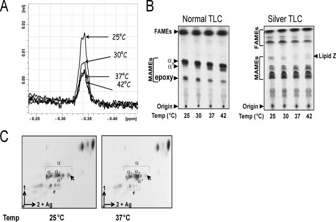
The cyclopropanation of mycolic acids in M. smegmatis mc2155 and its likely regulation by growth temperature were examined. The recently developed quantitative method of HR-MAS NMR (12) was used for the determination of the relative levels of mycolate cyclopropanation in intact cells of M. smegmatis. This method is based on the relative quantification of Ha protons of cyclopropane groups at δ −0.34 on 1H HR-MAS NMR spectra. We applied this method to directly observe and assess the content of cis-cyclopropane rings in M. smegmatis grown at different temperatures. As shown in Fig. 1A, cis-cyclopropane signals detected by 1H HR-MAS NMR increased gradually as an inverse function of the temperature, suggesting that lipid cyclopropanation is regulated by growth temperature in M. smegmatis. Indeed, differential integration of cyclopropane 1H NMR signals demonstrated that the total cyclopropane content increased by 150% in cells grown at 25 °C, compared with those grown at 42 °C.
FIGURE 1.
Regulation of mycolic acid cyclopropanation by growth temperature in M. smegmatis. Mycobacterial cultures grown at the indicated temperatures were processed for either in vivo relative quantification of cis-cyclopropane by 1H HR-MAS NMR (A) or for 14C-radiolabeling and extraction of mycolic acid (B and C). Mycolates were resolved on a normal TLC plate (B, left panel), a silver nitrate-impregnated TLC plate (B, right panel), or by two-dimensional TLC (C). FAME, fatty acid methyl ester. The new spot accumulating at low growth temperatures, termed lipid Z in B is indicated by an arrow. α1 and α2 correspond to the cis/cis and cis/trans di-unsaturated α-mycolic acids, respectively. e, epoxy mycolic acids.
Cyclopropanation of mycolates was assessed in M. smegmatis grown at different temperatures and subsequently metabolically labeled with [14C]acetate. Mycolic acids were extracted and analyzed by TLC using different solvent systems. A striking difference in the ratio of the α-, α′-, and epoxy-mycolate subclasses was observed at different temperatures (Fig. 1B, left panel). In particular, at 25 °C, the synthesis of α′-mycolates was almost completely abrogated, whereas a marked increase in the synthesis of epoxy-mycolates was observed at the lowest temperatures, consistent with the previous findings (22). Unexpectedly, when the same samples were separated on silver nitrate-impregnated TLC plates, we observed the presence of an additional band (referred to as lipid Z) appearing essentially at temperatures lower than 37 °C (Fig. 1B, right panel). Because silver nitrate retards migration of unsaturated lipids relative to cyclopropanated lipids, these results suggested that a new cyclopropanated lipid was produced during growth at low temperatures. Resolution on two-dimensional argentation TLC suggested that the radiolabeled spot accumulating at 25 °C corresponded to cyclopropanated mycolates belonging to the α-mycolate subclass (Fig. 1C). These data support the conclusion that cyclopropanation occurs in M. smegmatis and are strongly enhanced at growth temperatures below 37 °C.
MSMEG_1351 Mutant Fails to Produce the Lipid Accumulating at Low Temperature
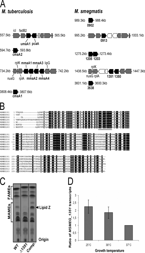
An in silico search for genes encoding mycolic acid methyltransferases in M. smegmatis mc2155 revealed the presence of at least seven putative coding regions (18). These seven open reading frames are localized to five different loci (Fig. 2A) and share 50–70% identity (Fig. 2B). Two of these, annotated MSMEG_1351 and MSMEG_1350, are present in the mmaA1–mmaA-containing region, although one, MSMEG_0913, is in the umaA1–pcaA region. In addition, M. smegmatis has four additional paralogues designated MSMEG_1205, MSMEG_1203, MSMEG_3538, and MSMEG_0902. The regions corresponding to cmaA1 and cmaA2 in M. tuberculosis are not found in M. smegmatis.
FIGURE 2.
Identification of the genes involved in the production of the cyclopropanated mycolates. A, schematic representation and genomic organization of the potential methyltransferases in M. smegmatis and comparison with M. tuberculosis. Black boxes indicate the mycolic acid methyltransferases; gray boxes indicate open reading frames encoding conserved proteins between the two species, and white boxes represent open reading frames unique to either M. smegmatis or M. tuberculosis. Coordinates of each gene cluster are indicated in kb. B, alignment of the M. smegmatis putative methyltransferases. The AdoMet-binding site as described previously (14) is underlined. C, argentation TLC of 14C-radiolabeled mycolic acids from the parental strain (1st lane), the ΔMSMEG_1351 mutant carrying either the empty pMV306 (2nd lane) or pMV306_1351 (3rd lane), all grown at 25 °C. The arrowhead indicates the position of lipid Z that is lacking in the ΔMSMEG_1351 mutant. D, temperature-regulated expression of MSMEG_1351. Wild-type M. smegmatis was grown at the indicated temperature, and the levels of MSMEG_1351 transcripts relative to those of the RNA polymerase σ-factor mysA gene (MSMEG_2758) were measured by quantitative reverse transcription-PCR. MSMEG_1351 transcript levels at the different temperatures were normalized to those from cells growing at 37 °C. Results are expressed as means ± S.D. from four independent experiments.
To validate M. smegmatis cyclopropanation activity, we generated M. smegmatis mutants in genes encoding putative methyltransferases by introducing a hygromycin resistance cassette into three different genes located within different loci (MSMEG_1351, MSMEG_1203, and MSMEG_0902) to generate independent, single mutant strains. Disruption of the open reading frames in each strain was confirmed by PCR analysis (data not shown) as well as by Southern blotting using a hygromycin probe (supplemental Fig. S2). Growth temperature-dependent cyclopropanation of cell wall mycolic acids in the three mutant strains was evaluated. Cultures were incubated either at 25 or 37 °C and labeled with [14C]acetate followed by mycolic acid analysis by two-dimensional TLC. There were no significant differences in mycolate profiles of the MSMEG_0902 or MSMEG_1203-deficient strains (supplemental Fig. S3). In contrast, as shown in Fig. 2C, the cyclopropanated lipid Z that accumulated at 25 °C in the parental strain was hardly detectable in the ΔMSMEG_1351 mutant strain. Except for this particular band, both parental and mutant strains exhibited a comparable mycolic acid profile. The absence of lipid Z in ΔMSMEG_1351 mutant affected neither the growth (supplemental Fig. S4) nor the colony morphology of the mutant (data not shown). Importantly, complementation with a plasmid-borne copy of the corresponding gene fully restored the production of this lipid. Interestingly, lipid Z reproducibly accumulated at higher amounts in the complemented strain compared with the parental strain (Fig. 2C). In the complementation construct, the MSMEG_1351 gene was placed under the control of its own promoter region, a 250-bp segment that may contain regulatory signals that allow expression to be induced at low temperatures. Restoration of the parental profile also ruled out any possible polar effect due to the introduction of the hygromycin cassette in the MSMEG_1351 locus. Taken together, these studies identify MSMEG_1351 as a gene necessary for the production of a cyclopropanated α-mycolate species that is synthesized at low temperature.
MSMEG_1351 Expression Is Regulated by Growth Temperature
Because the appearance of the cyclopropanated lipid Z was dependent on both the growth temperature and the presence of MSMEG_1351, we hypothesized that MSMEG_1351 gene expression is regulated by temperature. The transcript levels of MSMEG_1351 were estimated by quantitative PCR in M. smegmatis mc2155 grown at 25, 30, and 37 °C. Results presented in Fig. 2D clearly show a significant 2-fold up-regulation of the MSMEG_1351 mRNA in four independent experiments at 25 °C compared with 37 °C, when using the mysA gene (MSMEG_2758) as an internal standard. Similar results were obtained using rpsL as an internal control (data not shown). These data confirm that MSMEG_1351 is a thermally regulated gene and are in agreement with the synthesis of lipid Z at this temperature.
Structure of the MSMEG_1351-dependent Mycolic Acid
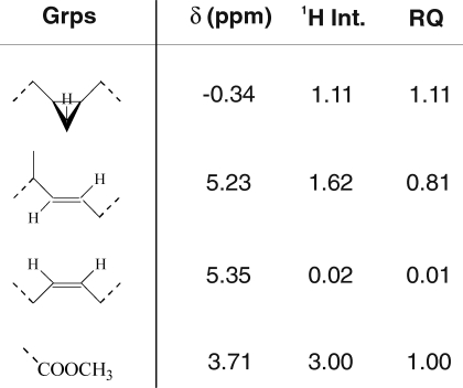
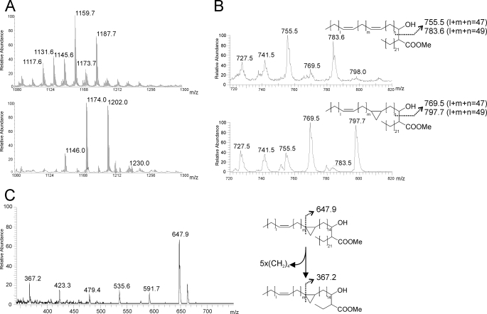
Cultures of the complemented strain grown at 25 °C were considered a good source of lipid Z, as it is produced in higher amounts in this strain compared with the mutant or parental strains (Fig. 2C). The extracted mycolic acids were purified as described under “Experimental Procedures” and subjected to a detailed structural analysis. First, functional groups corresponding to mycolates were identified by 1H NMR based on assignments of characteristic protons from trans-ethylenic groups (5.23 ppm), cis-ethylenic groups (5.35 ppm), carboxymethyl groups (3.71 ppm), and cis-cyclopropane groups (−0.34 ppm) (Table 1), in agreement with previously published parameters (12). Integration of individual 1H signals normalized on the –COOCH3 signal indicated an average of 1.11 cis-cyclopropane group and 0.81 trans-ethylenic group per molecule (Table 1), thus strongly suggesting that the purified mycolate is a mono-cyclopropanated α-type mycolate. No trans-cyclopropanes could be observed on the same spectrum. Furthermore, MALDI-TOF MS analysis of lipid Z showed two [M + Na]+ major signals at m/z 1174 and 1202, indicative of the presence of a family of C78 and C80 α-mycolic acids (Fig. 3A, lower panel) (34). By comparison, the α-mycolates devoid of lipid Z from M. smegmatis grown at 25 °C, as well as total α-mycolates purified from M. smegmatis grown at 42 °C (data not shown), exhibited a more complex pattern dominated by two major peaks at m/z 1160 and 1188, indicative of the presence of two major C77 and C79 α-mycolic acid subtypes (Fig. 3A, upper panel), as established previously in M. smegmatis (18). Comparison of high and low mobility α-mycolates on silver-impregnated TLC established that lipid Z was substituted on average by an additional –CH2– group, in accordance with the observation in NMR of a single cis-cyclopropane group per mycolate. Next, the location of the cyclopropane group on the meromycolyl chain was investigated by in-source pyrolysis of purified α-mycolates and analysis in electronic impact-mass spectrometry (EI-MS) (Fig. 3B). Indeed, the 14 mass unit increment observed by MALDI-MS on intact molecules was associated with a family of [M + H]+ signals at m/z 755/783 in α-mycolates devoid of lipid Z and to a family of signals at m/z 769/797 in lipid Z assigned to the meromycolyl chain of noncyclopropanated and mono-cyclopropanated α-mycolates, respectively. Accordingly, pyrolysis of both α-mycolates also generated a [M + H]+ signal at m/z 382 attributed to the fatty C24 acid chain, including the carboxyl group (data not shown). Finally, EI-MS analysis of intact lipid Z generated an intense primary fragment ion at m/z 648, originating from the cleavage of meromycolyl chain next to the cyclopropane group (Fig. 3C) (35). This fragment is further degraded by recurrent fragmentation of the fatty acyl chain of the α-mycolate that eventually generates a secondary fragment presenting a residual C3 fatty acid chain observed at m/z 367.2. Taken together, the data from NMR, MALDI-MS, and EI-MS establish that lipid Z is a mono-unsaturated, mono-cyclopropanated α-mycolate and strongly suggest that the cis-cyclopropane ring is located at the proximal position in the meromycolyl chain.
TABLE 1.
Identification and quantification of functional groups from lipid Z
δ indicates 1H NMR chemical shifts of indicative protons; 1H int. indicates integrations of 1H signals, and RQ indicates relative quantitation of functional groups.
FIGURE 3.
Structural analysis of lipid Z. A, MALDI-MS analysis of intact carboxymethylated mycolic acids from α-mycolic acids devoid of lipid Z (upper panel) and lipid Z (lower panel). B, EI-MS analysis of in-source pyrolysis fragment generated from a mixture of α-mycolates lacking lipid Z (upper panel) or containing lipid Z (lower panel). C, EI-MS analysis of fragment ions generated from carboxymethylated lipid Z. Cis- and trans-conformation of functional groups were omitted in schematic representations of mycolates for the sake of clarity.
MSMEG_1351 Can Replace pcaA in M. bovis BCG
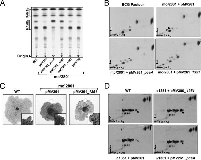
The above structural analyses of lipid Z suggest that MSMEG_1351 is a methyltransferase involved in cis-cyclopropanation of the proximal double bond in α-mycolates. In M. tuberculosis or M. bovis BCG, PcaA is required for the introduction of the proximal cis-cyclopropane ring of the α-mycolates (5). Therefore, we investigated whether MSMEG_1351 and PcaA are functionally equivalent by complementing M. bovis BCG mc22801 (BCG Pasteur pcaA::Tn5370) with MSMEG_1351, placed either under the control of its own promoter (pMV306_1351) or the constitutive hsp60 promoter (pMV261_1351). As shown in Fig. 4, A and B, M. bovis BCG mc22801 presented an altered α-mycolic acid profile as reported earlier (5), which could be restored by complementation through overexpression of pcaA. A similar mycolate profile was observed following overexpression of MSMEG_1351 in M. bovis BCG mc22801 via the constitutively strong promoter hsp60. When expressed from its native promoter, lower but reproducible levels of cis-cyclopropanated α-mycolic acids were obtained, which can be attributed to the fact that M. bovis BCG mc22801 was grown at 37 °C, a temperature where MSMEG_1351 gene expression is only mildly induced (Fig. 2D). Similarly, overexpression of pcaA in the ΔMSMEG_1351 mutant fully restored the production of the cyclopropanated α-mycolic acid lacking in this mutant (Fig. 4D). We conclude from these results that MSMEG_1351 and PcaA are functionally equivalent.
FIGURE 4.
Complementation of M. bovis BCG ΔpcaA M. smegmatis ΔMSMEG_1351 mutants. Complementation of M. bovis BCG ΔpcaA by MSMEG_1351. One-dimensional (A) and two-dimensional (B) argentation TLC of 14C-radiolabeled mycolic acids from M. bovis BCG Pasteur and BCG mc22801 (BCG Pasteur pcaA::Tn5370) transformed with various vectors and grown at 37 °C. The arrowhead indicates the position of the hybrid mycolate accumulating in the ΔpcaA mutant, with a cis double bond at the proximal position in place of a cis-cyclopropane present in the wild-type α-mycolate as determined previously (5). α, α-mycolates; K, keto-mycolates; WT, wild type. C, single BCG colonies grown on cord reading agar and photographed after 15 days of incubation at 37 °C. Magnification, ×100. D, complementation of M. smegmatis ΔMSMEG_1351 by pcaA. Two-dimensional TLC analysis of 14C-radiolabeled mycolic acids from M. smegmatis ΔMSMEG_1351 transformed either with pMV261, pMV306_1351, or pMV261_pcaA grown at 25 °C. Arrows indicate the position of the cyclopropanated mycolate missing in the ΔMSMEG_1351 mutant.
MSMEG_1351 Restores the Cording Phenotype of BCG ΔpcaA
BCG ΔpcaA mutant was first recognized for its defect in cord formation (5). This prompted us to investigate if MSMEG_1351 could also replace pcaA for the cording phenotype in BCG. As shown in Fig. 4C, transformation of M. bovis BCG mc22801 with MSMEG_1351 restored the ability to form the characteristic long serpentine cords.
DISCUSSION
Cyclopropanation is a well studied structural modification of mycolic acids typically seen in slow growing pathogenic mycobacteria. In contrast, fast growing species produce large amounts of unsaturated mycolic acids (14, 15, 36). In a recent study, Laval et al. (18) demonstrated the presence of cyclopropanated mycolates in the fast growing M. smegmatis by characterization of the methyltransferase MSMEG_0913 (UmaA1). It was demonstrated that a MSMEG_0913 deletion mutant no longer synthesized mycolates containing a methyl branch adjacent to either a trans-cyclopropyl group or a trans double bond at the proximal position of both α- and epoxy-mycolates. Surprisingly, complementation of this mutant with the putative M. tuberculosis mycolic acid methyltransferase UmaA1 did not restore the wild-type phenotype, indicating that umaA1 and MSMEG_0913 are not functionally orthologous, despite their synteny and high similarity (18). This study clearly indicates that MSMEG_1351 and PcaA (sharing 67% amino acid identity) are functionally orthologous, as MSMEG_1351 could complement a BCG ΔpcaA mutant and, conversely, pcaA restored a wild-type mycolic acid profile in the M. smegmatis ΔMSMEG_1351 mutant.
This study not only confirms the presence of cyclopropanation in M. smegmatis but also highlights the ability of this species to regulate cyclopropanation according to growth temperature. It is very likely that the inherent capacity of cyclopropanation has been overlooked in many earlier studies because of culturing M. smegmatis at 37 °C, where cyclopropanation is virtually absent. Here, we provide compelling evidence that M. smegmatis has a propensity to introduce cyclopropane rings in α-mycolates at growth temperatures below 37 °C and that this activity is catalyzed by MSMEG_1351. Inactivation of this gene in M. smegmatis abolished the production of cyclopropanated α-mycolates that accumulate at low temperatures, a phenotype that was reversed following complementation of the mutant with a functional MSMEG_1351 gene.
PcaA has been shown to be required for the cording phenotype in the tubercle bacillus and for its long term persistence in mice (5) and is thought to be absent in M. smegmatis. Only one gene, MSMEG_0913, is present in the genomic region of M. smegmatis that is “equivalent” to the umaA1-pcaA gene cluster in M. tuberculosis (Fig. 2A), and it has been assigned a clear function, distinct from the one catalyzed by PcaA (18). In M. tuberculosis, inactivation of pcaA is responsible for virulence attenuation, while stimulating a less severe granulomatous pathology in the mouse model (5). This was further validated by demonstrating that purified trehalose dimycolate from the ΔpcaA strain was hypo-inflammatory for macrophages (11). The presence of persistent bacteria is considered to be the major reason for a lengthy therapy (37). Therefore, genes such as pcaA have been proposed to be attractive targets for the development of drugs against persistent bacilli (5, 38). The determination of the crystal structure of PcaA has provided a foundation for rational drug design for the development of new inhibitors effective against persistent bacteria (39). Interestingly, subsequent work demonstrated that PcaA, along with other cyclopropane mycolic acid synthases, was extremely sensitive to inhibition by thiacetazone, a second line antitubercular drug (12). Another molecule dioctylamine, acts as a substrate mimic and inhibits the function of multiple cyclopropane mycolic acid synthases, including PcaA, resulting in loss of cyclopropanation, cell death, loss of acid fastness, and synergistic killing with isoniazid and ciprofloxacin (40).
The occurrence of an orthologue of a pathogenicity gene, pcaA, in a saprophytic species is rather intriguing. Although virulence factors typical of pathogenic species are thought to be absent or inactivated in nonpathogenic strains, this deserves to be re-examined. Our study revealed that expression of MSMEG_1351 was low at 37 °C, based on quantitative reverse transcription-PCR analysis as well as by the apparent lack of cyclopropanated α-mycolates. However, relative amounts of MSMEG_1351-specific transcripts increased significantly at lower growth temperatures, and this correlated with the accumulation of cis-cyclopropane rings, as indicated by biochemical and structural analyses and also directly on whole cells by HR-MAS NMR. However, in contrast to pknG (a virulence gene mediating intracellular survival of M. tuberculosis), which is not translated in M. smegmatis (41), expression of pcaA (MSMEG_1351) was temperature-dependent, and further work will be required to identify the regulatory region(s) upstream of MSMEG_1351. These results indicate that the pathogenicity-associated pcaA gene is differentially expressed in pathogenic and nonpathogenic mycobacteria.
The mycobacterial cell wall has a variable composition allowing the bacteria to maintain a constant viscosity in the face of changing environmental conditions (42, 43). Several mycobacterial species, including M. smegmatis (22, 24, 26), have been shown to respond to changing growth temperatures by adjusting the proportion of lipids found in the trans-configuration. These alterations in lipid structure are not in the plasma membrane lipids but rather in the mycolic acids, as would be expected because the cell wall represents the major permeability barrier in the mycobacteria. In M. smegmatis, this alteration results in changes in the geometry of the major olefinic mycolic acids, and in slow growing species, such as Mycobacterium avium, this alteration occurs through changes in the geometry about cyclopropane rings within the longer meromycolate chain. This alteration has been shown to directly correlate with changes in cell wall permeability to lipophilic agents (44). We therefore investigated whether proximal cyclopropanation alters cell wall permeability by testing the effect of various drugs, including rifampicin, isoniazid, erythromycin, and crystal violet, in the wild-type, the mutant, and the complemented strains. Comparable minimal inhibitory concentrations were obtained regardless of the strain (data not shown), suggesting that the loss or the gain of proximal cyclopropanation does not influence susceptibility to these drugs and cell wall permeability.
Because free mycolates have been shown to be important components of mycobacterial biofilms (45), and because M. smegmatis biofilms are formed at 30 °C (46), a temperature that is permissive to proximal cyclopropanation, we investigated whether proximal cyclopropanation affects biofilm formation. However, we did not observe any biofilm defect with the ΔMSMEG_1351 strain (data not shown).
The biological significance of mycolic acid cyclopropanation of saprophytic mycobacteria is currently not known, although one can envisage that it represents a mechanism allowing these species to cope with environmental factors and that cyclopropanation occurs when growing in their natural habitats. In contrast to M. tuberculosis, which is not exposed to temperature variations during infection in a human host, saprophytic mycobacteria have to face various environmental and temperature changes. M. smegmatis is present in genital secretions (smegma) and is also found in the soil, dust, and water. The presence of a large family of methyltransferase-encoding genes in the annotated genome indicates the possible requirement of these enzymes in adaptation to various habitats. Hence, ascribing a role to the proximal cyclopropanation of mycolates in thermal adaptation in M. smegmatis is an attractive hypothesis that requires further attention. Although the MSMEG_O902 and MSMEG_1203 mutants did not exhibit altered mycolic acid profiles compared with the parent stain, neither at 25 nor at 37 °C (supplemental Fig. S3), it remains possible that these genes (along with MSMEG_1350, MSMEG_1205, and MSMEG_3538) participate in other modifications of the meromycolyl chain that are apparent only under certain environmental conditions. Assignment of specific functions to these enzymes awaits further genetic and physiological studies. In this context, the noninvasive technique of HR-MAS NMR could be invaluable for estimating the levels of cis-cyclopropanes in intact whole cell basis. Finally, although M. smegmatis was used here as the prototype of nonpathogenic rapid growers, it is tempting to speculate that other nonpathogenic species have the capacity to cyclopropanate their mycolates.
Supplementary Material
Acknowledgments
We thank W. R. Jacobs and M. Glickman for the kind gift of BCG mc22801. We also thank Christophe Biot for help in structural analyses and Benedicte Gauriat-Deroy from Thermo Fisher Scientific Electron for EI analyses of pyrolysis fragments. The 800 MHz NMR spectrometer was funded by European Union (FEDER), Ministère Français de l'Enseignement Supérieur et de la Recherche, Région Nord-Pas de Calais, Université Lille1-Sciences et Technologies, and CNRS.
This work was supported in part by Très Grands Equipements Résonnance Magnétique Nucléaire Très Haut Champs Fr3050.

The on-line version of this article (available at http://www.jbc.org) contains supplemental Fig. S1, Tables S1 and S2, and additional references.
- AdoMet
- S-adenosylmethionine
- EI
- electronic impact
- HR-MAS
- high resolution magic angle spinning
- MALDI-TOF
- matrix-assisted laser desorption/ionization time-of-flight
- MAME
- mycolic acid methyl ester
- M. tuberculosis
- Mycobacterium tuberculosis
- MS
- mass spectrometry.
REFERENCES
- 1.Brennan P. J., Nikaido H. (1995) Annu. Rev. Biochem. 64, 29–63 [DOI] [PubMed] [Google Scholar]
- 2.Takayama K., Wang C., Besra G. S. (2005) Clin. Microbiol. Rev. 18, 81–101 [DOI] [PMC free article] [PubMed] [Google Scholar]
- 3.Kremer L., Baulard A. R., Besra G. S. (2000) in Molecular Genetics of Mycobacteria (Hatfull G. F., Jacobs W. R., Jr., eds) pp. 173–190, American Society for Microbiology, Washington, D. C. [Google Scholar]
- 4.Daffé M., Draper P. (1998) Adv. Microb. Physiol. 39, 131–203 [DOI] [PubMed] [Google Scholar]
- 5.Glickman M. S., Cox J. S., Jacobs W. R., Jr. (2000) Mol. Cell 5, 717–727 [DOI] [PubMed] [Google Scholar]
- 6.Glickman M. S. (2003) J. Biol. Chem. 278, 7844–7849 [DOI] [PubMed] [Google Scholar]
- 7.Glickman M. S., Cahill S. M., Jacobs W. R., Jr. (2001) J. Biol. Chem. 276, 2228–2233 [DOI] [PubMed] [Google Scholar]
- 8.Dubnau E., Chan J., Raynaud C., Mohan V. P., Lanéelle M. A., Yu K., Quémard A., Smith I., Daffé M. (2000) Mol. Microbiol. 36, 630–637 [DOI] [PubMed] [Google Scholar]
- 9.Behr M. A., Schroeder B. G., Brinkman J. N., Slayden R. A., Barry C. E., 3rd (2000) J. Bacteriol. 182, 3394–3399 [DOI] [PMC free article] [PubMed] [Google Scholar]
- 10.Dao D. N., Sweeney K., Hsu T., Gurcha S. S., Nascimento I. P., Roshevsky D., Besra G. S., Chan J., Porcelli S. A., Jacobs W. R. (2008) PLoS Pathog. 4, e1000081. [DOI] [PMC free article] [PubMed] [Google Scholar]
- 11.Rao V., Fujiwara N., Porcelli S. A., Glickman M. S. (2005) J. Exp. Med. 201, 535–543 [DOI] [PMC free article] [PubMed] [Google Scholar]
- 12.Alahari A., Trivelli X., Guérardel Y., Dover L. G., Besra G. S., Sacchettini J. C., Reynolds R. C., Coxon G. D., Kremer L. (2007) PLoS ONE 2, e1343. [DOI] [PMC free article] [PubMed] [Google Scholar]
- 13.Alahari A., Alibaud L., Trivelli X., Gupta R., Lamichhane G., Reynolds R. C., Bishai W. R., Guerardel Y., Kremer L. (2009) Mol. Microbiol. 71, 1263–1277 [DOI] [PubMed] [Google Scholar]
- 14.Barry C. E., 3rd, Lee R. E., Mdluli K., Sampson A. E., Schroeder B. G., Slayden R. A., Yuan Y. (1998) Prog. Lipid Res. 37, 143–179 [DOI] [PubMed] [Google Scholar]
- 15.Yuan Y., Lee R. E., Besra G. S., Belisle J. T., Barry C. E., 3rd (1995) Proc. Natl. Acad. Sci. U.S.A. 92, 6630–6634 [DOI] [PMC free article] [PubMed] [Google Scholar]
- 16.George K. M., Yuan Y., Sherman D. R., Barry C. E., 3rd (1995) J. Biol. Chem. 270, 27292–27298 [DOI] [PubMed] [Google Scholar]
- 17.Yuan Y., Barry C. E., 3rd (1996) Proc. Natl. Acad. Sci. U.S.A. 93, 12828–12833 [DOI] [PMC free article] [PubMed] [Google Scholar]
- 18.Laval F., Haites R., Movahedzadeh F., Lemassu A., Wong C. Y., Stoker N., Billman-Jacobe H., Daffé M. (2008) J. Biol. Chem. 283, 1419–1427 [DOI] [PubMed] [Google Scholar]
- 19.Iivanainen E., Martikainen P. J., Väänänen P., Katila M. L. (1999) J. Appl. Microbiol. 86, 673–681 [DOI] [PubMed] [Google Scholar]
- 20.Rao M., Streur T. L., Aldwell F. E., Cook G. M. (2001) Microbiology 147, 1017–1024 [DOI] [PubMed] [Google Scholar]
- 21.Cronan J. E., Jr. (2002) Curr. Opin. Microbiol. 5, 202–205 [DOI] [PubMed] [Google Scholar]
- 22.Baba T., Kaneda K., Kusunose E., Kusunose M., Yano I. (1989) J. Biochem. 106, 81–86 [DOI] [PubMed] [Google Scholar]
- 23.Tomiyasu I. (1982) J. Bacteriol. 151, 828–837 [DOI] [PMC free article] [PubMed] [Google Scholar]
- 24.Toriyama S., Yano I., Masui M., Kusunose M., Kusunose E. (1978) FEBS Lett. 95, 111–115 [DOI] [PubMed] [Google Scholar]
- 25.Toriyama S., Yano I., Masui M., Kusunose E., Kusunose M., Akimori N. (1980) J Biochem. 88, 211–221 [PubMed] [Google Scholar]
- 26.Kremer L., Guérardel Y., Gurcha S. S., Locht C., Besra G. S. (2002) Microbiology 148, 3145–3154 [DOI] [PubMed] [Google Scholar]
- 27.Stratton H. M., Brooks P. R., Carr E. L., Seviour R. J. (2003) Syst. Appl. Microbiol. 26, 165–171 [DOI] [PubMed] [Google Scholar]
- 28.Larsen M. H., Biermann K., Tandberg S., Hsu T., Jacobs W. R., Jr. (2007) Curr. Protoc. Microbiol. 10A.2, 1–21 [DOI] [PubMed] [Google Scholar]
- 29.Kremer L., Gurcha S. S., Bifani P., Hitchen P. G., Baulard A., Morris H. R., Dell A., Brennan P. J., Besra G. S. (2002) Biochem. J. 363, 437–447 [DOI] [PMC free article] [PubMed] [Google Scholar]
- 30.van Kessel J. C., Hatfull G. F. (2007) Nat. Methods 4, 147–152 [DOI] [PubMed] [Google Scholar]
- 31.Lutfalla G., Uze G. (2006) Methods Enzymol. 410, 386–400 [DOI] [PubMed] [Google Scholar]
- 32.Lorian V. (1966) Appl. Microbiol. 14, 603–607 [DOI] [PMC free article] [PubMed] [Google Scholar]
- 33.Lorian V. (1969) Appl. Microbiol. 17, 559–562 [DOI] [PMC free article] [PubMed] [Google Scholar]
- 34.Laval F., Lanéelle M. A., Déon C., Monsarrat B., Daffé M. (2001) Anal. Chem. 73, 4537–4544 [DOI] [PubMed] [Google Scholar]
- 35.Minnikin D. E. (1972) Lipids 7, 398–403 [Google Scholar]
- 36.Bhowruth V., Alderwick L. J., Brown A. K., Bhatt A., Besra G. S. (2008) Biochem. Soc. Trans. 36, 555–565 [DOI] [PubMed] [Google Scholar]
- 37.Mitchison D. A. (2004) Front. Biosci. 9, 1059–1072 [DOI] [PubMed] [Google Scholar]
- 38.Zhang Y., Post-Martens K., Denkin S. (2006) Drug Discov. Today 11, 21–27 [DOI] [PubMed] [Google Scholar]
- 39.Huang C. C., Smith C. V., Glickman M. S., Jacobs W. R., Jr., Sacchettini J. C. (2002) J. Biol. Chem. 277, 11559–11569 [DOI] [PubMed] [Google Scholar]
- 40.Barkan D., Liu Z., Sacchettini J. C., Glickman M. S. (2009) Chem. Biol. 16, 499–509 [DOI] [PMC free article] [PubMed] [Google Scholar]
- 41.Houben E. N., Walburger A., Ferrari G., Nguyen L., Thompson C. J., Miess C., Vogel G., Mueller B., Pieters J. (2009) Mol. Microbiol. 72, 41–52 [DOI] [PMC free article] [PubMed] [Google Scholar]
- 42.Barry C. E., 3rd, Mdluli K. (1996) Trends Microbiol. 4, 275–281 [DOI] [PubMed] [Google Scholar]
- 43.Sinensky M. (1974) Proc. Natl. Acad. Sci. U.S.A. 71, 522–525 [DOI] [PMC free article] [PubMed] [Google Scholar]
- 44.Liu J., Barry C. E., 3rd, Besra G. S., Nikaido H. (1996) J. Biol. Chem. 271, 29545–29551 [DOI] [PubMed] [Google Scholar]
- 45.Ojha A. K., Baughn A. D., Sambandan D., Hsu T., Trivelli X., Guerardel Y., Alahari A., Kremer L., Jacobs W. R., Jr., Hatfull G. F. (2008) Mol. Microbiol. 69, 164–174 [DOI] [PMC free article] [PubMed] [Google Scholar]
- 46.Ojha A., Anand M., Bhatt A., Kremer L., Jacobs W. R., Jr., Hatfull G. F. (2005) Cell 123, 861–873 [DOI] [PubMed] [Google Scholar]
Associated Data
This section collects any data citations, data availability statements, or supplementary materials included in this article.


