Abstract
Constitutive expression of the inflammatory cytokine tumor necrosis factor-α (TNF-α) is characteristic of malignant ovarian surface epithelium. We investigated the hypothesis that this autocrine action of TNF-α generates and sustains a network of other mediators that promote peritoneal cancer growth and spread. When compared with two ovarian cancer cell lines that did not make TNF-α, constitutive production of TNF-α was associated with greater release of the chemokines CCL2 and CXCL12, the cytokines interleukin-6 (IL-6) and macrophage migration-inhibitory factor (MIF), and the angiogenic factor vascular endothelial growth factor (VEGF). TNF-α production was associated also with increased peritoneal dissemination when the ovarian cancer cells were xenografted. We next used RNA interference to generate stable knockdown of TNF-α in ovarian cancer cells. Production of CCL2, CXCL12, VEGF, IL-6, and MIF was decreased significantly in these cells compared with wild-type or mock-transfected cells, but in vitro growth rates were unaltered. Tumor growth and dissemination in vivo were significantly reduced when stable knockdown of TNF-α was achieved. Tumors derived from TNF-α knockdown cells were noninvasive and well circumscribed and showed high levels of apoptosis, even in the smallest deposits. This was reflected in reduced vascularization of TNF-α knockdown tumors. Furthermore, culture supernatants from such cells failed to stimulate endothelial cell growth in vitro. We conclude that autocrine production of TNF-α by ovarian cancer cells stimulates a constitutive network of other cytokines, angiogenic factors, and chemokines that may act in an autocrine/paracrine manner to promote colonization of the peritoneum and neovascularization of developing tumor deposits.
Introduction
The inflammatory cytokine tumor necrosis factor (TNF)-α is an important tumor promoter in a variety of experimental animal models whether produced by initiated epithelial cells (1, 2) or by stromal components (3). This cytokine is also produced by the malignant cells of advanced cancers (4), its presence often being associated with poor prognostic factors (reviewed in refs. 5). Epithelial ovarian cancer is one cancer in which tumor cell TNF-α production has been described (6). In tissue culture, malignant ovarian epithelial cells secrete picogram quantities of TNF-α protein (7). In human ovarian cancer biopsies, epithelial TNF-α is associated with increased tumor grade (6) and expression of the chemokine receptor CXCR4 (8). Treatment of ovarian cancer cells with exogenous TNF-α in vitro enhances production of a range of other inflammatory cytokines and expression of CXCR4 (7, 8) and in a tumor xenograft model, TNF-α treatment converted ascitic ovarian xenograft tumors to peritoneal masses with well-developed stroma (9). Furthermore, inhibition of endogenous TNF-α protein, by RNA interference (RNAi) technology or neutralizing antibody, reduced expression of the chemokine receptor CXCR4 and the cytokine interleukin-6 (IL-6; refs. 7, 8) in ovarian cancer cells.
All these data led us to suggest that constitutive TNF-α production by tumor cells may generate and sustain a tumor-promoting cytokine network in the ovarian cancer microenvironment that would aid tumor growth and spread in vivo. To investigate this hypothesis, we compared release of a range of soluble mediators, and patterns of peritoneal tumor spread, in ovarian cancer cell lines that naturally produce TNF-α with ovarian cancer cell lines that do not. We then studied the effect of stable endogenous knockdown of TNF-α production in ovarian cancer cells using RNAi technology.
The results described in this article show that autocrine production of TNF-α by ovarian cancer cells does indeed stimulate a constitutive network of other cytokines, angiogenic factors, and chemokines without affecting growth of cells in tissue culture. We show that these soluble mediators act in an autocrine and/or paracrine manner to promote colonization of the peritoneum and neovascularization of developing tumor deposits.
Our results also suggest that TNF-α, or the intracellular pathways that it stimulates, is a target in ovarian cancer. Indeed, preliminary evidence from phase II clinical trials in patients with advanced cancer provides some evidence for this (10). We believe that the information provided in this article may aid in the design of future clinical trials of TNF-α antagonists, suggest suitable combinations with other targeted therapies, and identify biomarkers for patient selection and monitoring response to treatment.
Materials and Methods
Ovarian cancer cells
The human ovarian cancer cell line SKOV-3 was cultured in DMEM supplemented with 10% FCS. TOV112D, TOV21G (all from American Type Culture Collection, Rockville, MD), IGROV-1 (11), and IGROV-Mock or TNF-α RNAi IGROV were cultured in RPMI 1640 supplemented with 3.7 g/L NaHCO3 and 10% FCS. Before injection into mice, cells were removed using enzyme-free cell dissociation buffer (Life Technologies, Paisley, United Kingdom).
Cell motility assay
Cell motility was assayed using Falcon Transwells (24-well format, 8-μm pore; BD PharMingen). Cells (5 × 10⁵) were added to the upper chamber, and medium alone or supplemented with CXCL12 was added to the lower chamber. Following incubation for 18 h at 37°C in 5% CO2, migrated cells on the lower surface were stained using DiffQuik (Dade Behring, Düdingen, Switzerland). For each Transwell, the number of migrated cells in 10 medium power fields (× 20) was counted.
Cytokine ELISA
Cell culture supernatants were removed after 48 or 72 h of culture, and cytokine or growth factor concentrations were measured using Quantikine ELISA kits following the manufacturer’s instructions (R&D Systems). The sensitivity of the assays were 4.4 pg/mL TNF-α, 5 pg/mL CCL2, 0.017 ng/mL macrophage migration-inhibitory factor (MIF), 0.70 pg/mL IL-6, 18 pg/mL CXCL12, and 5 pg/mL vascular endothelial growth factor (VEGF).
Transfection of IGROV-1 cells
IGROV-1 cells were transfected with SUPER RNAi plasmids for TNF-α or a control plasmid containing scrambled RNA (IGROV-Mock) and isolated according to the protocols described (12). Cells were transfected using LipofectAMINE 2000 (Invitrogen, Paisley, United Kingdom) following the manufacturer’s instructions. Antibiotic selection for stable cell lines started after 48 to 72 h in 4 μg/mL puromycin (Sigma, St. Louis, MO) for 30 days. IGROV-Mock cells were a pool of clones, whereas individual clones of TNF-α RNAi–transfected cells were selected.
Lentiviral vector and infection of cells with luciferase reporter construct
Lentiviral vector containing a luciferase reporter construct was cloned as follows: restricted pHRSIN-CSGW-dlNotI (kindly provided by Dr. Y. Ikeda, Mayo Clinic, Rochester, MN) with BamHI-NotI, removing enhanced green fluorescent protein; inserted Luc+ gene removed from pGL3-Basic by restriction with BglII-EagI, forming the vector pHRSINC-SLuc+W. Lentivirus particles were generated by triplicate cotransient transfection of 293T cells (3 × 10⁶) with pMD.G, encoding the VSV-G envelope; with pCMV∆R8.1, encoding gag-pol genes and pHRSINCSLuc+W by the calcium phosphate precipitation method without osmotic shock. Medium was replaced the next day, and viral supernatant (6 mL) was collected every 12 h and replaced with fresh medium. After 3 days, the combined viral medium (30 mL) was filtered (0.45 μm) and ultracentrifuged in a Beckman XL90 (rotor 70 Ti: 19,500 rpm, 120 min, and 12°C). Lentivirus pellets were resuspended in a total of 200 μL DMEM (not supplemented), and aliquots of 10 μL were stored frozen (−80°C) until required.
IGROV-Mock or TNF-α RNAi IGROV cells were plated 1 to 200,000 per well in six-well plates. Medium was replaced with 0.5 mL DMEM [10% FCS, penicillin/streptomycin, and glutamine] containing polybrene (6 μg/mL). Concentrated lentivirus (5 μL) was then added and incubated for 6 h, at which point 2.0 mL DMEM (10% FCS, penicillin/streptomycin, and glutamine) was added, and incubation was continued overnight. Medium was replaced the next day, and cells were expanded the following day. In vitro luciferase activity was assayed in 0.5 × 10⁶ IGROV-Mock or TNF-α RNAi IGROV cells in triplicates according to the manufacturer’s instructions (Promega).
Cell proliferation assays
Cell proliferation assay was done using a Premix WST-1 Cell Proliferation Assay System (Roche Applied Science, United Kingdom). IGROV-1, IGROV-Mock, or TNF-α RNAi IGROV cells were seeded on 96-well plates at a density of 2 × 10³ per well in 100 μL culture medium with 10% FCS F ± 1 ng, 10 ng, or 100 ng/mL TNF-α (Peprotech, London, United Kingdom). To evaluate cell proliferation, cells were incubated for 1 to 4 days and subsequently exposed to 10 μL WST-1 reagent for 2 h. The absorbance of the treated samples against a blank control was measured at 450 nm as the detection wavelength and 670 nm as the reference wavelength for the assay. For proliferation assays, primary mouse lung endothelial cells were isolated and cultured as described previously (13), seeded on 96-well plates at a density of 2 × 10³ per well in 100 μL culture medium with 10% FCS. Twenty-four hours later, the medium was replaced with 100 μL of condition cell culture medium of IGROV-1, IGROV-Mock, or TNF-α RNAi IGROV cells in 1% FCS, exposed to 10 μL WST-1 reagent for 2 h at days 1 to 4, and the absorbance of the treated samples was measured as above. Alternatively, 2 × 10⁴ cells were plated in 24-well plates and cultured for 1 to 4 days. Cells were harvested by trypsinization and counted using trypan blue exclusion with a hemocytometer.
Growth of human ovarian cancer cell lines in vivo
Female nude mice from the Special Pathogen Free Unit (Cancer Research-UK, Clare Hall Laboratories, South Mimms, United Kingdom), 6 to 8 weeks of age, were used in all experiments. Mice were housed in sterile individually ventilated cages (IVC) at 20°C. All IVC supplies were sterilized and autoclaved before entering the cage. Mice were injected i.p. with either 5 × 10⁶ SKOV-3, TOV112D, TOV21G, IGROV-1, IGROV-Mock, or TNF-α RNAi × IGROV cells. Mice were observed daily for tumor growth and killed when peritoneal swelling reached Home Office limits (20% increase in abdominal girth).
Digital images were taken at postmortem, and organs and tumors from mice were fixed in formal saline. Paraffin-embedded sections were stained with hematoxylin and analyzed blind for tumor deposits.
Bioluminescence imaging
Mice were injected i.p. with 150 μg/g body weight D-luciferin in PBS, and bioluminescence imaging with a charge-coupled device camera (IVIS, Xenogen, Alameda, CA) was initiated 10 min after injection (Smith et al. 2004). Bioluminescence images were obtained with a 15-cm field of view, binning (resolution) factor of 8, 1/f stop, and open filter with an imaging time of 5 s. Data were analyzed using Living Image software (also from Xenogen) and presented as relative light units (RLU) of light emission/s/cm2 from ventral imaging and photon flux from a region of interest drawn over a mouse that was not given an injection of luciferin.
Quantification of tumor blood vessels
To visualize the architecture of blood vessels, animals were anesthetized with isoflurane and injected with FITC-conjugated Lycopersicon esculentum (tomato lectin; 100 μL, 2 mg/mL; Vector Laboratories, Burlingame, CA) via the tail vein 3 min before animals were perfused with 4% paraformaldehyde. Following fixation overnight in 4% paraformaldehyde, resected primary tumors were cryoprotected in 12%, 15%, and 18% sucrose for 1 h each. Tumors were subsequently snap frozen in ornithine carbamyl transferase compound (Sankura Finetek, Torrance, CA) and sectioned at 50-μm intervals. Vessels were visualized using confocal microscopy (Zeiss LSM S10 META) and microvessel density was quantified with Image Pro Plus software (Image Pro Plus, Media Cybernetics, Silver Spring, MD). Microvessel density was expressed as mean percentage of microvessel surface area.
Statistical analysis
Statistical analysis was evaluated using one-way ANOVA, χ2 test, or unpaired t test with Welch correction (GraphPad Prism version 3 software, San Diego, CA).
Results
Association between constitutive TNF-α production and release of other inflammatory mediators by ovarian cancer cells
In our first experiments, we used four ovarian cancer cell lines with differing constitutive production of TNF-α. After 48 h of culture, tissue culture medium from TOV112D and SKOV-3 cells did not contain measurable TNF-α, whereas TOV21G and IGROV-1 cells reproducibly released 15 to 20 pg/mL (Fig. 1; P < 0.0001). We then measured the production by these cell lines of six different factors known to be present in ovarian cancer biopsies and thought to be associated with ovarian cancer growth and spread: chemokines CCL2 (14) and CXCL12 (15); the angiogenic factor VEGF (16); and the cytokines IL-6 (17) and MIF (18). We also measured release of the growth factor fibroblast growth factor (FGF) 2 (19).
Figure 1.
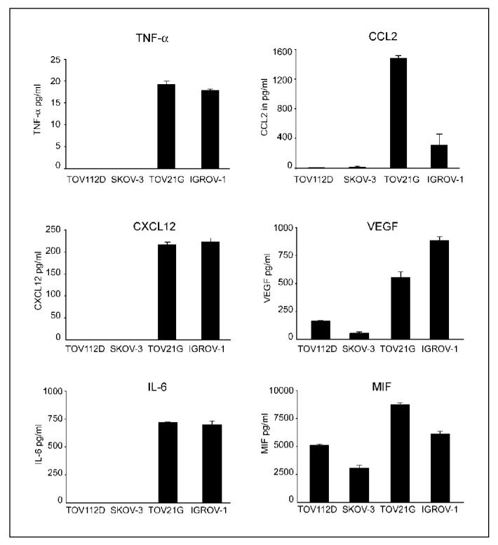
TNF-α release by ovarian cancer cell lines is associated with expression of other protein mediators. Proteins secreted by ovarian cancer cell lines were measured by ELISA after 48 h. Data are representative of three independent experiments done. Columns, mean of triplicate wells for TNF-α, CCL2, CXCL12, VEGF, IL-6, and MIF; bars, SD.
The TNF-α-producing TOV21G and IGROV-1 cells produced higher amounts of CCL2 (P = 0.017), CXCL12 (P < 0.0001), VEGF (P < 0.0001), IL-6 (P < 0.0001), and MIF (P = 0.002) compared with TOV112D and SKOV-3 cells that did not release measurable TNF-α (Fig. 1). IGROV-1 and TOV21G cells also had higher expression of CXCR4 as reported previously (8). In contrast, there was no association between TNF-α production and levels of FGF2 in the tissue culture medium; indeed, SKOV-3 was the only cell line that produced detectable amount of FGF2 (67 pg/mL) after 48 h (data not shown).
To investigate the hypothesis that these differences in endogenous cytokine network would influence growth and spread of the tumor cells, we injected the cell lines i.p. into nude mice and studied patterns of spread.
Association between tumor invasiveness and TNF-α production
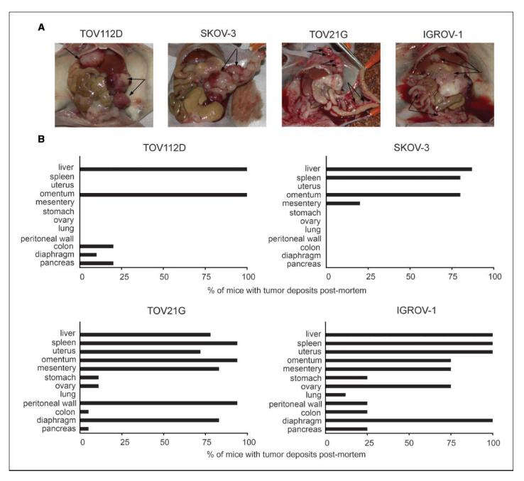
The TOV112D and SKOV-3 cell lines that did not produce TNF-α grew as well-defined encapsulated peritoneal masses, whereas the two cell lines that produced TNF-α, TOV21G, and IGROV-1 showed more widely dispersed tumors (Fig. 2A, arrows). This was confirmed by histologic examination of mouse organs at the survival end point (20% increase in abdominal girth). The distribution of both intraperitoneal and extraperitoneal deposits from the cell lines that did not produce TNF-α was much less than those that produced TNF-α (Fig. 2B). The TNF-α-producing ovarian cancer cells were capable of colonizing at least 12 different sites after i.p. injection into nude mice, whereas tumor colonies from those cell lines that did not produce TNF-α were only detected in five different sites.
Figure 2.
Appearance and location of tumor deposits in nude mice injected with ovarian cancer xenografts. A, gross patterns of peritoneal tumor spread of TOV112D, SKOV-3, TOV21G, and IGROV-1. B, location of tumor deposits in various organs as determined by histologic examination postmortem for TOV112D, SKOV-3, TOV21G, and IGROV-1.
If endogenous TNF-α was involved in tumor dissemination, we reasoned that TNF-α knockdown would inhibit this. We therefore established stable inhibition of TNF-α mRNA in TNF-α-producing IGROV-1 cells using RNAi technology.
Knockdown of TNF-α reduces production of other mediators by ovarian cancer cells
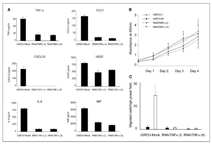
We transfected IGROV-1 cells with short hairpin RNA (shRNA) to TNF-α and established two clones of the IGROV-1 cell line with stable knockdown of TNF-α RNA (RNAi TNF-α I and II). The shRNA transfection resulted in reproducible, significant, and stable decreases in TNF-α release of 71% and 74% in two different TNF-α RNAi clones after 72 h (Fig. 3A; P = 0.0051 and 0.0034 compared with mock transfected). When supernatants from RNAi TNF-α cells were compared with those from cells transfected with shRNA with limited homology to any known sequences in the human, mouse, and rat genome (scrambled RNA, IGROV-Mock cells) release of CCL2, CXCL12, VEGF, IL-6, and MIF (Fig. 3A) was lower in the knockdown cells compared with mock-transfected cells. Silencing endogenous TNF-α decreased production of CCL2 by 86% and 90% in the two different TNF-α RNAi clones (P < 0.0001 and 0.0001 compared with mock transfected) and of VEGF by 40% and 29% (P = 0.016 and 0.008). IL-6 was reduced by 88% and 89% (P < 0.0001 and 0.0001) and MIF by 59% and 70% (P = 0.0015 and 0.0012). CXCL12 protein expression was completely inhibited in both the TNF-α RNAi IGROV clones. As described before, CXCR4 expression was also reduced by RNAi to TNF-α (8). None of the IGROV-1 cells released FGF nor did the cells release any IFN-γ whether they expressed shRNA constructs.
Figure 3.
In vitro effects of stable expression of shRNAto TNF-α. In all experiments, IGROV-Mock cells are compared with two independently isolated TNF-α shRNA clones RNAi TNF-α (I) and RNAi TNF-α (II). A, TNF-α protein secretion as measured by ELISA after 72 h. Columns, mean of triplicate wells; bars, SD. Silencing TNF-α expression in IGROV-1 cells also results in decreased protein secretion of CCL2, CXCL12, VEGF, IL-6, and MIF compared with IGROV-Mock as measured by ELISA after 48 h. Columns, mean of triplicate wells; bars, SD. B, proliferation of IGROV-1, IGROV-Mock, RNAi TNF-α (I), and RNAi TNF-α (II) cells in vitro as determined by WST-1 assay over a period of 4 d. Data are representative of three independent experiments. Points, mean for absorbance at 450 nm in triplicates; bars, SD. C, migration of IGROV-1 cells to CXCL12. IGROV-1 cells have low basal migration (black columns) but migrate toward CXCL12 (white columns). Results are representative of three experiments done Columns, mean of 10 determinations; bars, SD.
Knockdown of TNF-α and growth of cells in vitro
TNF-α did not, however, seem to be a growth factor for the ovarian cancer cells in tissue culture. The TNF-α RNAi cells grew at similar rates to IGROV-Mock cells and wild-type cells. Viable cell counts were assessed by trypan blue staining over 4 days of culture (data not shown) or when the WST-1 assay was used (Fig. 3B). Apoptosis was also assessed by fluorescence-activated cell sorting analysis using Annexin V and propidium iodide. Neither of these assays showed any difference in apoptosis between IGROV-Mock and TNF-α RNAi cells (data not shown). Addition of exogenous TNF-α at doses of 1, 10, and 100 ng did not alter growth rates of any of the lines (data not shown).
We therefore concluded that reduction of constitutive TNF-α production in ovarian cancer cells did not affect their ability to proliferate in vitro. However, knockdown of TNF-α was able to down-regulate several other soluble factors that could be important in growth and dissemination in vivo. To investigate this, we generated peritoneal xenografts of the cell lines, in which TNF-α production had been reduced.
Knockdown of TNF-α influences the growth and distribution of ovarian cancer xenografts
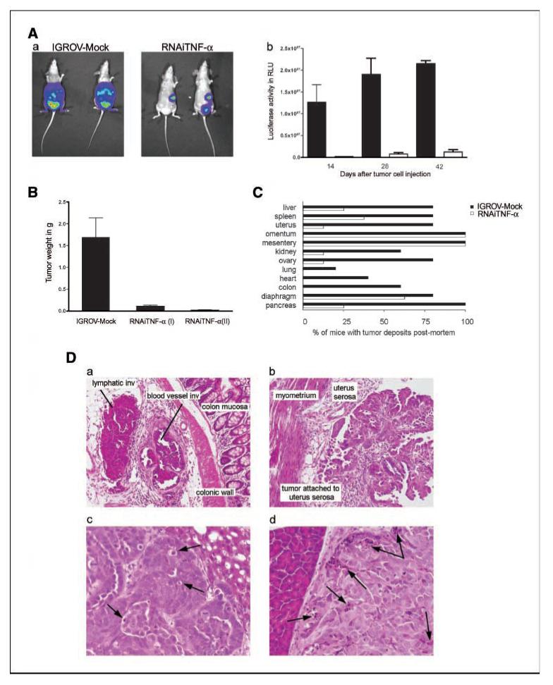
The influence of TNF-α knockdown on ovarian cancer xenograft growth and spread was measured in several ways. First, we measured the in vivo growth of IGROV-1 cells that expressed luciferase and were transfected with shRNA constructs. We selected representative mock-transfected and TNF-α RNAi clones that showed similar in vitro luciferase activity (46,000 ± 6,000 and 47,000 ± 2,000 RLU per 1 × 10⁴ cells, respectively). Cohorts of mice were investigated 14, 28, and 42 days after i.p. tumor cell injection. Both the tumor burden and the distribution were reduced in IGROV TNF-α RNAi cells compared with IGROV-Mock. Six weeks after IGROV-1 cell injection, IGROV-Mock cells were distributed widely in all areas of the peritoneum forming invasive masses in abdominal organs, but the IGROV RNAi TNF-α cells only localized to the peritoneal surface, spleen capsule, and uterine serosa (Fig. 4A, a). Quantitation of luciferase activity confirmed these imaging results (Fig. 4A, b; P < 0.0001).
Figure 4.
Effects of TNF-α knockdown on growth of cells in vivo. A, a, representative bioluminescence imaging in vivo of IGROV-Mock and RNAi TNF-α IGROV 42 d after i.p. injection. Bioluminescence is presented as a pseudocolor scale: red, highest photon flux; blue, lowest photon flux. b, quantification of bioluminescence from primary tumors (n = 6 each group) of images obtained on days 14, 28, and 42. B, tumor weight of dissectable tumors from each group (n = 5) are shown 6 wks after i.p. injection Columns, mean; bars, SD. C, dissemination of tumor deposits at 6 wks are presented as the percentage of mice with tumors at various anatomic sites. Because no differences between the RNAi TNF-α clones were seen, data from all mice injected with RNAi TNF-α cells were combined (n = 8). D, representative pictures of sections are shown from (a) IGROV-Mock and (b) RNAi TNF-α tumors (magnification, ×20). For detailed histologic analysis, images with higher magnification (magnification, ×40) from (c) IGROV-Mock and (d) RNAi TNF-α tumors are shown. Arrows, areas of apoptotic events.
In a separate experiment, we weighed all dissectable peritoneal tumor 6 weeks after mice had been injected with IGROV-Mock and RNAi TNF-α IGROV cell lines (Fig. 4B). The tumor burden was again significantly decreased when TNF-α production was reduced (P = 0.0033).
When organs from mice injected with mock or TNF-α RNAi cells were subjected to detailed histologic analysis 6 weeks after tumor cell injection, we found that knockdown of TNF-α significantly reduced the extent and invasiveness of tumor (Fig. 4C). There were tumor deposits in diaphragm, liver, uterus/fallopian tube, and spleen in 80% mice bearing IGROV-Mock–transfected cells in contrast to deposits 62.5%, 25%, 12.5%, and 37.5%, respectively, in the RNAi TNF-α–bearing mice. The pancreas was involved in all mice bearing IGROV-Mock tumors but in only 25% RNAi TNF-α-bearing mice. Involvement of bowel serosa (colon), heart, and lungs as well as pleura and mediastinum (data not shown) was only seen in mice bearing IGROV-Mock cells. The distribution of tumor deposits of the IGROV-Mock cells closely resembled that seen for the original IGROV cells at the survival end point (Fig. 2B). These differences at 6 weeks were also reflected in the times at which the mice had to be killed because they had reached ethical limits of abdominal swelling (20% increase in abdominal girth). Combining results from two separate experiments, injection of IGROV-Mock cells led to a median survival of 46 days (range, 39–62; n = 13) compared with 95 days (range, 54–272; n = 19) in mice bearing RNAi TNF-α I tumors and 210 days (range, 127–300; n = 13) in mice bearing RNAi TNF-α II tumors (P < 0.0001). TNF-α knockdown was sustained in vivo as measured by ELISA of tumor cell lysates at the survival end point. Lysates from IGROV-Mock tumors contained an average of 30 pg TNF-α/100 μg protein compared with 4 pg TNF-α/100 μg protein and 6 pg TNF-α/100 μg protein in lysates from RNAi TNF-α I and II tumors (P = 0.036 and 0.048, Mock compared with RNAi).
TNF-α knockdown influences tumor invasiveness
Tumors formed by IGROV-Mock cells were classified as invasive or invasive attached; none were well-circumscribed. In contrast, tumors derived from RNAi TNF-α cells were all classified as noninvasive and well-circumscribed tumors, although some were attached to the outer surface of abdominal organs. These differences in invasive patterns were statistically significant (P = 0.0038 comparing results from IGROV-Mock–injected mice to mice injected with RNAi TNF-α cells). Lymphovascular space invasion was present in all IGROV-Mock tumors but not in the RNAi TNF-α I and II tumors (Fig. 4D, a and b).
There was no difference in tumor differentiation between the lines in terms of grade or papillary versus solid pattern. All tumors had abnormal mitotic activity.
However, clusters of apoptotic cells were prominent in all tumors developing from the RNAi TNF-α lines even when the tumors were very small. In IGROV-Mock tumors, apoptotic activity invariably was low and seen only in isolated single cells (Fig. 4D, c and d).
TNF-α/CXCR4/CXCL12 and tumor dissemination
We have shown previously that knockdown of TNF-α in IGROV-1 cells results in down-regulation of CXCR4 (8). Therefore, one explanation for the reduction in tumor growth and dissemination could be the concurrent down-regulation of CXCR4 and its ligand CXCL12 (Fig. 3A) in these RNAi TNF-α lines. Expression of this chemokine receptor:ligand pair is a feature of malignant ovarian surface epithelium and has been implicated in cancer growth and spread (15). We therefore studied the ability of the cell lines to migrate to CXCL12. As shown in Fig. 3C, TNF-α knockdown abolished the ability of the IGROV-1 cells to migrate to CXCL12. The level of migration of the IGROV-Mock cells was essentially the same as the parental line (data not shown).
TNF-α knockdown influences tumor angiogenesis
A further explanation for differences in apoptosis, tumor size, and tumor dissemination could be that the RNAi TNF-α lines were not efficient in stimulating neovascularization. VEGF knockdown alone could have a profound effect on generation of blood vessels. In addition, recent data indicate that CXCL12 is important in attracting hemangiocytes from the bone marrow (20). We had noticed previously that angiogenesis was less in the SKOV-3-derived tumors than in IGROV-1-derived tumors, which suggested that TNF-α might be involved in the process. In addition, ascitic fluid never accumulated in the mice injected with TNF-α knockdown cells; however, it was a feature of animals injected with IGROV-1 parental and Mock-transfected cells. To investigate this further, we used confocal microscopy of peritoneal deposits to visualize and quantitate tumor blood vessels.
Silencing TNF-α in IGROV-1 cells markedly reduced tumor vascular area (Fig. 5A). For each group, mean vascular area of five tumors of matched size, taken from two different experiments, was assessed. IGROV-Mock tumors had a mean vascular area of 18% (range, 6–25%) compared with 5% (range, 1–17%) and 6% (range, 5–8%) for tumors from TNF-α RNAi clones I and II, respectively (Fig. 5C; P = 0.0027).
Figure 5.
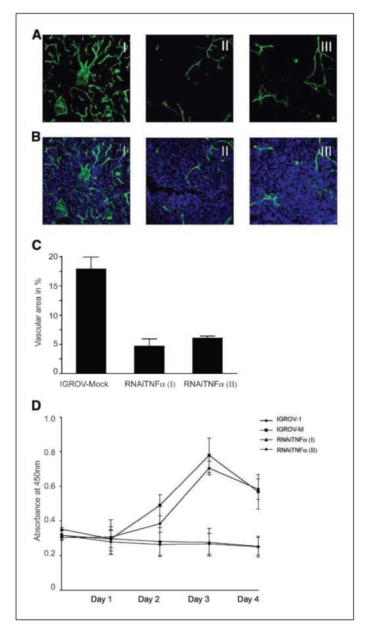
TNF-α knockdown influences tumor angiogenesis. In vivo angiogenesis was evaluated in mice 42 d after tumor cell injection. A, confocal images (magnification, ×20) of representative sections from (I) IGROV-Mock, (II) RNAi TNF-α I, and (III) RNAi TNF-α II tumors, after injection of FITC-conjugated lectin and in (B)4′,6-diamidino-2-phenylindole–stained cell nuclei are shown as control. C, columns, mean vascular area in each group quantified in 10 randomly selected areas of tumor sections (n = 5); bars, SD. D, proliferation of primary mouse lung endothelial cells in vitro with conditioned medium of IGROV-1, IGROV-Mock, and RNAi TNF-α cells. Representative of two independent experiments. Points, mean values for absorbance at 450 nm in triplicates; bars, SD.
To provide further proof that the network of soluble factors stimulated by TNF-α could stimulate tumor angiogenesis, we studied the influence of culture supernatants on endothelial cell growth in vitro. Culture supernatants from the original IGROV-1 cells and IGROV-Mock cells strongly stimulated the growth of primary mouse lung endothelial cells in vitro. In contrast, supernatants from the two clones of TNF-α RNAi cells had no such activity (Fig. 5D; P = 0.024 on day 3 and P = 0.037 on day 4).
Discussion
We have presented evidence to support our hypothesis about the role of constitutive production of TNF-α on the growth and spread of advanced ovarian cancer. From correlative studies and RNA knockdown, we show that autocrine action of TNF-α by ovarian cancer cells generates a constitutive network of other cytokines, angiogenic factors, and chemokines that may act in an autocrine/paracrine manner to promote colonization of the peritoneum and neovascularization of developing tumor deposits. The mechanisms of TNF-α action may include direct effects on tumor cell spread, via CXCR4; tumor cell survival, via CXCR4/CXCL12; but also stimulation of new blood vessels in the peritoneal tumor colonies, due to induction of CXCL12 and VEGF expression.
Many epithelial tumor cell lines release picogram quantities of TNF-α and the protein is detected in biopsies from advanced tumors (7). There also is some evidence that this TNF-α expression may be a downstream result of oncogenic mutation. For example, TNF-α is one of the targets of translational repression by the von Hippel Lindau factor gene, pVHL; mutation of this tumor suppressor gene in renal cell cancer increases stability of TNF-α protein in addition to its well-recognized effects on HIF-1α (21).
But is TNF-α a ‘master’ cytokine? Would silencing of other members of the autocrine network described in this article be equally effective in inhibiting tumor growth and spread? Although further experiments are required to formally prove this in ovarian cancer cell lines, there is other evidence that TNF-α can generate an ‘autocrine cascade’ in a single cell type (22). In a complex analysis (>8,000 proteins studied) of the effects of adding TNF-α to IFN-γ-pretreated cells (a sequence that generally induces apoptosis) and then ‘rescuing’ the cells with other growth factors, Janes et al. (22) concluded that epithelial cells can respond to TNF-α via sequential release of a range of TNF-α-stimulated cytokines and growth factors. Although the Janes article studied exogenous addition of cytokines, it is clear from our data that constitutive expression of TNF-α can also generate such an autocrine cascade and that, in the absence of a strong apoptotic signal, such a cascade can be advantageous to an epithelial tumor cell. It will be of interest to study a much larger number of cytokines, chemokines, and growth factors as well as activation of intracellular signaling pathways in the RNAi TNF-α cells we have generated.
A reduction in TNF-α levels in vivo had significant effects on development of new blood vessels in the peritoneal tumor deposits. There are close interactions with at least three of the mediators released by ovarian tumor cells. TNF-α is an inducer of VEGF, and VEGF in turn induces CXCL12 (20, 23). TNF-α also directly induces CXCL12 and CXCL12 and VEGF synergized in the stimulation of blood vessels in ovarian cancer (24).
A leucocyte infiltrate, especially of tumor-associated macrophages, may also be a prerequisite for angiogenesis (25, 26). In this respect, the decrease in CCL2 may also be relevant. This chemokine, like CXCL12, will cross the species barrier to act on mouse leucocytes. Likewise, TNF-α regulated MIF in the ovarian cancer cells. MIF is another major regulator of other inflammatory mediators and can also cross the species barrier from human to mouse. Hence, MIF production in ovarian cancer xenografts has the potential to regulate other cytokines, including TNF-α itself; nitric oxide; COX-2; and products of the arachidonic pathway and matrix metalloproteinases (27).
In fact, the only cytokine that may not act on the host in these experiments is IL-6 that may be an autocrine growth factor for ovarian cancer cells (28).
The issue of species specificity also relates to TNF-α itself. In vivo, mouse peritoneal TNF-α will be able to act on the human tumor cells via p55 TNF receptor 1, but this does not compensate for the reduction in constitutive production of the human cytokine. It seems that constitutive tumor cell–produced TNF-α is more important than paracrine cytokine, but we do not yet know the reason for this. These studies were conducted in immune compromised mice. It will be important to also study the effect of tumor cell TNF-α knockdown in syngeneic models in immunocompetent mice. We cannot predict the outcome of such experiments and this aspect is under investigation.
The results presented here suggest that direct targeting of tumor cell TNF-α may be a strategy that could influence the ability of tumor cells to disseminate in the peritoneum. Clinical trials are currently under way to test the action of TNF-α antagonists in patients with advanced cancer (29). Results published thus far would suggest that both neutralizing antibodies and soluble receptor fusion proteins are well tolerated in cancer patients and may have some disease-stabilizing action in ovarian cancer (10). Directed delivery of TNF-α inhibition using RNAi technology, or small molecular inhibitors of TNF-α signaling pathways that would serve to down-regulate a network of inflammatory cytokines, could be of interest, even if only given for short periods or with other more conventional therapies.
In summary, we have provided evidence that reduction of an autocrine inflammatory cytokine network in epithelial tumor cells can have major effects on their ability to grow and disseminate in vivo. This provides further evidence that targeting extracellular TNF-α or its intracellular pathways could be of use in advanced ovarian cancer.
Acknowledgments
Grant support: Cancer Research-UK, Barts and The London Cancer Research Committee and Joint Research Board; Deutsche Forschungsgemeinschaft grant HA 3565/11 (T. Hagemann); Association of International Cancer Research (J.L. Wilson); and European Union FP6 (R. Fatah) and Arthritis Research Campaign UK (D. Gould).
We thank George Elias (Cancer Research UK, London Research Institute, London, UK) for the help with the immunohistochemistry; Louise Reynolds (Cancer Research UK Clinical Centre, London, UK) for providing the primary mouse lung endothelial cells; Likengkeng Thokoa (International Society of Typographic Designers, Somerset, UK) for the graphic design; and George Wilbanks (University of South Florida, Tampa, FL), Ian Hart (Cancer Research UK Clinical Centre), and members of the Centre for Translational Oncology for their valuable discussion and proofreading of the manuscript.
Footnotes
The costs of publication of this article were defrayed in part by the payment of page charges. This article must therefore be hereby marked advertisement in accordance with 18 U.S.C. Section 1734 solely to indicate this fact.
References
- 1.Moore R, Owens D, Stamp G, et al. Tumour necrosis factor-α deficient mice are resistant to skin carcinogenesis. Nat Med. 1999;5:828–31. doi: 10.1038/10552. [DOI] [PubMed] [Google Scholar]
- 2.Arnott CH, Scott KA, Moore RJ, et al. Tumour necrosis factor-α mediates tumour promotion via a PKCα- AP-1-dependent pathway. Oncogene. 2002;21:4728–38. doi: 10.1038/sj.onc.1205588. [DOI] [PubMed] [Google Scholar]
- 3.Pikarsky E, Porat RM, Stein I, et al. NF-κB functions as a tumour promoter in inflammation-associated cancer. Nature. 2004;431:4461–6. doi: 10.1038/nature02924. [DOI] [PubMed] [Google Scholar]
- 4.Szlosarek PW, Grimshaw MJ, Kulbe H, et al. Expression and regulation of tumor necrosis factor-α in normal and malignant ovarian epithelium. Mol Cancer Ther. 2006;5:382–90. doi: 10.1158/1535-7163.MCT-05-0303. [DOI] [PubMed] [Google Scholar]
- 5.Szlosarek P, Balkwill F. Tumour necrosis factor-α: a potential target in the therapy of solid tumors. Lancet Oncol. 2003;4:565–73. doi: 10.1016/s1470-2045(03)01196-3. [DOI] [PubMed] [Google Scholar]
- 6.Naylor MS, Stamp GWH, Foulkes WD, et al. Tumor necrosis factor and its receptors in human ovarian cancer. J Clin Invest. 1993;91:2194–206. doi: 10.1172/JCI116446. [DOI] [PMC free article] [PubMed] [Google Scholar]
- 7.Szlosarek P, Charles KA, Balkwill FR. Tumour necrosis factor-α as a tumour promoter. Eur J Cancer. 2006;42:745–50. doi: 10.1016/j.ejca.2006.01.012. [DOI] [PubMed] [Google Scholar]
- 8.Kulbe H, Hagermann T, Szlosarek PW, et al. The inflammatory cytokine TNF-α upregulates chemokine receptor expression on ovarian cancer cells. Cancer Res. 2005;65:10355–62. doi: 10.1158/0008-5472.CAN-05-0957. [DOI] [PubMed] [Google Scholar]
- 9.Malik STA, Griffin DB, Fiers W, et al. Paradoxical, effects of tumour necrosis factor in experimental ovarian cancer. Int J Cancer. 1989;44:918–25. doi: 10.1002/ijc.2910440529. [DOI] [PubMed] [Google Scholar]
- 10.Madhusudan S, Muthuramalingam SR, Braybrooke JP, et al. A phase II study of ethanercept (ENBREL) a tumour necrosis factor-α inhibitor in recurrent ovarian cancer. J Clin Oncol. 2005;10:6528–34. doi: 10.1200/JCO.2005.04.127. [DOI] [PubMed] [Google Scholar]
- 11.Benard J, Da Silva J, De Blois MC, et al. Characterization of a human ovarian adenocarcinoma line, IGROV1, in tissue culture and in nude mice. Cancer Res. 1985;45:4970–9. [PubMed] [Google Scholar]
- 12.Brummelkamp TR, Bernards R, Agami R. A system for stable expression of short interfering RNAs in mammalian cells. Science. 2002;296:550–3. doi: 10.1126/science.1068999. [DOI] [PubMed] [Google Scholar]
- 13.Reynolds AR, Reynolds LE, Nagel TE, et al. Elevated Flk1 (vascular endothelial growth factor receptor 2) signaling mediates enhanced angiogenesis in β3-integrin-deficient mice. Cancer Res. 2004;64:8643–50. doi: 10.1158/0008-5472.CAN-04-2760. [DOI] [PubMed] [Google Scholar]
- 14.Negus RPM, Stamp GWH, Relf MG, et al. The detection and localization of monocyte chemoattractant protein-1 (MCP-1) in human ovarian cancer. J Clin Invest. 1995;95:2391–6. doi: 10.1172/JCI117933. [DOI] [PMC free article] [PubMed] [Google Scholar]
- 15.Scotton C, Milliken D, Wilson J, et al. Analysis of CC chemokine and chemokine receptor expression in solid ovarian tumours. Br J Cancer. 2001;85:891–7. doi: 10.1054/bjoc.2001.2020. [DOI] [PMC free article] [PubMed] [Google Scholar]
- 16.Rudlowski C, Pickart AK, Fuhljahn C, et al. Prognostic significance of vascular endothelial growth factor expression in ovarian cancer patients: a long-term follow-up. Int J Gynecol Cancer. 2006;16:183–9. doi: 10.1111/j.1525-1438.2006.00307.x. [DOI] [PubMed] [Google Scholar]
- 17.Kryczek I, Grybos M, Karabon L, et al. IL-6 production in ovarian carcinoma is associated with histiotype and biological characteristics of the tumour and influences local immunity. Br J Cancer. 2000;82:621–8. doi: 10.1054/bjoc.1999.0973. [DOI] [PMC free article] [PubMed] [Google Scholar]
- 18.Hagemann T, Wilson J, Kulbe H, et al. TNF-α dependent increased c-Jun and NF-κB activity in tumour cell lines upon co-cultivation with macrophages. J Immunol. 2005;175:1197–205. doi: 10.4049/jimmunol.175.2.1197. [DOI] [PubMed] [Google Scholar]
- 19.Whitworth MK, Backen AC, Clamp AR, et al. Regulation of fibroblast growth factor_2 activity by human ovarian cancer tumor endothelium. Clin Cancer Res. 2005;11:4282–8. doi: 10.1158/1078-0432.CCR-04-1386. [DOI] [PubMed] [Google Scholar]
- 20.Jin DK, Shido K, Kopp H-G, et al. Cytokine-mediated deployment of SDF-1 induces revascularization through recruitment of CXCR4+ hemangiocytes. Nat Med. 2006;12:557–67. doi: 10.1038/nm1400. [DOI] [PMC free article] [PubMed] [Google Scholar]
- 21.Galban S, Fan J, Martindale JL, et al. von Hippel-Lindau protein-mediated repression of tumor necrosis factor α translation revealed through use of cDNA arrays. Mol Cell Biol. 2003;23:2316–28. doi: 10.1128/MCB.23.7.2316-2328.2003. [DOI] [PMC free article] [PubMed] [Google Scholar]
- 22.Janes KA, Gaudet S, Albeck JG, et al. The response of human epithelial cells to TNF involves an inducible autocrine cascade. Cell. 2006;124:1225–39. doi: 10.1016/j.cell.2006.01.041. [DOI] [PubMed] [Google Scholar]
- 23.Kaplan RN, Riba RD, Zacharoulis S, et al. VEGFR1-positive haematopoietic bone marrow progenitors initiate the pre-metastatic niche. Nature. 2005;438:820–7. doi: 10.1038/nature04186. [DOI] [PMC free article] [PubMed] [Google Scholar]
- 24.Kryczek I, Lange A, Mottram P, et al. CXCL12 and vascular endothelial growth factor synergistically induce neoangiogenesis in human ovarian cancers. Cancer Res. 2005;65:465–72. [PubMed] [Google Scholar]
- 25.Mantovani A, Allavena P, Sica A. Tumour-associated macrophages as a prototypic type II polarised phagocyte population: role in tumour progression. Eur J Cancer. 2004;40:1660–7. doi: 10.1016/j.ejca.2004.03.016. [DOI] [PubMed] [Google Scholar]
- 26.Condeelis J, Pollard JW. Macrophages: obligate partners for tumor cell migration, invasion, and metastasis. Cell. 2006;124:263–6. doi: 10.1016/j.cell.2006.01.007. [DOI] [PubMed] [Google Scholar]
- 27.Calandra T, Roger T. Macrophage migration inhibitory factor: a regulator of innate immunity. Nat Rev Immunol. 2003;3:791–800. doi: 10.1038/nri1200. [DOI] [PMC free article] [PubMed] [Google Scholar]
- 28.Syed V, Ulinski G, Mok SC, et al. Reproductive hormone-induced, STAT3-mediated interleukin 6 action in normal and malignant human ovarian surface epithelial cells. J Natl Cancer Inst. 2002;94:617–29. doi: 10.1093/jnci/94.8.617. [DOI] [PubMed] [Google Scholar]
- 29.Yan L, Anderson GM, DeWitte M, et al. Therapeutic potential of cytokine and chemokine antagonists in cancer therapy. Eur J Cancer. 2006;42:793–802. doi: 10.1016/j.ejca.2006.01.013. [DOI] [PubMed] [Google Scholar]


