Abstract
Through chemical screening, we identified a pyrazolone that reversibly blocked the activation of phagocyte oxidase (phox) in human neutrophils in response to tumor necrosis factor (TNF) or formylated peptide. The pyrazolone spared activation of phox by phorbol ester or bacteria, bacterial killing, TNF-induced granule exocytosis and phox assembly, and endothelial transmigration. We traced the pyrazolone's mechanism of action to inhibition of TNF-induced intracellular Ca2+ elevations, and identified a nontransmembrane (“soluble”) adenylyl cyclase (sAC) in neutrophils as a Ca2+-sensing source of cAMP. A sAC inhibitor mimicked the pyrazolone's effect on phox. Both compounds blocked TNF-induced activation of Rap1A, a phox-associated guanosine triphosphatase that is regulated by cAMP. Thus, TNF turns on phox through a Ca2+-triggered, sAC-dependent process that may involve activation of Rap1A. This pathway may offer opportunities to suppress oxidative damage during inflammation without blocking antimicrobial function.
Neutrophils, the most abundant leukocytes in blood, provide a critical element of host defense. Defects in neutrophil numbers, migration across endothelium or bactericidal mechanisms lead to life-threatening infections. At the same time, neutrophil activation is a major contributor to inflammatory tissue damage (1). Thus, it is a goal of antiinflammatory therapy to target neutrophils, but it is a challenge to do so without impairing host defense.
Two families of neutrophil products play key roles in host defense and tissue damage. Activated neutrophils degranulate to release antibiotic proteins, including proteases that degrade connective tissue. Activated neutrophils also secrete reactive oxygen intermediates (ROIs), products of phagocyte oxidase (phox), a multicomponent enzyme whose catalytic flavocytochrome is a transmembrane protein of the granules. ROIs contribute directly to bacterial killing (2) but also promote proteolytic tissue damage by: triggering K+ influx into the phagolysosome, desorbing proteases from their proteoglycan bed (3), activating neutrophil proteases (4) and matrix metalloproteinases (5), inactivating antiproteases (1), and activating NF-κB and AP1, transcription factors for numerous genes with proinflammatory products, including chemokines that attract neutrophils. Thus, inhibition of neutrophil ROI production would be expected to reduce inflammatory proteolysis through multiple mechanisms.
Maximal neutrophil activation can be induced artificially with PMA or physiologically by inflammatory peptides and proteins, such as TNF (6). The soluble, physiologic agonists are far more effective if the neutrophils are adherent to extracellular matrix proteins by way of integrins (7). Adherence (8) and TNF (9) combine to trigger oscillatory elevations of intracellular Ca2+, which take the form of spatially restricted, moving waves in neutrophils responding to N-formylpeptide (10). However, it remains unclear how TNF activates neutrophils. To help answer this question, we decided to seek chemical inhibitors of TNF-induced neutrophil activation, and attempted to identify their mechanisms of action.
RESULTS AND DISCUSSION
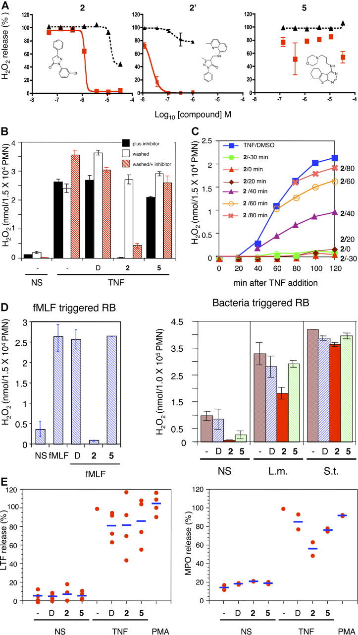
We screened 15,000 compounds for those that blocked H2O2 release by TNF-treated, adherent neutrophils, but spared H2O2 release when the same cells were triggered by PMA. The PMA counterscreen allowed us to exclude compounds that were toxic, inhibited phox or protein kinase C, or interfered with the assay. Four compounds were both selectively inhibitory and consistently effective with neutrophils from different donors. We chose compound 2 (2-[3-chloro-phenyl]-5-phenyl-2,4-dihydro-pyrazol-3-one) for analysis here. The others (1, 3, and 4) work in different ways, as will be reported elsewhere (Han et al., unpublished data). 5 ((3-morpholin-4-yl-propyl)-(5,6,7,8-tetrahydro-benzo[4,5]thieno[2,3-d]pyrimidin-4-yl)-amine) shares its core structure with 1 and 3, but had no effect on H2O2 release and was used as a control. Concentration-response curves of 2, using neutrophils from three donors, yielded a mean 50% inhibitory concentration (IC50) ± SEM of 1.6 ± 0.4 μM for H2O2 release (Fig. 1 A). A new screen of 1,100 congeners of 2 identified 2′ (5-methyl-4-[(2-methyl-quinolin-8-ylamino)-methyl]-2-phenyl-2,4-dihydro-pyrazol-3-one), with an IC50 of 24 ± 6 nM (Fig. 1 A). Because 2′ was identified after most of these studies were completed, we focus here on 2. At 5 μM, 2 yielded >95% inhibition of TNF-triggered H2O2 release with cells from all donors tested. This concentration was used for all further experiments.
Figure 1.

Identification of compounds that inhibit the neutrophil respiratory burst in response to TNF. (A) Chemical structures and concentration-response curves. Compounds were added at 37°C for 30 min before stimulation with TNF (red squares) or PMA (black triangles). H2O2 release at 90 min is displayed as percentage of H2O2 release seen with TNF or PMA in the presence of the vehicle, DMSO. (B) Reversibility. Neutrophils incubated with DMSO (D), 2, or 5 were left unmanipulated (black bars) or washed (open bars) with buffer before both sets were plated and stimulated with TNF. Other neutrophils from which the compounds had been washed off were reexposed to the compounds (red hatched bars). (C) Influence of 2 after the onset of respiratory burst. At indicated times, 2 was added to neutrophils already undergoing a TNF-triggered respiratory burst (RB). (D) Inhibition by 2 of the respiratory burst triggered by fMLF but not by bacteria. Neutrophils were preincubated with DMSO (D), 2, or 5 and not stimulated (NS) or stimulated with fMLF (100 nM), L. monocytogenes (L.m.) or S. enterica. (S.t.) (S.H2O2 release is depicted at the time it reached plateau. (A–C) means ± SEM for triplicates. (E) Impact on degranulation. Neutrophils were stimulated and their supernates were assayed for lactoferrin (LTF) and myeloperoxidase (MPO) as a percent of that released in the absence of compound. Each dot is the mean of duplicates in one experiment. Horizontal bars are group means. TNF and PMA were used at 100 ng/ml in all figures.
When neutrophils were treated with 2 and then washed, they regained responsiveness to TNF (Fig. 1 B). The cells remained susceptible to inhibition when 2 was added back (Fig. 1 B). Thus, inhibition by 2 was fully reversible. When we added 2 to neutrophils that already were releasing H2O2 in response to TNF, the respiratory burst slowed quickly (Fig. 1 C). Thus, the process inhibited by 2 was needed to launch the TNF-triggered respiratory burst and to sustain it.
Next, we tested the effects of 2 on neutrophils' responses to bacteria and to the major neutrophil stimulant released by Escherichia coli, formylated methionyl-leucyl-phenylalanine (fMLF). H2O2 release triggered by fMLF was blocked by 2. However, 2 did not inhibit H2O2 release triggered by Salmonella enterica var. Typhimurium, and only reduced slightly the respiratory burst that was elicited by Listeria monocytogenes (Fig. 1 D). 2 also did interfere with the killing of these pathogens by neutrophils (Fig. S1, available at http://www.jem.org/cgi/content/full/jem.20050778/DC1).
Degranulation delivers the phox flavocytochrome to phagosomal and plasma membranes (11) where the cytosolic components of phox are recruited. Therefore, we considered that 2 might block activation of phox by blocking degranulation. However, 2 did not block TNF-induced exocytosis of the specific granule marker, lactoferrin, and only inhibited slightly the release of the azurophil granule marker, myeloperoxidase (Fig. 1 E).
The large-scale respiratory burst triggered by TNF or fMLF requires that neutrophils spread on extracellular matrix (6, 7). Pretreatment with 2 allowed neutrophils to spread normally on serum-coated glass in response to PMA. With TNF as a stimulus in 2-pretreated cells, spreading was initiated, which suggested that 2 did not interfere with binding of TNF or transmission of some signals to the cytoskeleton. However, 2 caused TNF-triggered spreading to halt prematurely, which suggested inhibition of a later stage in TNF signaling (Fig. 2 A).
Figure 2.
Impact of inhibitors on neutrophil spreading. Neutrophils were plated on FBS-coated glass cover slips, incubated or not with each compound at 37°C for 30 min before stimulation with TNF, PMA, or an equal volume of buffer (no stimulus, NS). After 30 min, the cells were fixed and photographed with phase-contrast microscopy (100×).
Interference with spreading of neutrophils in response to TNF raised the question of whether 2 might block migration across endothelium—the process by which neutrophils enter tissues. However, pretreatment of neutrophils with 2 blocked neither the adhesion of the cells to, nor their migration across, monolayers of TNF-activated human umbilical vein endothelial cells (HUVECs; Fig. 2, available at http://www.jem.org/cgi/content/full/jem.20050778/DC1). Moreover, pretreatment of HUVECs with 2 did not influence the integrity of the monolayer nor the cells' ability to respond to TNF by supporting adhesion and transmigration of untreated neutrophils (unpublished data). Similarly, a 2-d incubation with 2 did not affect the morphology or adherence of primary mouse peritoneal exudate macrophages, nor the ability of TNF to synergize with IFN-γ in inducing them to release nitric oxide (unpublished data). These experiments demonstrated that 2 inhibited only a selective aspect(s) of TNF signal transduction.
Neutrophil responses to TNF require protein tyrosine phosphorylation involving Syk (12, 13); Src family kinases, Hck and Fgr (14); and Pyk2 (15, 16). However, treatment of adherent neutrophils with 2 had little impact on overall protein tyrosine phosphorylation induced by TNF (Fig. S3 A, available at http://www.jem.org/cgi/content/full/jem.20050778/DC1). Nor did 2 inhibit recombinant Src (Fig. S3 B), Syk (Fig. S3 C), or TNF-induced (auto)phosphorylation of Pyk2 on tyrosine 402 (Fig. S3 D), which is critical for Pyk2's kinase activity (17).
Cytosolic components of phox—p40phox, p47phox, and p67phox—form a M r ∼240,000 complex (18) in resting neutrophils, with p47phox serving as an essential adaptor. The complex translocates to the membrane upon activation. 2 had no effect on TNF-induced translocation of p47phox to the membrane (Fig. 3 A). Because 2 inhibited neither the assembly of phox (Fig. 3 A) nor its catalytic activity when triggered by PMA (Fig. 1 A), we hypothesized that 2 might interfere with a later stage in phox activation, namely, TNF-dependent activation of one of the guanosine triphosphatases bound to phox, Rac2 (19) or Rap1A (20). Rac2-deficient neutrophils have severe migration defects (21), whereas 2-treated neutrophils migrated normally (Fig. S2). Thus, Rac2 was unlikely to be the locus of regulation by 2, and we focused on Rap1A.
Figure 3.
Impact of inhibitors on components of the phox complex. (A) Translocation of p47phox to membranes. Neutrophils were incubated with DMSO (D) or 2 at 37°C for 30 min and stimulated with TNF (T), PMA (P), or buffer (NS). After 40 min, cells were lysed and membrane fractions collected by ultracentrifugation, separated by SDS-PAGE, and Western-blotted with anti-p47phox antibody. (B) Activation of Rap1A. Lysates of neutrophils that had been pretreated for 30 min with DMSO (D), 2, KH7 (25 μM), or the tmAC inhibitor ddAdo (25 μM), and then stimulated for 30 min with TNF or buffer (NS) alone were incubated with agarose beads coupled to recombinant RalGDS-Rap binding domain to affinity purify GTP-bound Rap1A. Beads were boiled in SDS sample buffer and the supernate subjected to SDS-PAGE and Western blot with anti-Rap1A antibody (top row). Western blot of total cell lysates (bottom row) served as a control.
Rap1A is bound stoichiometrically to the phox flavocytochrome in a guanosine triphosphate (GTP)– and phosphorylation-dependent manner (22). Treatment of neutrophils with fMLF or GM-CSF activated Rap1A (i.e., converted it to the GTP-bound state; reference 23). We found that TNF treatment of adherent neutrophils also activated Rap1A, and 2 blocked TNF-induced Rap1A activation (Fig. 3 B).
Rap1A can be activated by a guanine nucleotide exchange protein, exchange protein directly activated by cAMP (Epac; references 24, 25). The cAMP that activates Epac can arise from the long-studied G protein–regulated, transmembrane adenylyl cyclases (tmACs) or from a recently discovered nontransmembrane (“soluble”) adenylyl cyclase, sAC, which is regulated by intracellular Ca2+ and bicarbonate (26, 27). We considered tmACs unlikely candidates for neutrophil activation because their pharmacologic stimulation inhibits most neutrophil functions, including TNF-induced activation of phox (28). Thus, we asked if neutrophils contain sAC.
Immunoblot revealed sAC in highly purified neutrophil preparations (Fig. 4 A). Neutrophils were the source of the immunoreactivity because they were stained uniformly and specifically by three mAbs, each directed against a different epitope of sAC, as shown for one mAb in Fig. 4 B. The granular pattern of staining raised the possibility that sAC may reside near phox, but definitive analysis of sAC's subcellular localization awaits immuno-electron microscopy.
Figure 4.


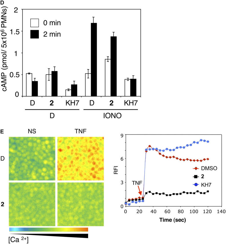
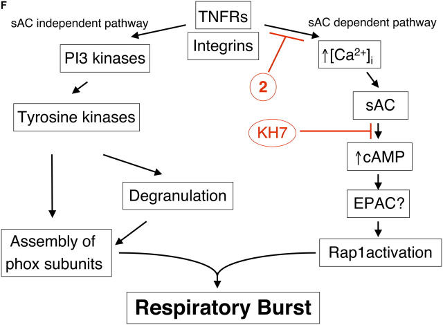
sAC as a critical element of TNF signaling in neutrophils. (A) Western blot. Neutrophils were lysed and subjected to SDS-PAGE/Western blot with anti-sAC mAb R21. Lane 1 (control): lysate of Chinese hamster ovary cells transfected with sperm isoform of sAC. Lanes 2, 3: neutrophil lysates treated without (lane 2) or with (lane 3) protease inhibitor diisopropylfluorophosphate. (B) Immunofluorescence microscopy. Neutrophils were stained with or without biotinylated anti-sAC mAb R41 followed by streptavidin-Alexafluor 594. (C) Effects of sAC inhibitor KH7 and tmAC inhibitor ddAdo on the respiratory burst. Compounds were added to adherent neutrophils at 37°C for 30 min before stimulation with TNF (red squares) or PMA (black triangles). (D) sAC activity in neutrophils (5 × 10⁶/well) preincubated 30 min with the phosphodiesterase inhibitor, isobutylmethylxanthine (100 μM) and DMSO (D), 2, or KH7, then stimulated with ionomycin (1 μM). cAMP accumulation was measured at 0 min and 2 min after stimulation. (E) Impact on TNF-triggered Ca2+ elevation in Fluo-3/AM-loaded neutrophils. Left panels are pseudocolored images of adherent neutrophils pretreated with DMSO (D) or 2 for 30–60 min and then treated with TNF or no stimulus (NS). Average relative fluorescence intensity (RFI) for all cells in similar microscopic fields is plotted as a function of time on the right. Arrow marks addition of TNF. Some cells were pretreated with KH7 (25 μM) as indicated. (F) Speculative model.
Next, we selected pharmacologic reagents to distinguish between possible sources of cAMP in a mammalian cell. A chemical screen identified a specific inhibitor of recombinant human sAC, 2-(1H-benzoimidazole-2-ylsulfanyl)-propionic acid (5-bromo-2-hydroxy-benzylidene)-hydrazide (KH7) that spares tmACs (29). Reciprocally, we determined a concentration range in which 2′,5′ dideoxyadenosine (ddAdo) acts as a selective inhibitor of tmACs, sparing sAC. Like 2, KH7 inhibited TNF-triggered activation of Rap1A (Fig. 3 B), and suppressed TNF-triggered H2O2 release with an IC50 ± SEM of 4.7 ± 0.3 μM while sparing PMA-triggered H2O2 release (Fig. 4 C). In contrast, the selective inhibitor of tmACs, ddAdo, had no effect on the activation of Rap1 (Fig. 3 B) or on the TNF-induced respiratory burst (Fig. 4 C). KH7 seemed to elicit a small degree of Rap1A activation by itself, the basis of which is not known. These findings suggest that TNF-triggered activation of Rap1 is mediated by sAC, not by tmACs.
Unlike KH7, 2 did not inhibit recombinant sAC (data not shown). Thus, we hypothesized that 2 might block a step upstream of sAC, such as TNF-induced elevation of intracellular Ca2+. We first demonstrated that neutrophil sAC could respond to elevations of intracellular Ca2+. Ionomycin, a Ca2+ ionophore, induced production of cAMP in neutrophils, and the ionomycin-induced cAMP production was blocked by the sAC inhibitor, KH7 (Fig. 4 D); this indicated that sAC was responsible for the increase in cAMP. 2 had no effect on the ionomycin-induced production of cAMP, which confirmed that 2 does not inhibit endogenous sAC. To test whether 2 blocked TNF-induced Ca2+ elevation, we loaded adherent neutrophils with the Ca2+-sensitive fluorochrome, Fluo3/AM, and studied their fluorescence by way of videomicroscopy. TNF triggered an almost instantaneous elevation of intracellular Ca2+ that persisted throughout the observation period (∼2 min; Fig. 4 E). The elevation seemed to be nearly constant when expressed as average fluorescence intensity per microscopic field in fields that contained hundreds of cells. However, inspection of individual cells revealed asynchronous, irregular oscillations, as reported (30). Pretreatment of the cells with 2 substantially suppressed the TNF-induced increase in intracellular Ca2+ (Fig. 4 E), although 2 had no effect on Ca2+ elevation that was induced artificially by ionomycin (not depicted). In contrast, pretreatment with KH7 did not suppress the TNF-induced increase in intracellular Ca2+ (Fig. 4 E). We propose that 2 be named “neucalcin-1” for neutrophil calcium inhibitor, and 2′ be named “neucalcin-2.”
These findings outline a pathway for TNF signal transduction that is distinct from the known routes that lead to transcriptional regulation and apoptosis. Fig. 4 F outlines a speculative model: exposure of adherent neutrophils to TNF leads to elevation of intracellular Ca2+ (9, 30), which stimulates sAC (26, 27) to generate cAMP. The elevated cAMP causes activation of Rap1A, perhaps by way of Epac. Activated Rap1A may turn on phox, whose components have been assembled in response to TNF, fMLF, and other soluble, physiologic agonists. This proposed pathway is independent of the previously described TNF signaling cascade involving phosphatidylinositol 3-kinase and tyrosine kinases that mediates degranulation and the degranulation-dependent assembly of phox (13, 16). The sAC-dependent and sAC-independent pathways seem to converge on phox. The two chemically distinct inhibitors used in this study, neucalcin and KH7, seem to act at two different points in the sAC-dependent pathway (Fig. 4 F). However, further work is needed to identify the molecular target of neucalcins, the means by which TNF triggers the elevation of intracellular Ca2+, the role (if any) of Epac, and the participation and mechanism of Rap1A in the activation of phox.
It may seem paradoxical that in previous studies, direct measurement of cAMP levels in TNF-treated neutrophils did not reveal an elevation; on the contrary, TNF prolonged a decline in cAMP that was induced when neutrophils adhered to biologic surfaces (28). Those observations can be reconciled with our new observations by proposing that the elevations of cAMP that are required to sustain the activation of phox by TNF are spatially confined and quantitatively insufficient to nullify the decrease in cAMP that proceeds at the same time in a larger cellular compartment.
Opposing effects of cAMP have been reported on protein kinase B, depending on whether the cAMP effector is protein kinase A or Epac (31). A similar situation may pertain to Rap1A. Pharmacologically induced, global, sustained elevations in cAMP that suppress neutrophil functions are likely to activate protein kinase A, which can phosphorylate Rap1A and block its association with phox (22). In contrast, because TNF-induced Ca2+ elevations in neutrophils are oscillatory (9, 30) and probably localized (10), sAC-mediated production of cAMP and the ensuing activation of Rap1A also may be oscillatory and localized. It has been argued that Rap1A must undergo cycles of association with, and dissociation from, phox to sustain phox activity (32, 33). The kinetic studies in Fig. 1 C are consistent with the concept that sustained H2O2 release in response to TNF may require repetitive Rap1A activation.
Thus, we have identified a new enzyme in neutrophils, sAC; a new pathway for TNF signal transduction, centered on sAC; the ability of endogenous sAC to sense Ca2+ elevations; a possible functional role for the Ca2+ elevations in adherent, TNF-stimulated neutrophils; evidence that cAMP can not only inhibit but also activate neutrophils; and chemical agents, neucalcins, that may serve as tools to discover how TNF leads to Ca2+ elevation. These findings help to generate a list of targets whose inhibition might allow selective intervention in neutrophil functions.
Blocking nonbacterial activation of phox might be a more effective way to reduce neutrophil-dependent proteolysis of tissue than attempting to inhibit individual proteases. The latter approach suffers from the redundancy of proteases released by neutrophils, and those produced by other cells but activated, in part, by neutrophils. In contrast, selective interference with the inflammatory activation of phox may retard the release of neutrophil proteases from their proteoglycan matrix in the lysosomes (3); prevent the oxidative activation of neutrophil proteases (4); blunt the oxidative activation of matrix metalloproteinases (5); diminish the oxidative inactivation of the major antiproteases secretory leukocyte protease inhibitor, α1-antiprotease and α2 macroglobulin (1); and diminish the oxidative contribution to NF-κB– and AP1-dependent expression of chemokines and other inflammatory mediators.
In sum, this work shows that it is feasible to block the activation of phox by inflammatory factors without interfering with neutrophil migration or phox-dependent killing of ingested bacteria.
MATERIALS AND METHODS
Cells.
All cells were used under Institutional Review Board–approved protocols. Neutrophils were isolated from heparinized (10 U/ml) blood of healthy, consenting adult donors to >95% purity using Polymorphprep (Axis-Shield PoC AS) according to the manufacturer's instructions. Contaminating erythrocytes were lysed by hypotonic shock for 45 s with 0.2% saline. Neutrophils were resuspended in Krebs Ringer phosphate with glucose (KRPG) formulated as described (34). HUVECs were isolated from umbilical cords and cultured as described (35). Peritoneal macrophages were isolated from C57BL/6 mice 4 d after injection of 2 ml of thioglycollate broth (4%) and cultured as described (34).
H2O2 release and degranulation.
The neutrophil respiratory burst was measured as described (34). In brief, 96-well flat-bottomed plates (Primaria, Falcon) were coated with 50 μl/well of FBS in 5% CO2 in air at 37°C for at least 1 h and washed three times with 0.9% saline. 1.5 × 10⁴ neutrophils were added to triplicate wells containing 100 μl of reaction mixture (2.4 nmol scopoletin, 0.5 μg horseradish peroxidase, and 1 mM NaN3) and stimulated with either buffer control, TNF (Preprotech) or PMA, each at 100 ng/ml. The reduction of scopoletin by H2O2 was recorded every 15 min on a plate-reading fluorometer until H2O2 release reached plateau. The amount of H2O2 released was calculated as described (34). The supernate from the H2O2 release assay with 1.5 × 10⁴ neutrophils was used to measure degranulation using lactoferrin or myeloperoxidase ELISA kit (Oxis International, Inc.). Spontaneous cell death from the same supernate was measured using Cytotoxicity Detection Kit (Roche Molecular Biochemicals), an assay for lactate dehydrogenase. The total cell content of lactoferrin, myeloperoxidase, and lactate dehydrogenase was determined after lysing cells with 1% Triton X-100.
Automated assay screening.
A collection of 15,000 compounds with drug-like characteristics from ChemDiv Labs was screened robotically in the High Throughput Screening Facility at The Rockefeller University to identify specific inhibitors of TNF-triggered H2O2 release in human neutrophils. KH7 was identified in the same library during an independent screen for inhibition of sAC. H2O2 release was measured as above with the following modifications. Black instead of clear, 96-well tissue culture plates (Falcon, catalog number 353945) were used to reduce background fluorescence as measured in a Perkin-Elmer Fusion microplate reader. A Titertek Multidrop 96/384 dispenser was used for bulk reagent dispensing, Bi-Tek Elx 405 Select system was used for plate washing, and a PerkinElmer MiniTrakV liquid handling dispenser was used for the delivery of compound aliquots to each well. Percent inhibition by each compound was calculated using the formula: ([T0- T90] × 100)/(Tc0-Tc90), where T0 and T90 are fluorescence readings at each well at 0 and 90 min, respectively, and Tc0 and Tc90 are the mean fluorescence readings in the compound-free control wells at 0 and 90 min, respectively. Compounds that showed >90% inhibition of the TNF-triggered respiratory burst were retested with TNF and PMA as stimuli to eliminate nonspecific, toxic chemicals and protein kinase C inhibitors.
Test of reversibility of inhibition.
Neutrophils were incubated with each compound at 37°C for 30 min in FBS-coated tubes (Falcon), washed twice with cold KRPG (5 min each at 4°C), and plated in 96-well plates before immediate stimulation with TNF or an equivalent volume of KRPG buffer as a control. On a separate plate, compounds were added back to the neutrophils from which compounds had been washed off, and cells were stimulated to evaluate the neutrophils' capacity to respond. Neutrophils incubated with each compound without washing also were stimulated for comparison.
Bacteria-triggered respiratory burst.
Neutrophils were incubated with DMSO or each compound for 30 min at 37°C and exposed to 10% autologous serum-opsonized S. enterica var. Typhimurium (14028s; American Type Culture Collection) or L. monocytogenes (104035; American Type Culture Collection) at a multiplicity of infection of 0.5 bacteria per neutrophil. H2O2 release was measured as described before.
Bacterial killing.
Neutrophils were preincubated with or without 2 for 30 min at 37°C and exposed to 10% autologous serum-opsonized S. enterica var. Typhimurium (14028s; American Type Culture Collection) or L. monocytogenes (15323; American Type Culture Collection) at a multiplicity of infection of 0.5 bacteria per neutrophil. Neutrophils were lysed at the indicated times with 1% sodium deoxycholate for assays with Salmonella or 1% Triton X-100 for assays with Listeria. Bacteria recovered from each condition were grown in Luria Bertani agar plates at 37°C overnight, and the colonies were counted.
Morphology.
Acid-washed glass cover slips were placed in a 12-well tissue culture plate, coated with FBS in 5% CO2 at 37°C for at least 1 h, and washed three times with 0.9% saline. Neutrophils (2 × 10⁶) were added to each well in 1 ml of reaction mixture, and incubated or not with each compound at 37°C for 30 min before stimulation with TNF (100 ng/ml), PMA (100 ng/ml), or an equal volume of KRPG. Cells were fixed with 2% paraformaldehyde and 3.7% formaldehyde buffer, and photographed through a phase-contrast microscope. Results are typical of those based on inspecting living cells at 15-min intervals over 2 h.
Transendothelial migration assay.
HUVECs were isolated and cultured in M199 medium (GIBCO BRL) supplemented with 20% normal human serum, penicillin, and streptomycin as described (35). Experiments used cells at passage two that were cultured on hydrated type I collagen gels in 96-well plates. Where shown, HUVECs were stimulated with TNF (50 pg/ml) in 5% CO2 at 37°C for the final 12–18 h. Neutrophils were isolated from the peripheral blood of healthy adult volunteers by density gradient sedimentation in a discontinuous gradient of Ficoll (GE Healthcare) and Histopaque (Sigma-Aldrich), washed in Hanks' balanced salt solution with 0.1% human serum albumin, resuspended to 0.5 × 10⁶ cells/ml, and added to HUVEC monolayers. Test compounds were incubated with the neutrophils at room temperature for 30 min before transendothelial migration, and allowed to remain during the duration of the assay. After neutrophils were allowed to transmigrate for 30 min at 37°C, nonadherent neutrophils were washed off with PBS, and the remaining adherent and transmigrated cells were fixed with the endothelial monolayer by incubation overnight in 2.5% glutaraldehyde (Electron Microscopy Sciences) in 0.1 sodium cacodylate buffer. HUVECs and neutrophils were stained differentially with Wright-Giemsa stain, and adherent and transmigrated cells were counted in multiple fields from the six replicates of each condition tested. Total adhesion was calculated as the total number of adherent and transmigrated cells per high-powered field. The percentage of adherent cells that transmigrated below the endothelial layer was called percentage of transendothelial migration.
Immunoprecipitation and Western blot.
Tissue culture plates (Primaria, Falcon) were coated with 3 ml of FBS in 5% CO2 at 37°C for at least 1 h and washed twice with 0.9% saline. Neutrophils (15 × 106) were added to each plate containing 4 ml of reaction mixture, incubated with each compound at 37°C for 30 min, and stimulated with buffer control or TNF (100 ng/ml). When cells were spread fully (20–40 min), they were treated with 5 mM diisopropylfluorophosphate to inhibit serine proteases, and lysed with 125 μl of SDS lysis buffer (10 mM Tris-HCl, pH 7.6; 150 mM NaCl; 1% SDS; 1 mM PMSF; 1 mM Na pyrophosphate; 1 mM NaF; 1 mM vanadate; 5 μg/ml each of aprotinin, leupeptin, chymostatin, pepstatin A) for Western blot or with 200 μl of nondenaturing modified radioimmunoprecipitation assay buffer (10 mM Tris-HCl, pH 7.6; 150 mM NaCl; 1% Triton X-100; 1 mM PMSF; 1 mM Na pyrophosphate; 1 mM NaF; 1 mM vanadate; 5 μg/ml each of aprotinin, leupeptin, chymostatin, pepstatin A) for immunoprecipitation. The cell lysate was passed through a 26-gauge needle six times to shear DNA, and centrifuged at 20,000 g for 15 min to remove cell debris and DNA. The protein concentration was determined using a Bio-Rad kit. Cell lysates were separated by SDS-PAGE and transferred electrophoretically to nitrocellulose membranes (Schleicher & Schuell, Inc.). The membranes were incubated with 5% milk in TBST (100 mM Tris-HCl, pH 7.5; 9% NaCl; 0.1% Tween-20) for 1 h at 37°C, and then overnight at 4°C with antiphosphotyrosine antibody (Santa Cruz Biotechnology, Inc.), anti-Syk antibody (Transduction Laboratories), antiphospho-specific Pyk2 antibodies (Biosource International), or anti-Pyk2 antibody (Upstate Biotechnology). sAC protein was immunoblotted with monoclonal antibody R21 and immunostained with monoclonal antibodies R21, R41, and R52 produced in the laboratories of two of us (L. Levin and J. Buck). Membranes were washed with TBST and incubated with secondary antibody conjugated with horseradish peroxidase in 5% milk in TBST for 1 h at 37°C. After further washing with TBST, bound antibody was detected by enhanced chemiluminescence (Pierce Chemical Co.).
In vitro kinase assay for Src and Syk.
Kinase assays were performed according to the instructions provided by UBI, from which recombinant Src and Syk were purchased. In brief, after incubation of recombinant Src (40 U) with each compound at room temperature for 30 min, Src kinase reaction buffer (100 mM Tris-HCl, pH 7.2; 125 mM MgCl2; 25 mM MnCl2; 2 mM EGTA; 0.25 mM NaVO4; 2 mM DTT), Src kinase substrate peptide (375 μM) and [γ-32P]ATP were added to the reaction mixture. After incubation at 30°C for 10 min, the reaction was stopped by addition of 40% TCA. After transferring each sample to a P81 paper square (UBI), unincorporated radioactivity was washed off with 0.75% phosphoric acid and acetone, and the assay square was read in a scintillation counter. For Syk kinase assay, reaction buffer contained 50 mM Tris-HCl, pH 7.5; 0.1 mM EGTA; 0.1 mM NaVO4; 0.15 M 2-mercaptoethanol and substrate was poly (Glu4-Tyr); 4:1; (CSI Biointernational). Each condition was done in duplicate.
Translocation of p47phox.
Neutrophil lysates were prepared as above with 2.5% Triton X-100 lysis buffer (final concentration of 1% Triton X-100 after dilution in plate with residual KRPG). The cell lysate was passed through a 26-gauge needle six times to shear DNA and centrifuged at 20,000 g for 15 min to remove cell debris and DNA. The supernatant was centrifuged again at 100,000 g for 1 h to pellet membrane fraction. The pellet was washed with PBS and resuspended in 2% Triton X-100 lysis buffer. Membrane fraction from each condition was separated by SDS-PAGE and Western blotted with anti-p47phox Ab (a gift from T. Leto, National Institutes of Health, Bethesda, MD; reference 36).
cAMP accumulation assay.
Neutrophils (5 × 10⁶/well) were plated in FBS-coated 24-well tissue culture plates (Corning) in 500 μl of KRPG. The cells were pretreated with 3-isobutyl-1-methylxanthine (100 μM), and 2 (5 μM) or KH7 (25 μM) for 30 min. Ionomycin (1 μM, Sigma-Aldrich) was added to each well to stimulate calcium release, and cAMP was allowed to accumulate for 0 or 2 min at 37°C. The assay was stopped, cells were lysed by adding 500 μl of 0.2 N HCl, and cAMP concentration was determined using the Correlate-EIA Direct cAMP Kit (Assay Designs, Inc.).
Rap1 activation assay.
Lysates of neutrophils that had been pretreated for 30 min with DMSO, 2, KH7 (25 μM), or ddAdo (25 μM; Calbiochem; reference 37) and then stimulated for 30 min with TNF or buffer alone were incubated with agarose beads coupled to recombinant RalGDS-Rap binding domain to affinity purify GTP-bound Rap1. Beads were boiled in SDS sample buffer and the supernate was subjected to SDS-PAGE and Western blot with anti Rap1 antibody (Santa Cruz Biotechnology, Inc.). Western blot of total cell lysates served as a control for equal input into the Rap binding domain affinity purification.
Immunofluorescence staining of sAC in human neutrophils.
Neutrophils were fixed and permeabilized with 3.3% paraformaldehyde, 0.05% glutaraldehyde, and 0.25 mg/ml saponin in PBS for 5 min at room temperature. The reactions were stopped with the same volume of 20 mM glycine buffer. The cells were washed three times with PBS. After permeabilization, cells were blocked in 2% BSA for at least 1 h. Cells were stained with biotin-conjugated anti-sAC (R21, R41, or R52) monoclonal antibody (1:250) overnight in 2% BSA, 0.01% Triton X-100; washed five times in 2% BSA; stained for 45 min with streptavidin Alexa Fluor 594 (Molecular Probes) in 2% BSA, 0.01% Triton X-100; and mounted with gelvatol/diaminobicyclooctane (Sigma-Aldrich). Fluorescent images were recorded by a digital camera (Hamamatsu) connected to an inverted epifluorescent microscope (Nikon).
Determination of neutrophil intracellular free Ca2+ ([Ca2+]i).
The Ca2+-sensitive fluorescent probe Fluo-3/AM (Molecular Probes) was used for determination of changes in [Ca2+]i. Neutrophils were incubated in the dark for 40 min at room temperature with Fluo-3/AM (10 μM) in M199 (GIBCO BRL) containing 0.025% pluronic acid, 2.5 mM probenecid, 0.5% human serum albumin, and 20 mM Hepes. Cells were washed twice and incubated with M199 containing 2.5 mM probenecid, 0.5% human serum albumin, and 20 mM Hepes in the dark for 20 min at room temperature to allow hydrolysis of the dye ester. 105 PMN with DMSO, 2, or KH7 were plated in 35-mm glass-bottomed coverslip dishes (Mat Tek Corporation) precoated with FBS, and allowed to settle for 10 min. Changes in neutrophil [Ca2+]i in response to TNF (100 ng/ml) or ionomycin (1 μM) were measured using a Zeiss Axiovert 200M wide-field microscope. Digitized images were captured every 5 s before and after TNF stimulation through a charge-coupled device camera controlled by MetaMorph software (Universal Imaging). Quantitative analysis of images was performed with MetaMorph software. In brief, after background subtraction, the total averaged intensity per field was measured per time point. Relative fluorescence intensities per field for each time point were plotted.
Online supplemental material.
Figs. S1–S3 illustrate the impact of 2 on bacterial killing by neutrophils, the interaction of neutrophils with TNF-activated HUVEC monolayers, and the impact of inhibitors on biochemical events involved in TNF-induced activation of adherent neutrophils. Online supplemental material is available at http://www.jem.org/cgi/content/full/jem.20050778/DC1.
Acknowledgments
We thank M. Fink for exploratory experiments, R. Liebman for venepuncture, M. Fuortes for advice, and J. Zippin for insights into sAC stimulation by intracellular Ca2+.
This research was supported by National Institutes of Health grant nos. AI46382 (to C. Nathan); HD38722, GM62328, and HD42060 (to L.R. Levin/J. Buck); and HL46849 and HL64774 (to W.A. Muller). L.R. Levin is supported by The Hirschl Foundation. J. Buck and C. Nathan are scholars of the Ellison Medical Foundation. The Department of Microbiology and Immunology is supported by the William Randolph Hearst Foundation.
The authors have no conflicting financial interests.
References
- 1.Weiss, S.J. 1989. Tissue destruction by neutrophils. N. Engl. J. Med. 320:365–376. [DOI] [PubMed] [Google Scholar]
- 2.Fang, F.C. 2004. Antimicrobial reactive oxygen and nitrogen species: concepts and controversies. Nat. Rev. Microbiol. 2:820–832. [DOI] [PubMed] [Google Scholar]
- 3.Reeves, E.P., H. Lu, H.L. Jacobs, C.G. Messina, S. Bolsover, G. Gabella, E.O. Potma, A. Warley, J. Roes, and A.W. Segal. 2002. Killing activity of neutrophils is mediated through activation of proteases by K+ flux. Nature. 416:291–297. [DOI] [PubMed] [Google Scholar]
- 4.Weiss, S.J., G. Peppin, X. Ortiz, C. Ragsdale, and S.T. Test. 1985. Oxidative autoactivation of latent collagenase by human neutrophils. Science. 227:747–749. [DOI] [PubMed] [Google Scholar]
- 5.Peppin, G.J., and S.J. Weiss. 1986. Activation of the endogenous metalloproteinase, gelatinase, by triggered human neutrophils. Proc. Natl. Acad. Sci. USA. 83:4322–4326. [DOI] [PMC free article] [PubMed] [Google Scholar]
- 6.Nathan, C.F. 1987. Neutrophil activation on biological surfaces. Massive secretion of hydrogen peroxide in response to products of macrophages and lymphocytes. J. Clin. Invest. 80:1550–1560. [DOI] [PMC free article] [PubMed] [Google Scholar]
- 7.Nathan, C., S. Srimal, C. Farber, E. Sanchez, L. Kabbash, A. Asch, J. Gailit, and S.D. Wright. 1989. Cytokine-induced respiratory burst of human neutrophils: dependence on extracellular matrix proteins and CD11/CD18 integrins. J. Cell Biol. 109:1341–1349. [DOI] [PMC free article] [PubMed] [Google Scholar]
- 8.Kruskal, B.A., S. Shak, and F.R. Maxfield. 1986. Spreading of human neutrophils is immediately preceded by a large increase in cytoplasmic free calcium. Proc. Natl. Acad. Sci. USA. 83:2919–2923. [DOI] [PMC free article] [PubMed] [Google Scholar]
- 9.Richter, J., J. Ng-Sikorski, I. Olsson, and T. Andersson. 1990. Tumor necrosis factor-induced degranulation in adherent human neutrophils is dependent on CD11b/CD18-integrin-triggered oscillations of cytosolic free Ca2+. Proc. Natl. Acad. Sci. USA. 87:9472–9476. [DOI] [PMC free article] [PubMed] [Google Scholar]
- 10.Kindzelskii, A.L., and H.R. Petty. 2003. Intracellular calcium waves accompany neutrophil polarization, formylmethionylleucylphenylalanine stimulation, and phagocytosis: a high speed microscopy study. J. Immunol. 170:64–72. [DOI] [PubMed] [Google Scholar]
- 11.Bjerrum, O.W., and N. Borregaard. 1989. Dual granule localization of the dormant NADPH oxidase and cytochrome b559 in human neutrophils. Eur. J. Haematol. 43:67–77. [DOI] [PubMed] [Google Scholar]
- 12.Yan, S.R., M. Huang, and G. Berton. 1997. Signaling by adhesion in human neutrophils: activation of the p72syk tyrosine kinase and formation of protein complexes containing p72syk and Src family kinases in neutrophils spreading over fibrinogen. J. Immunol. 158:1902–1910. [PubMed] [Google Scholar]
- 13.Mocsai, A., M. Zhou, F. Meng, V.L. Tybulewicz, and C.A. Lowell. 2002. Syk is required for integrin signaling in neutrophils. Immunity. 16:547–558. [DOI] [PubMed] [Google Scholar]
- 14.Lowell, C.A., L. Fumagalli, and G. Berton. 1996. Deficiency of Src family kinases p59/61hck and p58c-fgr results in defective adhesion-dependent neutrophil functions. J. Cell Biol. 133:895–910. [DOI] [PMC free article] [PubMed] [Google Scholar]
- 15.Han, H., M. Fuortes, and C. Nathan. 2003. Critical role of the carboxyl terminus of proline-rich tyrosine kinase (Pyk2) in the activation of human neutrophils by tumor necrosis factor: separation of signals for the respiratory burst and degranulation. J. Exp. Med. 197:63–75. [DOI] [PMC free article] [PubMed] [Google Scholar]
- 16.Fuortes, M., M. Melchior, H. Han, G.J. Lyon, and C. Nathan. 1999. Role of the tyrosine kinase pyk2 in the integrin-dependent activation of human neutrophils by TNF. J. Clin. Invest. 104:327–335. [DOI] [PMC free article] [PubMed] [Google Scholar]
- 17.Sieg, D.J., D. Ilic, K.C. Jones, C.H. Damsky, T. Hunter, and D.D. Schlaepfer. 1998. Pyk2 and Src-family protein-tyrosine kinases compensate for the loss of FAK in fibronectin-stimulated signaling events but Pyk2 does not fully function to enhance FAK- cell migration. EMBO J. 17:5933–5947. [DOI] [PMC free article] [PubMed] [Google Scholar]
- 18.Park, J.W., M. Ma, J.M. Ruedi, R.M. Smith, and B.M. Babior. 1992. The cytosolic components of the respiratory burst oxidase exist as a M(r) approximately 240,000 complex that acquires a membrane-binding site during activation of the oxidase in a cell-free system. J. Biol. Chem. 267:17327–17332. [PubMed] [Google Scholar]
- 19.Knaus, U.G., P.G. Heyworth, T. Evans, J.T. Curnutte, and G.M. Bokoch. 1991. Regulation of phagocyte oxygen radical production by the GTP-binding protein Rac 2. Science. 254:1512–1515. [DOI] [PubMed] [Google Scholar]
- 20.Quinn, M.T., C.A. Parkos, L. Walker, S.H. Orkin, M.C. Dinauer, and A.J. Jesaitis. 1989. Association of a Ras-related protein with cytochrome b of human neutrophils. Nature. 342:198–200. [DOI] [PubMed] [Google Scholar]
- 21.Ambruso, D.R., C. Knall, A.N. Abell, J. Panepinto, A. Kurkchubasche, G. Thurman, C. Gonzalez-Aller, A. Hiester, M. deBoer, R.J. Harbeck, et al. 2000. Human neutrophil immunodeficiency syndrome is associated with an inhibitory Rac2 mutation. Proc. Natl. Acad. Sci. USA. 97:4654–4659. [DOI] [PMC free article] [PubMed] [Google Scholar]
- 22.Bokoch, G.M., L.A. Quilliam, B.P. Bohl, A.J. Jesaitis, and M.T. Quinn. 1991. Inhibition of Rap1A binding to cytochrome b558 of NADPH oxidase by phosphorylation of Rap1A. Science. 254:1794–1796. [DOI] [PubMed] [Google Scholar]
- 23.M'Rabet, L., P. Coffer, F. Zwartkruis, B. Franke, A.W. Segal, L. Koenderman, and J.L. Bos. 1998. Activation of the small GTPase rap1 in human neutrophils. Blood. 92:2133–2140. [PubMed] [Google Scholar]
- 24.de Rooij, J., F.J. Zwartkruis, M.H. Verheijen, R.H. Cool, S.M. Nijman, A. Wittinghofer, and J.L. Bos. 1998. Epac is a Rap1 guanine-nucleotide-exchange factor directly activated by cyclic AMP. Nature. 396:474–477. [DOI] [PubMed] [Google Scholar]
- 25.Kawasaki, H., G.M. Springett, N. Mochizuki, S. Toki, M. Nakaya, M. Matsuda, D.E. Housman, and A.M. Graybiel. 1998. A family of cAMP-binding proteins that directly activate Rap1. Science. 282:2275–2279. [DOI] [PubMed] [Google Scholar]
- 26.Litvin, T.N., M. Kamenetsky, A. Zarifyan, J. Buck, and L.R. Levin. 2003. Kinetic properties of “soluble” adenylyl cyclase. Synergism between calcium and bicarbonate. J. Biol. Chem. 278:15922–15926. [DOI] [PubMed] [Google Scholar]
- 27.Jaiswal, B.S., and M. Conti. 2003. Calcium regulation of the soluble adenylyl cyclase expressed in mammalian spermatozoa. Proc. Natl. Acad. Sci. USA. 100:10676–10681. [DOI] [PMC free article] [PubMed] [Google Scholar]
- 28.Nathan, C., and E. Sanchez. 1990. Tumor necrosis factor and CD11/CD18 (beta 2) integrins act synergistically to lower cAMP in human neutrophils. J. Cell Biol. 111:2171–2181. [DOI] [PMC free article] [PubMed] [Google Scholar]
- 29.Hess, K.C., B.H. Jones, B. Marquez, Y. Chen, T.S. Ord, M. Kamenetsky, C. Miyamoto, J.H. Zippin, G.S. Kopf, S.S. Suarez, L.R. Levin, C.J. Williams, J. Buck, and S.B. Moss. 2005. The “soluble” adenylyl cyclase in sperm mediates multiple signaling events in sperm required for fertilization. Develpomental Cell. In press. [DOI] [PMC free article] [PubMed] [Google Scholar]
- 30.Schumann, M.A., P. Gardner, and T.A. Raffin. 1993. Recombinant human tumor necrosis factor alpha induces calcium oscillation and calcium-activated chloride current in human neutrophils. The role of calcium/calmodulin-dependent protein kinase. J. Biol. Chem. 268:2134–2140. [PubMed] [Google Scholar]
- 31.Mei, F.C., J. Qiao, O.M. Tsygankova, J.L. Meinkoth, L.A. Quilliam, and X. Cheng. 2002. Differential signaling of cyclic AMP: opposing effects of exchange protein directly activated by cyclic AMP and cAMP-dependent protein kinase on protein kinase B activation. J. Biol. Chem. 277:11497–11504. [DOI] [PubMed] [Google Scholar]
- 32.Gabig, T.G., C.D. Crean, P.L. Mantel, and R. Rosli. 1995. Function of wild-type or mutant Rac2 and Rap1a GTPases in differentiated HL60 cell NADPH oxidase activation. Blood. 85:804–811. [PubMed] [Google Scholar]
- 33.Maly, F.E., L.A. Quilliam, O. Dorseuil, C.J. Der, and G.M. Bokoch. 1994. Activated or dominant inhibitory mutants of Rap1A decrease the oxidative burst of Epstein-Barr virus-transformed human B lymphocytes. J. Biol. Chem. 269:18743–18746. [PubMed] [Google Scholar]
- 34.De la Harpe, J., and C.F. Nathan. 1985. A semi-automated micro-assay for H2O2 release by human blood monocytes and mouse peritoneal macrophages. J. Immunol. Methods. 78:323–336. [DOI] [PubMed] [Google Scholar]
- 35.Muller, W.A., C.M. Ratti, S.L. McDonnell, and Z.A. Cohn. 1989. A human endothelial cell-restricted, externally disposed plasmalemmal protein enriched in intercellular junctions. J. Exp. Med. 170:399–414. [DOI] [PMC free article] [PubMed] [Google Scholar]
- 36.Leto, T.L., M.C. Garrett, H. Fujii, and H. Nunoi. 1991. Characterization of neutrophil NADPH oxidase factors p47-phox and p67-phox from recombinant baculoviruses. J. Biol. Chem. 266:19812–19818. [PubMed] [Google Scholar]
- 37.Onda, T., Y. Hashimoto, M. Nagai, H. Kuramochi, S. Saito, H. Yamazaki, Y. Toya, I. Sakai, C.J. Homcy, K. Nishikawa, and Y. Ishikawa. 2001. Type-specific regulation of adenylyl cyclase. Selective pharmacological stimulation and inhibition of adenylyl cyclase isoforms. J. Biol. Chem. 276:47785–47793. [DOI] [PubMed] [Google Scholar]


