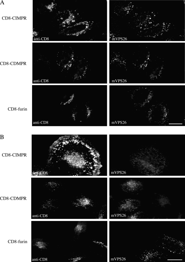
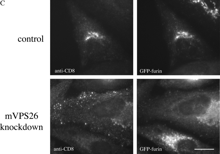
Abstract
fEndosome-to-Golgi retrieval of the mannose 6-phosphate receptor (MPR) is required for lysosome biogenesis. Currently, this pathway is poorly understood. Analyses in yeast identified a complex of proteins called “retromer” that is essential for endosome-to-Golgi retrieval of the carboxypeptidase Y receptor Vps10p. Retromer comprises five distinct proteins: Vps35p, 29p, 26p, 17p, and 5p, which are conserved in mammals. Here, we show that retromer is required for the efficient retrieval of the cation-independent MPR (CI-MPR). Cells lacking mammalian VPS26 fail to retrieve the CI-MPR, resulting in either rapid degradation of or mislocalization to the plasma membrane. We have localized mVPS26 to multivesicular body endosomes by electron microscopy, and through the use of CD8 reporter protein constructs have examined the effect of loss of mVPS26 upon the trafficking of membrane proteins that cycle between the endosome and the Golgi. The data presented here support the hypothesis that retromer performs a selective function in endosome-to-Golgi transport, mediating retrieval of the CI-MPR, but not furin.
Keywords: retromer; vesicle; retrieval; endosome; Golgi
Introduction
Delivery of acid hydrolases to the lysosome/vacuole is initiated in the TGN or late Golgi and is mediated by specific receptors. In mammalian cells, mannose 6-phosphate receptors (MPRs) recognize a phosphorylated carbohydrate tag in the TGN and are then sorted into clathrin-coated vesicles for delivery to a prelysosomal endosome (Kornfeld and Mellman, 1989; Kornfeld, 1992). Although the mannose 6-phosphate tag is not used in simple eukaryotes such as yeast, Saccharomyces cerevisiae uses a similar mechanism to sort vacuolar hydrolases to the vacuole. The Vps10 protein binds vacuolar hydrolases such as carboxypeptidase Y (CPY) via the pro-domain in the late Golgi. Receptor and ligand are then sorted into vesicles for delivery to the prevacuolar compartment (Marcusson et al., 1994; Cooper and Stevens, 1996).
Two types of MPR exist in mammalian cells, the cation-independent MPR (CI-MPR) and the cation-dependent MPR (CD-MPR). Both are type 1 transmembrane proteins that share some sequence similarity in their luminal domains (Kornfeld, 1992). Although there is no sequence homology between Vps10p and the MPRs, fundamentally they perform an identical task, namely that of sorting newly synthesized hydrolases into a pathway that will ultimately deliver the enzymes to the lysosome/vacuole. Recent evidence now supports a role for the conserved Golgi-associated, γ ear–containing ARF-binding proteins (GGAs) in the sorting of both MPRs and Vps10p at the TGN/late Golgi (Robinson and Bonifacino, 2001). The VHS domains of GGA proteins recognize acidic cluster–dileucine signals in the cytoplasmic tails of MPRs and Vps10p, and can also bind clathrin via their hinge regions (Mullins and Bonifacino, 2001; Misra et al., 2002; Shiba et al., 2002). These interactions form the basis of the sorting of MPRs and Vps10p and their cargo of hydrolases into vesicles for eventual delivery to lysosomes/vacuoles.
To maintain efficient sorting and transport of lysosomal/vacuolar hydrolases, the receptors have to be retrieved from the endosome. In contrast to the exit of receptor ligands from the TGN, the process of retrieval is currently poorly understood at the molecular level. Analyses in yeast have identified a complex of five proteins that is necessary for the endosome-to-Golgi retrieval of Vps10p. This complex was dubbed “retromer” and comprises the Vps35p, 29p, 26p, 17p, and 5p proteins (Seaman et al., 1997, 1998). Phenotypic analysis of the respective mutants along with biochemical analyses led to the hypothesis that retromer was a candidate vesicle–coat protein complex that mediates endosome-to-Golgi retrieval in yeast.
Characterization of yeast retromer has provided many insights into the assembly of the complex and the respective roles of the individual components. Several lines of evidence, both genetic and biochemical, favor a role in cargo selection for Vps35p (Nothwehr et al., 1999, 2000). Vps29p is essential for the assembly of the retromer complex. Vps5p and Vps17p are members of the sorting nexin (Snx) family of proteins, and due to the intrinsic self-assembly activity of Vps5p, it was suggested that theVps5p–Vps17p complex may promote vesicle budding (Seaman et al., 1998). Vps26p plays a crucial role in directing the interactions of Vps35p and helps stabilize the retromer complex (Reddy and Seaman, 2001).
Significantly, retromer is highly conserved and an analogous complex has been identified in mammalian cells (Renfrew-Haft et al., 2000). SNX1, the mammalian homologue of Vps5p, associates with the cytoplasmic tails of several proteins that traffic in the endocytic system, including the EGF receptor and the transferrin receptor (TfnR; Kurten et al., 1996; Renfrew-Haft et al., 1998). Does this mean that mammalian retromer mediates endosome-to-Golgi retrieval? This question has yet to be addressed directly, but it has been proposed that mammalian retromer is likely to function in an endosome-to-Golgi retrieval pathway with cargoes yet unknown (Pfeffer, 2001). Aside from retromer, other candidate molecules that could mediate the retrieval of the MPRs are TIP47 (Diaz and Pfeffer, 1998) with rab9 (Riederer et al., 1994) and the clathrin adaptor, AP-1 (Meyer et al., 2000).
Here, we have addressed the specific question of the role of retromer in endosome-to-Golgi retrieval. Using cells derived from transgenic mice that are deleted for mammalian VPS26 (mVPS26) and through the application of small interfering RNA (siRNA) to knock down expression of mVPS26, we show that absence of mVPS26 (and therefore functional retromer) results in a range of phenotypes consistent with a defect in endosome-to-Golgi retrieval.
Results
mVPS26 localizes to endosomes
To examine the role of retromer in endosome-to-Golgi retrieval, we have attempted to address two specific questions. First, where is retromer localized? And second, which proteins are trafficked in a retromer-dependent pathway? To aid in answering both these questions, we have created stably transfected HeLaM cell lines expressing CD8 reporter proteins in which the cytoplasmic tail of CD8 is replaced with the cytoplasmic tail of a protein of interest. CD8 is an excellent candidate for this purpose, as it is a cell surface protein normally expressed in cytotoxic T lymphocytes and is not expressed in HeLaM cells. It is a type I transmembrane protein and does not appear to have intrinsic targeting information in its luminal or transmembrane domain. There are also many commercially available antibodies against CD8 that recognize the luminal domain. Therefore, reporter constructs were created in which the cytoplasmic tail of CD8 was replaced with the cytoplasmic tails of the CI-MPR, the CD-MPR, and furin.
In Fig. 1 A, HeLaM cells stably transfected with these constructs were fixed and double labeled with antibodies against CD8 (left panels) and mVPS26 (right panels). The three different CD8 reporters all have slightly different steady-state localizations. For example, the CD8-furin construct localizes to a perinuclear structure that colocalizes with TGN46 (unpublished data). The CD8–CD-MPR chimera has a more punctate localization consistent with an endosomal localization. The CD8–CI-MPR reporter has both endosomal and TGN localization. Extensive colocalization between mVPS26 and the CD8–CD-MPR construct was observed and some colocalization between mVPS26 and the CD8–CI-MPR construct was also apparent, but there was no significant colocalization between mVPS26 and CD8-furin. Additionally, we find that antibodies against Snx1 label very similar structures to those labeled with anti-mVPS26 antibodies, and there is a similar degree of colocalization between Snx1 and the three CD8 reporters as was observed for mVPS26 (unpublished data).
Figure 1.
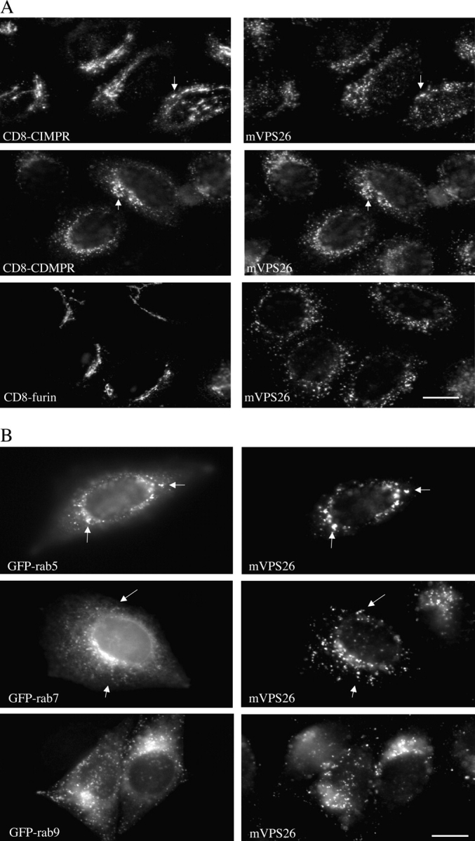
Immunofluorescence localization of mVPS26. (A) HeLaM cells stably transfected with the respective CD8 reporter were fixed and labeled with polyclonal anti-mVPS26 and monoclonal anti-CD8 followed by secondary antibodies. Bar, 20 μm. (B) HeLaM cells stably expressing GFP-rab proteins were fixed and labeled with antisera against mVPS26 followed by fluorescently labeled secondary antibodies. Bar, 20 μm. Arrows indicate coincident labeling.
The rab family of small GTPases regulates membrane docking and fusion events throughout the secretory and endocytic pathways. Therefore, rab proteins can be used as markers for particular membranes. For example, rab5 is localized to early endosomes, whereas rabs 7 and 9 are present on late endosomes (and some lysosomes) (Pfeffer, 2003). Using GFP-tagged rab proteins stably expressed in HeLaM cells, we have compared the localization of mVPS26 to rabs 5, 7, and 9. In Fig. 1 B, we find that there is significant colocalization between mVPS26 and GFP-rab5 and modest colocalization between mVPS26 and GFP-rab7, but very little overlap between mVPS26 and GFP-rab9.
We have also determined the localization of mVPS26 and Snx1 by EM. Attempts to use conventional cryo-EM to localize mVPS26 were unsuccessful. Therefore, we developed a method to allow the pre-embedding labeling of mVPS26. In Fig. 2 , a montage of images is shown. Antibodies against both mVPS26 (a–c) and Snx1 (d and e) labeled structures that had morphology typical of endosomes. Arrows indicate where tubular–vesicular elements are labeled with the anti-Snx1 antibody.
Figure 2.
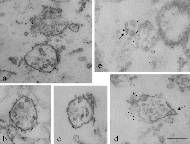
Electron microscopic localization of mVPS26 and Snx1. HeLaM cells grown in 6-cm dishes were permeabilized by rapid freeze/thaw. After fixation, mVPS26 or Snx1 was labeled with polyclonal antisera followed by 10-nm colloidal gold anti–rabbit. The cells were embedded in Epon and then sectioned to produce ∼50-nm-thick sections. mVPS26 (a–c) and Snx1 (d and e) are found on multivesicular bodies characteristic of endosomal compartments. Bar, 250 nm. Arrows indicate Snx1 localization to tubular–vesicular structures.
Loss of mVPS26 expression results in altered CI-MPR localization
The localization data are consistent with a role for retromer in endosome-to-Golgi retrieval. To examine this possibility more directly, we used cell lines lacking mVPS26 to determine the role of retromer in membrane trafficking. Cells derived from transgenic mVPS26−/− mice were analyzed along with cells from homozygous wild-type mice. Additionally, we used siRNA to knock down the expression of mVPS26 in HeLaM cells. In Fig. 3 A, the expression of mVPS26 is examined and compared with a control protein (actin). No expression of mVPS26 was detected in the mVPS26−/− cells. Similarly, siRNA treatment of the HeLaM cells results in complete abolition of mVPS26 expression.
Figure 3.
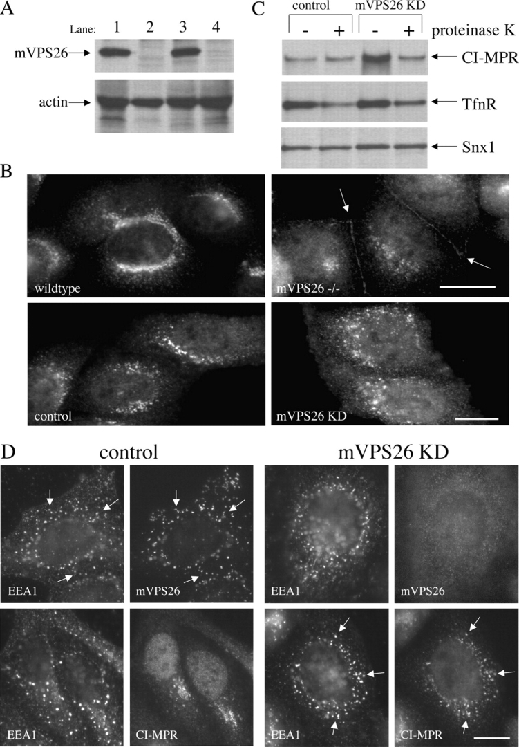
Loss of mVPS26 expression results in increased cell surface CI-MPR. (A) Cells grown in 6-cm dishes were labeled with [35S]methionine continuously for 3 h before lysis. mVPS26 or actin was immunoprecipitated and then subjected to SDS-PAGE and fluorography. Lane 1, wild-type cells; lane 2, mVPS26−/− cells; lane 3, control cells; lane 4, mVPS26 knock-down cells. (B) Cells were fixed and labeled with antibodies against the CI-MPR. Arrows indicate CI-MPR localization to the plasma membrane. Bars, 20 μm. (C) Cells grown in 9-cm dishes were labeled overnight with [35S]methionine. After removal from the dish, the cells were treated with proteinase K and then were precipitated and lysed. The CI-MPR, TfnR, and Snx1 were recovered from the resulting lysates and subjected to SDS-PAGE and fluorography. (D) Control or mVPS26 knock-down cells were fixed and labeled with antibodies against EEA1, mVPS26, or the CI-MPR. Arrows indicate coincident labeling. Bar, 20 μm.
Loss of Vps26p in yeast results in a defect in endosome-to-Golgi retrieval of Vps10p. If mVPS26 functions in an analogous pathway, the trafficking of the CI-MPR would be predicted to be affected after loss of mVPS26. Accordingly, we investigated the distribution of the CI-MPR in mVPS26−/− cells and also in mVPS26 knock-down cells by immunofluorescence. In Fig. 3 B, cells labeled with anti-CI-MPR are shown. There is a dramatic change in the CI-MPR localization in the mVPS26−/− cells compared with wild-type cells with much reduced intensity of labeling and apparently some cell surface labeling of the CI-MPR (indicated by arrows). In the mVPS26 knock-down cells the change in CI-MPR localization is more subtle, with CI-MPR labeling being detected in more dispersed structures no longer restricted to the perinuclear region. To determine if there is increased cell surface CI-MPR in the mVPS26 knock-down cells, a more quantitative method was adopted. Cells labeled with [35S]methionine were treated with exogenous protease. After lysis, the CI-MPR, TfnR, and an intracellular protein (Snx1) were immunoprecipitated. In Fig. 3 C, the CI-MPR becomes susceptible to exogenous protease after mVPS26 knock down, indicating that there is more cell surface CI-MPR. The TfnR is no more susceptible to proteolysis after mVPS26 knock down and the intracellular protein Snx1 is unaffected by addition of the protease, showing that proteolysis was restricted to only cell surface proteins.
Increased cell surface localization of the CI-MPR after mVPS26 knock down would also be expected to result in increased colocalization of the CI-MPR with early endosomal markers such as EEA1. This was investigated by double labeling control and mVPS26 knock-down cells with antibodies against the CI-MPR and EEA1. In Fig. 4 D, the control cells display extensive overlap in the labeling patterns of EEA1 and mVPS26 (indicated by arrows), but relatively little coincidence between the CI-MPR and EEA1. After siRNA knock down of mVPS26, the EEA1 distribution is largely unchanged, but the CI-MPR appears to be redistributed to EEA1-positive structures (indicated by arrows). These data are consistent with increased cycling of the CI-MPR between the cell surface and early endosomes after mVPS26 knock down.
Figure 4.
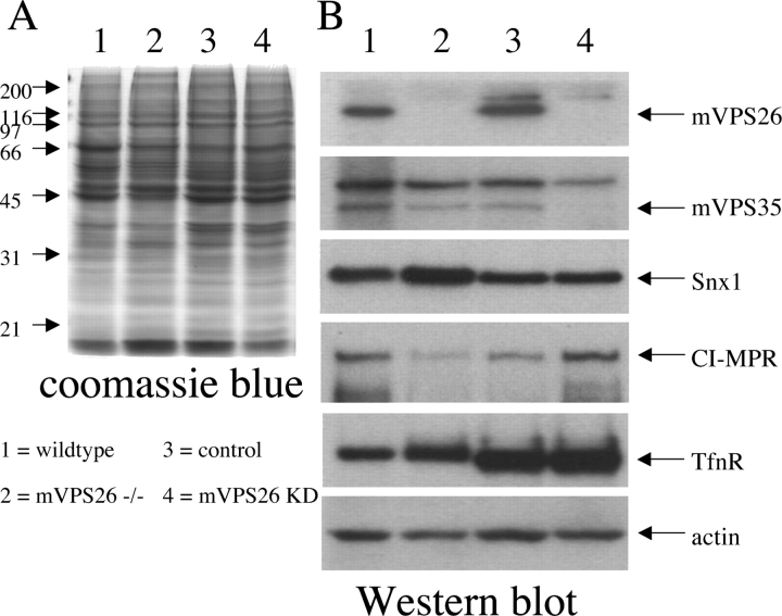
Steady-state levels of the CI-MPR are affected after loss of mVPS26. (A) Lysates from the wild-type and mVPS26−/− mouse cells and lysates from the control and mVPS26 knock-down HeLaM cells were prepared and electrophoresed on a 10% polyacrylamide gel. The gel was stained with Coomassie blue. (B) Similar gels to the one shown in A were transferred onto nitrocellulose and then were Western blotted with various antibodies followed by 125I-protein A. Lane 1, wild-type cells; lane 2, mVPS26−/− cells; lane 3, control cells; lane 4, mVPS26 knock-down cells.
To examine the steady-state levels of the CI-MPR and other proteins after loss of mVPS26, lysates prepared from unlabeled cells were subjected to SDS-PAGE and the resulting gel was stained with Coomassie blue (Fig. 4 A). Steady-state levels of various proteins were determined by Western blotting. In Fig. 4 B, consistent with the data from the metabolic labeling experiment (Fig. 3 A), mVPS26 was undetectable in the mVPS26−/− cells and HeLaM knock-down cells. Levels of mVPS35 (bottom band) were reduced in the mVPS26−/− cells, but appeared virtually undetectable in the knock-down sample. Most interesting is the effect on levels of the CI-MPR. In the mVPS26−/− sample it is severely reduced, whereas in the knock-down sample it appears elevated. Another membrane protein, the TfnR, does not appear to be significantly affected in the mVPS26−/− or knock-down samples. Using a short (15 min) pulse of [35S]methionine followed by a 5-min chase, we found that there was a 1.4-fold increase in the expression of the CI-MPR in mVPS26 knock-down cells relative to a control protein, actin (unpublished data).
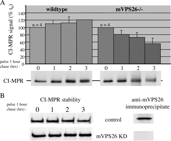
Increased expression of the CI-MPR in the mVPS26 knock-down cells could account for the apparent increased steady-state levels of CI-MPR, but could also have been masking an increased turnover of the CI-MPR in the knock-down cells. The apparent loss of the CI-MPR in mVPS26−/− cells indicates that there is a defect in endosome-to-Golgi retrieval, resulting in the degradation of the CI-MPR in lysosomes. Therefore, we investigated the stability of the CI-MPR in both the mVPS26−/− cells and in the mVPS26 knock-down cells by a pulse–chase experiment. Cells were labeled with [35S]methionine and then chased over a 3-h period. The CI-MPR was recovered from lysates prepared at various time points by immunoprecipitation. After analysis of CI-MPR levels by SDS-PAGE and fluorography, the data were converted to graph form (Fig. 5 A). The CI-MPR was rapidly degraded in mVPS26−/− cells with a half-life of ∼3 h; however, to our surprise, we have found that there was no increased turnover of the CI-MPR in the mVPS26 knock-down cells (Fig. 5 B).
Figure 5.
The CI-MPR is unstable in the mVPS26 −/− cells. (A) Cells grown in 3-cm dishes were pulse labeled with [35S]methionine for 1 h and then chased for 0, 1, 2, or 3 h. The cells were lysed and the CI-MPR was recovered by immunoprecipitation and then subjected to SDS-PAGE and fluorography. The signal on the resulting film was quantified and expressed as a percentage of the signal at time 0. The data from four experiments were averaged together and are shown in the graph. The error bars are SDs. The bottom panels are representative of the data obtained and show the instability of the CI-MPR in the mVPS26−/− cells. (B) The CI-MPR stability experiment was repeated as described above using control and mVPS26 knock-down cells. There is no apparent instability of the CI-MPR after mVPS26 knock down.
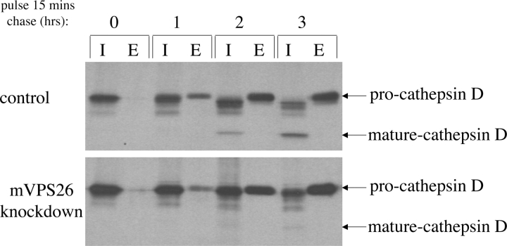
A defect in retrieval of the CI-MPR would rapidly lead to a loss of CI-MPR in the TGN. This could manifest itself as a loss of proper sorting of lysosomal hydrolases. This is indeed the case in yeast when CPY sorting in retromer mutants is measured. A well-characterized lysosomal hydrolase in mammalian cells is cathepsin D. Sorting of cathepsin D in the TGN requires the MPRs. Therefore, we measured cathepsin D maturation in control and mVPS26 knock-down cells. As shown in Fig. 6 , mVPS26 knock down results in a severe defect in cathepsin D maturation, with almost no mature cathepsin D detectable after a 3-h chase.
Figure 6.
Loss of mVPS26 results in a defect in cathepsin D maturation. Cells grown in 3-cm dishes were pulse labeled with [35S]methionine for 15 min and then chased for 0, 1, 2, or 3 h. At the end of the chase, the media was removed and the cells were lysed. Cathepsin D was immunoprecipitated from both the intracellular (I) and extracellular (E) (media) fractions and subjected to SDS-PAGE and fluorography.
Loss of mVPS26 causes a selective endosome-to-Golgi retrieval defect
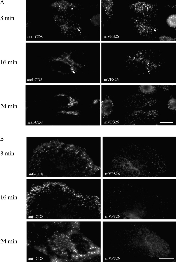
The lack of retrieval of the CI-MPR in mVPS26 knock-down cells, which is indicated by the increased cell surface levels of the CI-MPR and the lack of cathepsin D maturation, was further investigated using the CD8 reporter constructs. Stably transfected cells expressing the CD8–CI-MPR, CD8–CD-MPR, and CD8-furin were treated with the mVPS26 siRNA. Cells were fixed and labeled with antibodies against CD8 and mVPS26. As shown in Fig. 7 A, siRNA knock down of mVPS26 results in a very significant redistribution of the CD8–CI-MPR from a predominantly perinuclear localization to peripheral structures (compare Fig. 7 A with Fig. 1 A). The distribution of the CD8–CD-MPR was not significantly affected, and although the appearance of the CD8-furin chimera was altered, its localization was still predominantly perinuclear. Indeed, it appeared that the CD8-furin labeling and also the perinuclear CD8–CI-MPR labeling was that of a fragmented structure. This is particularly noticeable when occasional unaffected cells are found next to cells in which the knock down of mVPS26 has occurred. Subsequently, we decided to investigate the state of the Golgi complex in cells lacking mVPS26 using the Golgi marker protein GM130 and the TGN marker TGN46 as a means of labeling Golgi and TGN membranes, respectively. In Fig. 7 B, the immunofluoresence patterns of GM130 and TGN46 are shown. Loss of mVPS26 expression results in fragmentation of the Golgi complex. This is particularly evident in the mVPS26 knock-down cells, but is also apparent in the mVPS26−/− cells (unpublished data). By double labeling with antibodies against TGN46, we find that the CD8-furin construct in mVPS26 knock-down cells remains TGN localized, but the TGN is now fragmented (Fig. S3 B, available at http://www.jcb.org/cgi/content/full/jcb.200312034/DC1).
Figure 7.
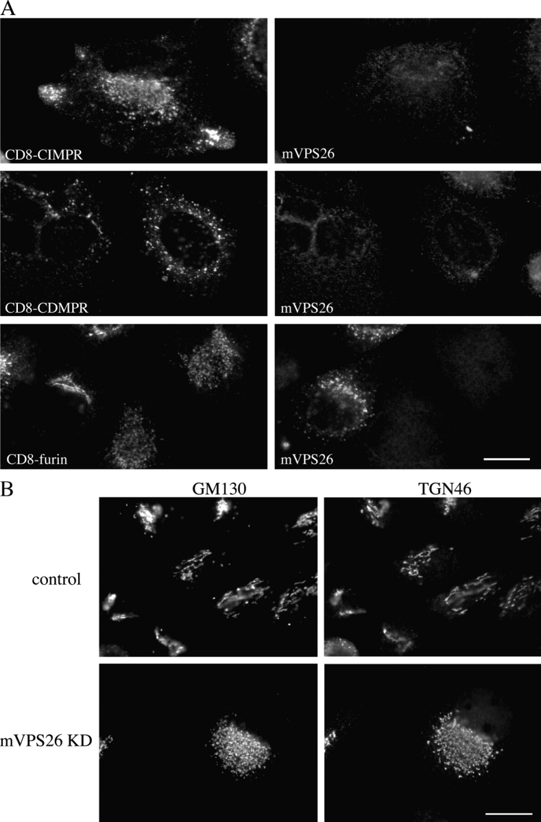
CD8 reporter localization and Golgi fragmentation after loss of mVPS26 expression. (A) Cells that had been treated with mVPS26 knock-down siRNA were fixed and labeled with antibodies against CD8 and mVPS26 (as in Fig. 1 A). Bar, 20 μm. (B) Control cells and mVPS26 knock-down cells labeled with anti-GM130 and anti-TGN46. Bar, 20 μm.
Although the steady-state localization of the CI-MPR and CD8 reporter constructs strongly suggests a defect in endosome-to-TGN retrieval, a more direct assay would be to follow the movement of proteins from endosomes to the TGN. Using the CD8 reporter constructs, we can examine trafficking from the cell surface to the TGN. In Fig. 8 A, cells expressing the CD8–CI-MPR construct were labeled with the anti-CD8 antibody at 4°C to label only the protein at the surface. After washing away unbound antibody, the cells were warmed to 37°C and the antibody was chased for 8, 16, and 24 min before the cells were washed again and fixed. After 8 min, there was extensive colocalization between the CD8 antibody and mVPS26. By 16 min, much of the antibody (and therefore the CD8–CI-MPR) had reached the perinuclear region, although some colocalization between the mVPS26 and the CD8 antibody remained. After 24 min of chase, the antibody had reached the perinuclear region of the cell and had a typical TGN appearance. By double labeling cells with anti-CD8 and anti-TGN46, we confirmed that after 24 min of chase, the anti-CD8 antibody had reached the TGN (Fig. 8 C, top panels).
Figure 8.
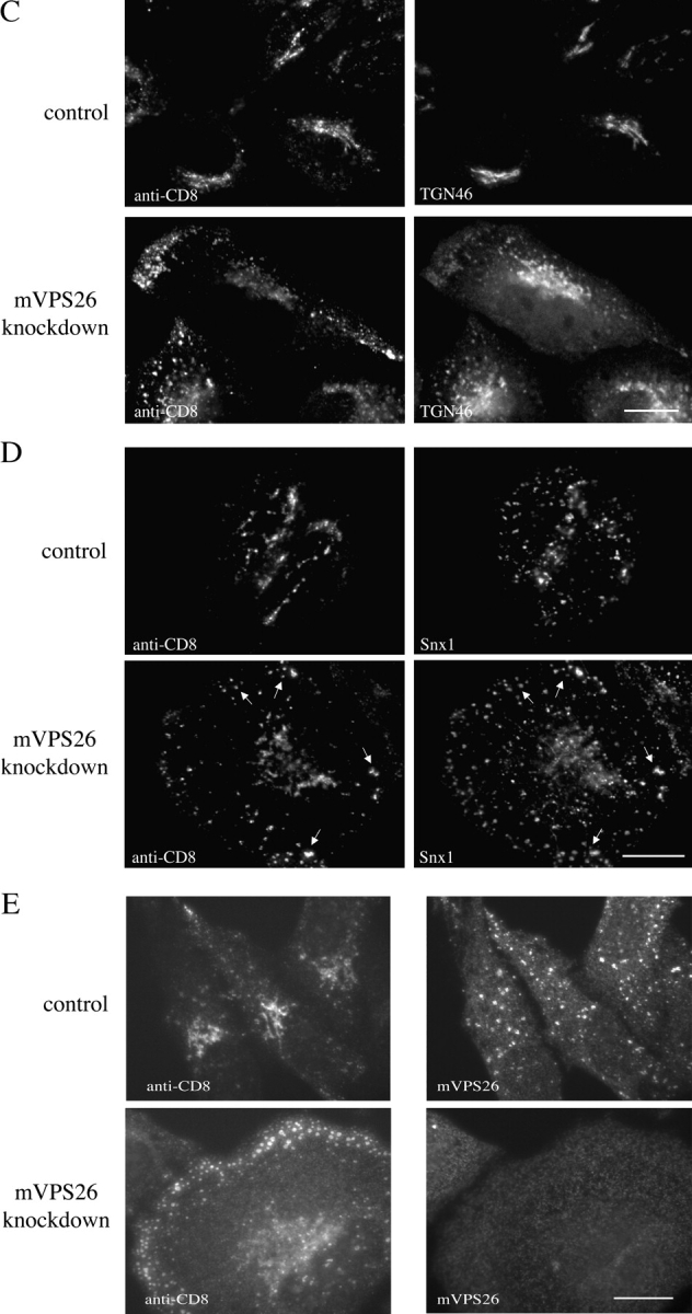
Antibody uptake experiments demonstrate a defect in the kinetics of endosome-to-TGN retrieval after mVPS26 knock down. (A) Control cells expressing the CD8–CI-MPR were incubated with anti-CD8 antibodies, washed, and then warmed to 37°C for 8, 16, or 24 min before fixation. The cells were then labeled with antibodies against mVPS26 followed by secondary antibodies. (B) mVPS26 knock-down cells were treated as in A. (C) Cells from A and B that had been chased for 24 min were labeled with antibodies against TGN46. (D) Cells from A and B that had been chased for 24 min were labeled with antibodies against Snx1. (E) CD8-sortilin–expressing cells were subjected to the anti-CD8 uptake assay. After 24 min of chase, the cells were fixed and labeled with antibodies against mVPS26 followed by secondary antibodies. Arrows in A and D indicate coincident labeling. Bars, 20 μm.
Therefore, the movement of the CD8–CI-MPR from the cell surface to the TGN involved passage through an mVPS26-positive compartment. In Fig. 8 B, the experiment is repeated in mVPS26 knock-down cells. Loss of mVPS26 expression resulted in the CD8–CI-MPR accumulating in peripheral structures close to the plasma membrane. After 24 min, some of the CD8 antibody had reached the perinuclear region, but a significant proportion remained in peripheral structures. The perinuclear localized anti-CD8 is indeed a region of the TGN, as confirmed by double labeling with anti-TGN46 (Fig. 8 C, bottom panels), but the TGN had become fragmented in the mVPS26 knock-down cells, resulting in altered morphology of the TGN. The peripheral structures containing the anti-CD8 antibody were found to be positive for the Snx1 protein (Fig. 8 D), indicating that the CD8–CI-MPR was retained in endosomes after mVPS26 knock down.
Loss of mVPS26 results in a defect in the retrieval of endogenous CI-MPR and also the CD8–CI-MPR reporter. Are there other proteins that cycle between the endosome and TGN in a retromer-dependent manner? To answer this question, we made another CD8 reporter, this time with the tail of sortilin. Sortilin is a homologue of the Vps10 protein (Petersen et al., 1997) and is believed to have a similar trafficking itinerary to the CI-MPR. In Fig. 8 E, the anti-CD8 uptake assay was performed on cells stably transfected with CD8-sortilin. In control cells (top panels), the antibody is endocytosed and (after 24 min of chase) delivered to the TGN. After mVPS26 knock down, the endocytosed antibody accumulates in peripheral structures that have a similar morphology to those observed in CD8–CI-MPR cells (compare Fig. 8 E with Fig. 8 B).
Therefore, loss of mVPS26 expression results in a significant effect upon the endosome-to-Golgi retrieval of the CD8–CI-MPR chimera. The effect on the trafficking of the CD8–CD-MPR and CD8-furin chimeras was also examined using the anti-CD8 antibody uptake assay, although this time by continuous incubation with antibody over 3 h. Over the 3-h period, the CD8 reporter construct with bound antibody will reach a steady-state localization. In Fig. 9 A, control cells were incubated with anti-CD8 for 3 h at 37°C. After washes, the cells were fixed and mVPS26 was labeled along with the anti-CD8 antibody. The distribution of the anti-CD8 antibody in the cells expressing the three CD8 reporter constructs is virtually the same as shown for the steady-state distribution of the three chimeras in Fig. 1 A. There is some significant colocalization between the CD8–CI-MPR and mVPS26 and very extensive colocalization between the CD8–CD-MPR and mVPS26, but very little colocalization between CD8-furin and mVPS26. After mVPS26 knock down (Fig. 9 B), endocytosed anti-CD8 antibody is found in peripheral and some perinuclear structures in CD8–CI-MPR expressing cells. The CD8–CD-MPR expressing cells appear to endocytose more anti-CD8 antibody after knock down of mVPS26. This is apparent when some unaffected cells are compared with cells in which the mVPS26 knock down has occurred. The overall localization of the endocytosed anti-CD8 antibody in CD8–CD-MPR expressing cells is not any different after mVPS26 knock down. In cells expressing the CD8-furin chimera, endocytosed anti-CD8 reaches a perinuclear compartment that, although fragmented, does correspond to the TGN and colocalizes with anti-TGN46 (unpublished data).
Figure 9.
mVPS26 knock down results in a selective endosome-to-Golgi retrieval defect. (A) Control cells expressing the CD8 reporter chimeras were incubated at 37°C continuously for 3 h with the anti-CD8 antibody. After washes, the cells were fixed and then labeled as in Fig. 8, A and B. (B) As in A, but with mVPS26 knock-down cells. (C) Cells expressing both GFP-furin and CD8–CI-MPR were used in an antibody uptake experiment similar to those in Fig. 8. After 24 min of chase, the cells were fixed and labeled with secondary antibodies. Bars, 20 μm.
This experiment indicates that although the CD8–CI-MPR reporter requires mVPS26 for its retrieval, CD8-furin does not. This possibility was investigated further using a cell line with both furin and CI-MPR reporter proteins. By replacing the CD8-coding region of the CD8-furin reporter with GFP, a GFP-furin reporter was generated. This was found to localize to the TGN in an identical fashion to the CD8-furin reporter. Cells expressing GFP-furin were transfected with CD8–CI-MPR on a different vector. The GFP-furin/CD8–CI-MPR transfected cells were subjected to the anti-CD8 uptake experiment after siRNA knock down of mVPS26. In Fig. 9 C, in control cells, the anti-CD8 (i.e., CD8–CI-MPR) is endocytosed and delivered to the TGN where it colocalizes precisely with GFP-furin (top panels). After mVPS26 knock down (bottom panels), the endocytosed anti-CD8 is found in peripheral structures and no longer colocalizes with the GFP-furin (bottom panels). Together, these data provide compelling evidence that mVPS26 (and therefore retromer) is required for efficient retrieval of the CI-MPR from endosomes to the Golgi.
Discussion
This work has attempted to address the question of whether retromer is required for endosome-to-Golgi retrieval in mammalian cells. Using cells derived from transgenic mVPS26−/− mice or HeLaM cells in which mVPS26 expression was ablated by siRNA transfection, we have investigated the trafficking of both endogenous proteins such as the CI-MPR and chimeric constructs using the luminal domain of CD8 as a reporter.
Key to understanding the role that retromer plays in mammalian cells is the localization of retromer itself. We find that mVPS26 is localized to endosomal membranes that are positive for rab5 and EEA1. These data are consistent with the published colocalization between Snx1 and EEA1 (Chin et al., 2001). The EM analysis of mVPS26 and Snx1 localization confirms that mVPS26 is present on multivesicular bodies.
CD8 has been extensively and successfully used in previous works as a reporter protein for type I transmembrane proteins such as furin, TGN38, and endolyn (Chapman and Munro, 1994; Ponnambalam et al., 1994; Ihrke et al., 2000). The CD8 reporter constructs used here have proven to be invaluable for these experiments, as they each have a slightly different steady-state localization. The CD8–CI-MPR appears to reside in both TGN and endosomal compartments, whereas the CD8–CD-MPR and CD8-furin constructs are restricted to endosomal or TGN localization, respectively. This difference in steady-state localization of the three chimeras can only be due to the intrinsic signals present in the three different cytoplasmic tails of the CD8 reporter constructs, as the transmembrane and luminal domains are identical in each case. As the cytoplasmic tails of the three chimeras are responsible for the trafficking/localization of the protein, it is logical to assert that interactions with cytoplasmic machinery such as vesicle coat proteins are critical to determine the trafficking of the chimeras. So although we do not claim that the trafficking of the CD8–CI-MPR is exactly identical to the endogenous CI-MPR, the chimeras do, nevertheless, provide a means of examining the trafficking of various type I transmembrane proteins present in the TGN/endocytic system.
The EM localization and colocalization of mVPS26 with rab5 and EEA1 establishes that mVPS26 is in the “right place” to mediate endosome-to-TGN retrieval (see also Fig. S2, available at http://www.jcb.org/cgi/content/full/jcb.200312034/DC1). To investigate whether retromer has a role in endosome-to-TGN retrieval, we have used a cell line derived from mVPS26−/− transgenic mice (Lee et al., 1992), and we have succeeded in knocking down mVPS26 expression by RNA interference. The siRNA knock down of mVPS26 in the HeLaM cells can achieve an almost complete loss of mVPS26 expression such that by Western blotting and metabolic labeling, levels of mVPS26 protein are undetectable and therefore no different to levels of mVPS26 protein in the mVPS26−/− cells. Only occasionally are mVPS26-positive cells observed by immunofluorescence. Loss of mVPS26 expression has a profound effect upon the trafficking of the CI-MPR, resulting in a significant increase in the amount of CI-MPR present at the cell surface and extensive overlap in the immunofluorescence labeling patterns of the CI-MPR and EEA1.
If retromer is required for the endosome-to-Golgi retrieval of the CI-MPR, then redistribution to the cell surface and increased cycling between the plasma membrane and an early endosome is one of two possible fates of the CI-MPR in retromer-deficient cells. The other possible destination is the lysosome, which would result in increased turnover of the CI-MPR. This does appear to be the case for the mVPS26−/− cells, which have significantly reduced steady-state levels of CI-MPR and degrade their CI-MPR with a half-life of ∼3 h. In the mVPS26 knock-down cells there doesn't appear to be any increased turnover of the CI-MPR. A possible reason why there is a lack of turnover of the CI-MPR in the mVPS26 knock-down cells is that there may be more trafficking of the CI-MPR between endosomes and the plasma membrane and less delivery of the CI-MPR to lysosomes relative to the mVPS26−/− cells. Recent reports by Maxfield and colleagues have indicated that a significant proportion of the CI-MPR cycles between the plasma membrane and early endosomes after being endocytosed (Lin et al., 2004). Additionally, experiments performed over many years have shown that the CI-MPR localization can differ considerably between cell types being either predominantly TGN localized or endosomally/prelysosomally localized (Brown et al., 1986; Brown and Farquhar, 1987; Griffiths et al., 1988; Griffiths, 1989).
Even though there is no apparent instability of the CI-MPR after mVPS26 knock down, there is in fact a significant endosome-to-TGN retrieval defect resulting in rerouting of the CI-MPR into other pathways (e.g., endosome to plasma membrane). This assertion is supported by the fact that maturation of cathepsin D is completely abolished after mVPS26 knock down. Again, this is consistent with the yeast retromer deletion mutants, which all display strong CPY sorting defects.
When the steady-state localization of the CD8 reporter constructs was examined after mVPS26 knock down, we found that the CD8–CI-MPR construct was partially redistributed to peripheral structures, but retained some perinuclear labeling in a compartment that was fragmented. The distribution of the CD8–CD-MPR chimera was not significantly affected, but the CD8-furin construct was now also in a fragmented perinuclear compartment. This fragmented perinuclear compartment is likely to be the Golgi/TGN, as labeling with antibodies against GM130 and TGN46 showed that the Golgi complex fragments as a consequence of the loss of mVPS26 (see also Fig. S1, available at http://www.jcb.org/cgi/content/full/jcb.200312034/DC1). It is worth noting that although fragmented, the TGN/Golgi remains very much restricted to the perinuclear region of the cell. The cause of this fragmentation is not clear, but may be due to a defect in endosome-to-TGN retrieval of SNARE proteins that are required for homotypic Golgi membrane fusion. This is currently being investigated. Golgi fragmentation appeared less severe in the mVPS26−/− cells possibly due to some form of adaptation that has occurred after prolonged culture.
The steady-state distributions of the CD8 reporters after mVPS26 knock down suggested that loss of mVPS26 could have a major impact on endosome-to-Golgi retrieval. However, the dynamic nature of the TGN/endocytic system requires that the kinetics of transport also be investigated. Using an antibody uptake assay, we followed the trafficking of the CD8–CI-MPR from the cell surface to the TGN. In control cells, the process is rapid and occurs with kinetics consistent with previously published reports on CI-MPR retrieval (Hirst et al., 1998). CD8–CI-MPR at the cell surface is efficiently endocytosed, and after an 8-min chase is present in a compartment positive for mVPS26. After 24 min, the CD8–CI-MPR has reached the TGN and colocalizes with TGN46. In mVPS26 knock-down cells, the CD8–CI-MPR appears to be efficiently endocytosed, but it is not rapidly returned to the TGN. Instead, the CD8–CI-MPR appears to accumulate in structures close to the plasma membrane. After 24 min, a small minority of CD8–CI-MPR does appear to be delivered to the fragmented perinuclear structures that are positive for TGN46, but the vast majority of internalized CD8–CI-MPR remains in peripheral structures that are positive for Snx1. A CD8-sortilin reporter behaved in a similar fashion after mVPS26 knock down. As sortilin is a mammalian homologue of the yeast Vps10 protein (Petersen et al., 1997), it would perhaps be expected to traffic in a similar fashion to Vps10p (i.e., retromer dependent). Additionally, it has been observed that the tail of Vps10p can functionally substitute for the tail of the CI-MPR (Dennes et al., 2002). As yeast retromer mediates retrieval of Vps10p, mammalian retromer performs the same task for the CI-MPR.
Although the kinetics of the CD8–CI-MPR retrieval in control cells appeared to be rapid, the CD8-furin chimera took much longer to reach the TGN, requiring a chase of ∼60 min (unpublished data). It was also difficult to examine retrieval of the CD8-furin chimera, as very little of the chimera is present at the cell surface at any one time. Therefore, prolonged incubation with the anti-CD8 antibody was used to determine the extent of uptake and to allow the antibody to reach its steady-state distribution. Loss of mVPS26 results in an accumulation of the CD8 antibody in peripheral endosomal structures in the CD8–CI-MPR chimera-expressing cells. However, the CD8-furin chimera is endocytosed and delivered to the fragmented compartment positive for TGN46 (unpublished data).
Therefore, these data suggest that mVPS26 knock down can affect the trafficking of some proteins far more than others. The retrieval of the CD8–CI-MPR (and CD8-sortilin) reporter is greatly affected, whereas the CD8-furin protein is only marginally affected, if at all. This hypothesis was confirmed by following the uptake of the CD8–CI-MPR reporter in cells expressing a GFP-furin chimera. These data are in line with the current hypothesis that there are two distinct endosome-to-Golgi retrieval pathways in mammalian cells (Mallet and Maxfield, 1999). This can also explain why there is in fact some retrieval of the CD8–CI-MPR to the TGN after mVPS26 knock down. A parallel pathway, possibly using AP-1 (Meyer et al., 2000), could provide for partial (perhaps kinetically slower) retrieval of the CI-MPR (and CD8–CI-MPR).
In summary, loss of mVPS26 results in a severe defect in endosome-to-TGN retrieval of the CI-MPR. mVPS26 could mediate retrieval of the CI-MPR from both early and late endosomes. This would account for the partial overlap in the labeling patterns of mVPS26 and GFP-rab7, and may suggest that retromer-mediated retrieval is part of the process of endosome maturation. In this scenario, retromer could initiate endosome-to-TGN retrieval of the CI-MPR at an early endosome, but retrieval would continue as the endosome matures. The apparent lack of colocalization between mVPS26 and GFP-rab9 may indicate that an additional trafficking step is required for the retrieval of the CI-MPR from endosomes. This additional step would require TIP47 (Diaz and Pfeffer, 1998) and rab9 (Riederer et al., 1994). Alternatively, retromer and TIP47/rab9 could function sequentially in endosome-to-TGN retrieval.
The CD8 reporter constructs have been used to investigate which proteins may be retrieved in a retromer-dependent fashion. The retrieval of the CD8–CI-MPR chimera depends upon retromer, whereas the trafficking of the CD8-furin construct does not, probably because of the existence of a parallel endosome-to-TGN retrieval pathway. An obvious candidate to mediate this pathway is AP-1 in concert with the furin-interacting “adaptor” PACS-1 (Crump et al., 2001; Folsch et al., 2001).
Materials and methods
Laboratory reagents
Most laboratory reagents including tissue culture media were purchased from Sigma-Aldrich. Restriction enzymes and other molecular biology reagents were obtained from New England Biolabs, Inc. Radioisotopes were purchased from Amersham Biosciences.
Antibodies
The monoclonal anti-CD8 was obtained from Qbiogene. Anti-GM130 and anti-EEA1 were purchased from Becton Dickinson, and the anti-TfnR antibody was obtained from Zymed Laboratories. Polyclonal anti-actin was purchased from Sigma-Aldrich, and the polyclonal anti-CI-MPR (Reaves et al., 1996) was a gift from J. Paul Luzio (University of Cambridge, Cambridge, UK). Anti-cathepsin D antiserum was obtained from DakoCytomation, and anti-TGN46 was supplied by Serotec. Polyclonal anti-mVPS26, anti-Snx1, and anti-mVPS35 were produced in house using GST fusion proteins as antigens. The antisera were subsequently affinity purified using the respective antigens coupled to CNBr-Sepharose.
Microscopy
Immunofluorescence microscopy was performed using an epifluorescence microscope (Axioplan; Carl Zeiss MicroImaging, Inc.) with a Plan-Apochromat 63× oil immersion lens (Carl Zeiss MicroImaging, Inc.) at RT. The secondary antibodies were obtained from Molecular Probes, Inc. and were conjugated to Alexa Fluor® 488 or 594. The image was obtained using a CCD camera (Princeton Instruments) that was controlled via IP Lab software. Images were cropped using Adobe Photoshop® software.
Cell culture, wild-type, and mVPS26−/− mouse cells
HeLaM cells (Tiwari et al., 1987) were provided by M.S. Robinson (University of Cambridge, Cambridge, UK). Embryonic stem cells derived from either homozygous wild-type or homozygous mVPS26−/− mouse embryos (Robertson et al., 1992) were provided by F. Constantini (Columbia University, New York, NY). The embryonic stem cells were produced as part of an experiment to identify genes important for early embryogenesis (Radice et al., 1991). Upon receipt, the cells were cultured in DME supplemented with 10% FCS, 2 mM glutamine, and 1 mM pyruvate, and containing 0.1 mM β-mercaptoethanol and penicillin/streptomycin. Wild-type and mVPS26−/− cells were treated identically and were grown over a period of several months, passaging as necessary.
SDS-PAGE and Western blotting
SDS-PAGE and Western blotting were performed as described in Reddy and Seaman (2001).
GFP-rabs and CD8 reporter constructs
EST cDNA encoding rab5c, rab7, and rab9 were obtained from the IMAGE Consortium. The rab cDNAs were amplified by PCR using primers to add a BglII and an SalI site to the 5′ and 3′ ends, respectively. The PCR products were then cloned into the pEGFP-C1 vector (CLONTECH Laboratories, Inc.) for expression as a GFP fusion in mammalian cells. The CD8–CD-MPR, CD8-furin, and CD8-sortilin constructs were made in a similar fashion. ESTs for the human cDNAs of the CD-MPR, furin, and sortilin were obtained from the IMAGE Consortium. Primers were designed to PCR through the region encoding the cytoplasmic tails of the respective proteins. The 5′ primers incorporated an AflII site; the 3′ primers were designed to anneal 3′ to the stop codon. The PCR products were first cloned using the pCR blunt vector (Invitrogen). After digestion with AflII and either SpeI or NotI (depending on orientation), the insert was cloned into CD8 in pBlueScript® that had been digested with AflII and either XbaI or NotI to excise the endogenous CD8 cytoplasmic tail. Constructs were sequenced and then subcloned from pBlueScript® into the mammalian expression vector pIRESneo2 (CLONTECH Laboratories, Inc.). To generate the GFP-furin chimera, the luminal domain of CD8 (downstream of the signal sequence) was replaced with GFP. To facilitate production of this construct, a BamHI site was engineered into the CD8 cDNA 3′ to the signal sequence. The luminal region of CD8 was excised by digestion with BamHI and EcoRV and replaced with GFP, which had a BamHI site introduced at the 5′ end by PCR.
The CD8–CI-MPR reporter was constructed by subcloning the cDNA encoding the cytoplasmic tail of the bovine CI-MPR into CD8-pIRESneo2. The bovine CI-MPR cDNA (provided by H. Davidson, University of Colorado, Denver, CO) was digested with BsrGI, which cuts precisely at the junction between the transmembrane domain and the cytoplasmic tail. After BsrGI digestion, the overhang was filled by T4 polymerase and then the CI-MPR cDNA was cut with NotI. The fragment corresponding to the tail was gel isolated and cloned into CD8-pIRESneo2, which had been digested with AflII, blunted, and then digested with NotI. This construct was also sequenced to confirm the success of the cloning. The constructs in pIRESneo2 or pEGFP-C1 were transfected into HeLaM cells using FuGENE™ 6 (Roche). Stably transfected cells were selected using 500 μg/ml G418 (Life Technologies) and were screened for expression of the CD8 reporter constructs by immunofluorescence.
Pre-embedding labeling EM
Cells grown in 6-cm dishes were washed once with cold PBS and then once with cold glutamate lysis buffer (25 mM Hepes-KOH, pH 7.4, 25 mM KCl, 2.5 mM magnesium acetate, 5 mM EGTA, and 150 mM potassium glutamate) before draining on ice for 5 min. The cells were then snap frozen by immersion in liquid nitrogen and quick thawed by placing the dish on the bench for ∼60 s. The dish was then returned to ice and the cells were washed once with 2 ml glutamate lysis buffer. Excess buffer was removed and replaced with 2 ml of 4% PFA in PBS. The cells were then fixed at RT for 30 min with one change of fixative after 10 min. The cells were then washed for 5 min each time with 2 ml of the following: PBS containing 1 mg/ml sodium borohydride, PBS containing 5 mg/ml BSA, and then PBS containing 1 mg/ml BSA. After these washes, the cells were incubated (60 min) with 2 ml PBS containing 30 mg/ml BSA and either anti-mVPS26 or anti-Snx1 diluted 1:200. After washes with PBS/BSA, the cells were incubated (60 min) with 10-nm colloidal gold anti–rabbit diluted 1:50. After washes, the cells were fixed again with 2% PFA and 2.5% glutaraldehyde in 0.1 M cacodylate, pH 7.4, containing 30 mM CaCl2. The cells were then scraped up, serially dehydrated, and embedded in Epon before sectioning.
siRNA knock downs
siRNA oligos to knock down mVPS26 expression (AAUGAUGGGGAAACCAGGAAA) were obtained from Dharmacon and were resuspended into water according to the manufacturer's instructions. HeLaM cells were grown in 6-well dishes to a confluency of ∼60–70%. Transfections with siRNA were performed as in Motley et al. (2003) using one-fifth the amounts of Opti-MEM®, Oligofectamine™, and siRNA per well as described.
Metabolic labeling experiments
Cells grown in tissue culture dishes were incubated with methionine-free DME for 1 h before labeling. After washes, the cells were pulse labeled with [35S]methionine in methionine-free DME and then chased with normal DME plus additions that had been supplemented with 5 mM methionine/1 mM cysteine. The duration of the labeling and chase varied depending on what type of experiment was being performed. After the chase (in most cases), the cells were washed with ice-cold PBS and then lysed using the following buffer: 50 mM Tris-HCl, pH 7.4, 150 mM NaCl, 1 mM EDTA, 1% Triton X-100, 0.1% SDS, and 0.2% sodium azide. The lysate was cleared by centrifugation at 13,000 g for 5 min and then was transferred to a fresh tube. Appropriate antibodies were added and allowed to incubate overnight at 4°C on a rotating wheel. The antibody was then captured using protein A–Sepharose or protein G–Sepharose (Amersham Biosciences), washed, dried down, and then resuspended into SDS-PAGE buffer for electrophoresis. The determination of the amount of cell surface CI-MPR required a slight modification as follows: cells labeled overnight with [35S]methionine in 9-cm dishes were gently removed using a nonenzymatic cell dissociation solution. The cells were recovered by centrifugation, resuspended into 2 ml of serum-free DME, equally divided between two tubes, and then placed in a shaking water bath at 30°C. 10 μl proteinase K (20 mg/ml) was added to one of each pair and allowed to incubate for 15 min. The cells were then transferred to a tube on ice containing 100 μl of 100% TCA. After precipitation, the pellet was washed with acetone, dried, solubilized in 100 μl of 50 mM Tris-HCl, pH 7.4, 6 M urea, and 1% SDS buffer, and was diluted with the lysis buffer above before clearing by centrifugation. Antibodies were added to the resulting supernatant to immunoprecipitate the CI-MPR, TfnR, or Snx1. Immunoprecipitates were washed with 1-ml aliquots of four different buffers as described in Reddy and Seaman (2001) before desiccation. After resuspension in SDS-PAGE loading buffer, the samples were subjected to SDS-PAGE and fluorography.
Anti-CD8 uptake assay
HeLaM cells expressing the CD8 reporter constructs were grown to ∼70% confluency on 22-mm coverslips. After washing the cells with ice-cold PBS, the coverslips were placed in the well of a 6-well dish containing 3 ml chilled media and allowed to incubate for 15 min at 4°C. This ensured that all trafficking had stopped at the time of the antibody incubations. The coverslips were then washed again in ice-cold PBS, blotted dry, and then placed over a 100-μl drop of DME containing 1 μg anti-CD8 antibody. The coverslip was incubated with the antibody for 30 min at 4°C before being washed again with cold PBS and then transferred to well of a 6-well dish containing 3 ml prewarmed media at 37°C. After incubation for either 8, 16, or 24 min, the coverslips were washed and fixed with ice-cold methanol/acetone. The continuous uptake assay was performed in a similar manner, except that the cells were incubated for 3 h continuously with the anti-CD8 antibody and there were no preincubations at 4°C.
Online supplemental material
Fig. S1 A shows lysates from control and knock-down cells subjected to SDS-PAGE and then Western blotted with antibodies against mVPS26. The mVPS26b knock down was less effective at eliminating mVPS26 expression. After knock down of mVPS35, mVPS26 becomes unstable. Fig. S1 B shows control and knock-down cells labeled with antibodies against mVPS26 and GM130. Golgi fragmentation was observed after mVPS26b knock down and mVPS35 knock down, but with less frequency. Fig. S2 shows control and mVPS26 knock-down cells incubated with serum-free media for 30 min before incubation with fluorescent transferrin for 10 min. The cells were then fixed and labeled with antibodies against mVPS26. After mVPS26 knock down, transferrin uptake appears normal. Fig. S3 (A and B) shows control, mVPS26 knock-down, and mVPS35 knock-down cells expressing either CD8–CI-MPR (A) or CD8-furin (B) fixed and labeled with antibodies against CD8 and TGN46. Knock-down of mVPS26 or mVPS35 results in a defect in the retrieval of the CD8–CI-MPR but not the CD8-furin reporter. Online supplemental material available at http://www.jcb.org/cgi/content/full/jcb.200312034/DC1.
Acknowledgments
We are indebted to Frank Constantini for the gift of the wild-type and mVPS26−/− cells. The EM shown here was performed by Abi Stewart. We would like to thank Paul Luzio, Scottie Robinson, Jenny Hirst, and Suzanne Gokool for critical reading of the manuscript.
This work was supported by a Medical Research Council senior research fellowship awarded to M.N.J. Seaman.
The online version of this article includes supplemental material.
Abbreviations used in this paper: CD-MPR, cation-dependent mannose 6-phosphate receptor; CI-MPR, cation-independent mannose 6-phosphate receptor; CPY, carboxypeptidase Y; MPR, mannose 6-phosphate receptor; siRNA, small interfering RNA; Snx, sorting nexin; TfnR, transferrin receptor.
References
- Brown, W.J., and M.G. Farquhar. 1987. The distribution of the 215-kilodalton mannose 6-phosphate receptors within cis (heavy) and trans (light) Golgi subfractions varies in different cell types. Proc. Natl. Acad. Sci. USA. 84:9001–9005. [DOI] [PMC free article] [PubMed] [Google Scholar]
- Brown, W.J., J. Goodhous, and M.G. Farquhar. 1986. Mannose-6-phosphate receptors for lysosomal enzymes cycle between the Golgi complex and endosomes. J. Cell Biol. 103:1235–1247. [DOI] [PMC free article] [PubMed] [Google Scholar]
- Chapman, R.E., and S. Munro. 1994. Retrieval of TGN proteins from the cell surface requires endosomal acidification. EMBO J. 13:2305–2312. [DOI] [PMC free article] [PubMed] [Google Scholar]
- Chin, L.-S., M.C. Raynor, X. Wei, H.-Q. Chen, and L. Li. 2001. Hrs interacts with sorting nexin 1 and regulates degradation of epidermal growth factor receptor. J. Biol. Chem. 276:7069–7078. [DOI] [PubMed] [Google Scholar]
- Cooper, A.A., and T.H. Stevens. 1996. Vps10p cycles between the late-Golgi and prevacuolar compartments in its function as the sorting receptor for multiple yeast vacuolar hydrolases. J. Cell Biol. 133:529–542. [DOI] [PMC free article] [PubMed] [Google Scholar]
- Crump, C.M., Y. Xiang, L. Thomas, F. Gu, C. Austin, S.A. Tooze, and G. Thomas. 2001. PACS-1 binding to adaptors is required for acidic cluster motif-mediated protein traffic. EMBO J. 20:2191–2201. [DOI] [PMC free article] [PubMed] [Google Scholar]
- Dennes, A., P. Madsen, M.S. Nielsen, C.M. Petersen, and R. Pohlmann. 2002. The yeast Vps10p cytoplasmic tail mediates lysosomal sorting in mammalian cells and interacts with human GGAs. J. Biol. Chem. 277:12288–12293. [DOI] [PubMed] [Google Scholar]
- Diaz, E., and S.R. Pfeffer. 1998. TIP47: a cargo selection device for mannose 6-phosphate receptor trafficking. Cell. 93:433–443. [DOI] [PubMed] [Google Scholar]
- Folsch, H., M. Pypaert, P. Schu, and I. Mellman. 2001. Distribution and function of AP-1 clathrin adaptor complexes in polarized epithelial cells. J. Cell Biol. 152:595–606. [DOI] [PMC free article] [PubMed] [Google Scholar]
- Griffiths, G. 1989. The structure and function of a mannose 6-phosphate receptor enriched, pre-lysosomal compartment in animal cells. J. Cell Sci. Suppl. 11:139–147. [DOI] [PubMed] [Google Scholar]
- Griffiths, G., B. Hoflack, K. Simons, I. Mellman, and S. Kornfeld. 1988. The mannose 6-phosphate receptor and the biogenesis of lysosomes. Cell. 52:329–341. [DOI] [PubMed] [Google Scholar]
- Hirst, J., C.E. Futter, and C.R. Hopkins. 1998. The kinetics of mannose 6-phosphate receptor trafficking in the endocytic pathway in HEp-2 cells: The receptor enters and rapidly leaves multivesicular endosomes without accumulating in a prelysosomal compartment. Mol. Biol. Cell. 9:809–816. [DOI] [PMC free article] [PubMed] [Google Scholar]
- Ihrke, G., S.R. Gray, and J.P. Luzio. 2000. Endolyn is a mucin-like type I membrane protein targeted to lysosomes by its cytoplasmic tail. Biochem. J. 345:287–296. [PMC free article] [PubMed] [Google Scholar]
- Kornfeld, S. 1992. Structure and function of the mannose 6-phosphate/insulin-like growth factor II receptors. Annu. Rev. Biochem. 61:307–330. [DOI] [PubMed] [Google Scholar]
- Kornfeld, S., and I. Mellman. 1989. The biogenesis of lysosomes. Annu. Rev. Cell Biol. 5:483–525. [DOI] [PubMed] [Google Scholar]
- Kurten, R.C., D.L. Cadena, and G.N. Gill. 1996. Enhanced degradation of EGF receptors by a sorting nexin, SNX1. Science. 272:1008–1010. [DOI] [PubMed] [Google Scholar]
- Lee, J.J., G. Radice, C. Perkins, and F. Costantini. 1992. Identification and characterization of a novel, evolutionary conserved gene disrupted by the murine Hβ58 embryonic lethal transgene insertion. Development. 115:277–288. [DOI] [PubMed] [Google Scholar]
- Lin, S.X., W.G. Mallet, A.Y. Huang, and F.R. Maxfield. 2004. Endocytosed cation-independent mannose 6-phosphate receptor traffics via the endocytic recycling compartment en route to the trans-Golgi network and a subpopulation of late endosomes. Mol. Biol. Cell. 15:721–733. [DOI] [PMC free article] [PubMed] [Google Scholar]
- Mallet, W.G., and F.R. Maxfield. 1999. Chimeric forms of furin and TGN38 are transported from the plasma membrane to the trans-Golgi network via distinct endosomal pathways. J. Cell Biol. 146:345–359. [DOI] [PMC free article] [PubMed] [Google Scholar]
- Marcusson, E.G., B.F. Horazdovsky, J.-L. Cereghino, E. Gharakhanian, and S.D. Emr. 1994. The sorting receptor for yeast vacuolar carboxypeptidase Y is encoded by the VPS10 gene. Cell. 77:579–586. [DOI] [PubMed] [Google Scholar]
- Meyer, C., D. Zizoli, S. Lausmann, E.-L. Eskelinen, J. Hamann, P. Saftig, K. Von Figura, and P. Schu. 2000. m1A-adaptin-deficient mice: Lethality, loss of AP-1 binding and rerouting of mannose 6-phosphate receptors. EMBO J. 19:2193–2203. [DOI] [PMC free article] [PubMed] [Google Scholar]
- Misra, S., R. Puertollano, Y. Kato, J.S. Bonifacino, and J.H. Hurley. 2002. Structural basis for acidic-cluster-dileucine sorting-signal recognition by VHS domains. Nature. 415:933–937. [DOI] [PubMed] [Google Scholar]
- Motley, A., N.A. Bright, M.N.J. Seaman, and M.S. Robinson. 2003. Clathrin-mediated endocytosis in AP-2-depleted cells. J. Cell Biol. 162:909–918. [DOI] [PMC free article] [PubMed] [Google Scholar]
- Mullins, C., and J.S. Bonifacino. 2001. Structural requirements for function of yeast GGAs in vacuolar protein sorting, α-factor maturation, and interactions with clathrin. Mol. Cell Biol. 21:7981–7994. [DOI] [PMC free article] [PubMed] [Google Scholar]
- Nothwehr, S.F., P. Bruinsma, and L.S. Strawn. 1999. Distinct domains within Vps35p mediate retrieval of two different cargo proteins from the yeast prevacuolar/endosomal compartment. Mol. Biol. Cell. 10:875–890. [DOI] [PMC free article] [PubMed] [Google Scholar]
- Nothwehr, S.F., S.-A. Ha, and P. Bruinsma. 2000. Sorting of yeast membrane proteins into an endosomal-to-Golgi pathway involves direct interaction of their cytosolic domains with Vps35p. J. Cell Biol. 151:297–309. [DOI] [PMC free article] [PubMed] [Google Scholar]
- Petersen, C.M., M.S. Nielsen, A. Nykjaer, L. Jacobsen, N. Tommerup, H.H. Rasmussen, H. Roigaard, J. Gliemann, P. Madsen, and S.K. Moestrup. 1997. Molecular identification of a novel candidate sorting receptor purified from human brain by receptor-associated protein affinity chromatography. J. Biol. Chem. 272:3599–3605. [DOI] [PubMed] [Google Scholar]
- Pfeffer, S.R. 2001. Membrane transport: retromer to the rescue. Curr. Biol. 11:R109–R111. [DOI] [PubMed] [Google Scholar]
- Pfeffer, S. 2003. Membrane domains in the secretory and endocytic pathways. Cell. 112:507–517. [DOI] [PubMed] [Google Scholar]
- Ponnambalam, S. C. Rabouille, J.P. Luzio, T. Nilsson, and G. Warren. 1994. The TGN38 glycoprotein contains two non-overlapping signals that mediate localization to the trans-Golgi network. J. Cell Biol. 125:253–268. [DOI] [PMC free article] [PubMed] [Google Scholar]
- Radice, G., J.J. Lee, and F. Costantini. 1991. Hβ58, an insertional mutation affecting early postimplantation development of the mouse embryo. Development. 111:801–811. [DOI] [PubMed] [Google Scholar]
- Reaves, B.J., N.A. Bright, B.M. Mullock, and J.P. Luzio. 1996. The effect of wortmannin on the localisation of lysosomal type I integral membrane glycoproteins suggests a role for phosphoinositide 3-kinase activity in regulating membrane traffic late in the endocytic pathway. J. Cell Sci. 109:749–762. [DOI] [PubMed] [Google Scholar]
- Reddy, J.V., and M.N.J. Seaman. 2001. Vps26p, a component of retromer, directs the interactions of Vps35p in endosome-to-Golgi retrieval. Mol. Biol. Cell. 12:3242–3256. [DOI] [PMC free article] [PubMed] [Google Scholar]
- Renfrew-Haft, C., M. Sierra, V.A. Barr, D.H. Haft, and S.I. Taylor. 1998. Identification of a family of sorting nexin molecules and characterisation of their association with receptors. Mol. Cell. Biol. 18:7278–7287. [DOI] [PMC free article] [PubMed] [Google Scholar]
- Renfrew-Haft, C., M. Sierra, R. Bafford, M.A. Lesniak, V.A. Barr, and S.I. Taylor. 2000. Human orthologs of yeast vacuolar protein sorting proteins Vps26, 29 and 35: assembly into multimeric complexes. Mol. Biol. Cell. 11:4105–4116. [DOI] [PMC free article] [PubMed] [Google Scholar]
- Riederer, M.A., T. Soldati, A.D. Shapiro, J. Lin, and S.R. Pfeffer. 1994. Lysosome biogenesis requires Rab9 function and receptor recycling from endosome to the trans-Golgi network. J. Cell Biol. 125:573–582. [DOI] [PMC free article] [PubMed] [Google Scholar]
- Robertson, E.J., F.L. Conlon, K.S. Barth, F. Constantini, and J.J. Lee. 1992. Use of embryonic stem cells to study mutations affecting postimplantation development in the mouse. Ciba Found. Symp. 165:237–255. [DOI] [PubMed] [Google Scholar]
- Robinson, M.S., and J.S. Bonifacino. 2001. Adaptor-related proteins. Curr. Opin. Cell Biol. 13:444–453. [DOI] [PubMed] [Google Scholar]
- Seaman, M.N.J., E.G. Marcusson, J.-L. Cereghino, and S.D. Emr. 1997. Endosome to Golgi retrieval of the vacuolar protein sorting receptor, Vps10p, requires the function of VPS29, VPS30 and VPS35 gene products. J. Cell Biol. 137:79–92. [DOI] [PMC free article] [PubMed] [Google Scholar]
- Seaman, M.N., J.M. McCaffery, and S.D. Emr. 1998. A membrane coat complex essential for endosome-to-Golgi retrograde transport in yeast. J. Cell Biol. 142:665–681. [DOI] [PMC free article] [PubMed] [Google Scholar]
- Shiba, T., H. Takatsu, T. Nogi, N. Matsugaki, M. Kawasaki, N. Igarashi, M. Suzuki, R. Kato, T. Earnest, K. Nakayama, and S. Wakatsuki. 2002. Structural basis for the recognition of acidic-cluster dileucine sequence by GGA1. Nature. 415:937–941. [DOI] [PubMed] [Google Scholar]
- Tiwari, R.K., J. Kusari, and G.C. Sen. 1987. Functional equivalents of interferon-mediated signals needed for induction of an mRNA can be generated by double-stranded RNA and growth factors. EMBO J. 6:3373–3378. [DOI] [PMC free article] [PubMed] [Google Scholar]


