Abstract
Several lines of evidence have implicated matrix metalloproteinase 9 (MMP-9) as a protease inducing an angiogenic switch critical for tumor progression. Among MMP-9-expressing cell types, including cancer cells and tumor-associated leukocytes, inflammatory neutrophils appear to provide an important source of MMP-9 for tumor angiogenesis. However, delivery of MMP-9 by neutrophils has not been mechanistically linked to its catalytic activity at the angiogenic site. By using a modified angiogenic model, allowing for a direct analysis of exogenously added cells and their products in collagen onplants grafted on the chorioallantoic membrane of the chicken embryo, we demonstrate that intact human neutrophils and their granule contents are highly angiogenic. Furthermore, purified neutrophil MMP-9, isolated from the released granules as a zymogen (proMMP-9), constitutes a distinctly potent proangiogenic moiety inducing angiogenesis at subnanogram levels. The angiogenic response induced by neutrophil proMMP-9 required activation of the tissue inhibitor of metalloproteinases (TIMP)-free zymogen and the catalytic activity of the activated enzyme. That the high angiogenic potency of neutrophil proMMP-9 is associated with its unique TIMP-free status was confirmed when a generated and purified stoichiometric complex of neutrophil proMMP-9 with TIMP-1 failed to induce angiogenesis. Recombinant human proMMP-9, operationally free of TIMP-1, also induced angiogenesis at subnanomolar levels, but lost its proangiogenic potential when stoichiometrically complexed with TIMP-1. Similar proMMP-9/TIMP-1 complexes, but naturally produced by human monocytic U937 cells and HT-1080 fibrosarcoma cells, did not stimulate angiogenesis. These findings provide biochemical evidence that infiltrating neutrophils, in contrast to other cell types, deliver a potent proangiogenic moiety, i.e., the unencumbered TIMP-free MMP-9.
Keywords: inflammation, chick embryo model
Angiogenesis, the generation of new blood vessels from preexisting vessels, is a progressive physiological process in response to tissue insult and a critical step in the pathology of tumor progression (1, 2). Specific mechanisms, which are induced to overcome the natural angiostatic state characteristic of the vasculature in most adult tissues, lead to the angiogenic switch (3, 4). The angiogenic switch involves in part the selective remodeling of the tissue's extracellular matrix (ECM) and/or basement membrane before the endothelium can sprout and reorganize to form the new blood vessels. This tissue remodeling involves a number of proteolytic systems, including matrix metalloproteinases (MMPs) (5, 6). In addition to matrix remodeling, MMPs have been shown to process and generate angiogenic regulatory molecules and release or activate sequestered growth factors and cytokines (7).
Growing evidence indicates that inflammatory cells, including monocyte/macrophages and neutrophils, constitute a major cellular source of the proangiogenic proteases in acutely or chronically inflamed tissues (8–10). The association between inflammation and pathologic angiogenesis has become more apparent as a number of chronic inflammatory diseases have been linked to malignancy (11). Monocytes/macrophages are persistently present in the sites of pathological angiogenesis and have been closely linked to tumor expansion (12–14). Neutrophils, acting alone or in concert with macrophages, also have been linked to tumor progression by rapidly releasing their secretory granules filled with prestored cytokines and proteases (8, 15). Indeed, the ablation of neutrophils with antigranulocyte antibodies attenuated both growth factor- and tumor-induced angiogenesis in mouse model systems (16, 17). In addition, experimental studies using tumor transplant or transgenic models have indicated a distinct role for neutrophils and, specifically, for neutrophil MMP-9 in carcinogenesis and cancer progression (17–19). Reconstitution experiments with MMP-9 null mice have demonstrated that MMP-9 produced by bone marrow-derived inflammatory leukocytes, including neutrophils, appears to be a significant contributor to tumor development and angiogenesis (13, 14, 20–22).
MMP-9 is one of two major gelatinases in the MMP family, which in addition to efficiently and rapidly cleaving unfolded collagen (gelatin) has been reported to cleave other matrix and nonmatrix components (23–25). Some of the reported extracellular catalytic actions of MMP-9 include generation of tumstatin from type IV collagen (26) and release of soluble Kit ligand (21, 27) or bioactive VEGF (20). The apparent release of VEGF from the ECM has often been attributed to the functionality of MMP-9 during the angiogenic switch (20, 21), even though the substrate target of MMP-9 has not been conclusively identified. Thus, although MMP-9 has been strongly associated with angiogenesis, its direct catalytic reactions in vivo and regulation of its activity in angiogenic processes remain largely undefined. Furthermore, when, where, and how neutrophil MMP-9 exerts its proangiogenic action also has not been well established.
We have previously demonstrated the functional role of distinct inflammatory cells and their MMPs during physiologic and tumor-induced angiogenesis by using a quantitative in vivo angiogenesis model (28), modified from the original assay described by Folkman and colleagues (29). In the model, angiogenesis is induced in 3D collagen rafts grafted on the chorioallantoic membrane (CAM) of the developing chicken embryo. We have shown that angiogenesis involves the influx of inflammatory cells, including avian heterophils and monocytes/macrophages, and is facilitated by MMPs released or produced by these cells, i.e., heterophil MMP-9 and monocyte MMP-13, suggesting that both MMPs act as proangiogenic proteinases (30, 31). This model system uniquely allows for the functional analysis of exogenously added molecules or cells without species-specific restrictions. Taking advantage of this distinct feature, we have probed the regulatory mechanisms determining the proangiogenic status of neutrophil MMP-9 and demonstrate a direct functional role of human neutrophils and neutrophil MMP-9 in physiologic angiogenesis. The potent angiogenic characteristics of neutrophil MMP-9 can be attributed directly to its tissue inhibitor of metalloproteinases (TIMP)-free nature and also its immediate availability upon release from stimulated neutrophils. Overall, the findings of this study provide mechanistic reasons for the unique proangiogenic function of neutrophil-derived MMP-9.
Results
Avian Heterophils and Human Neutrophils Induce Physiologic Angiogenesis.
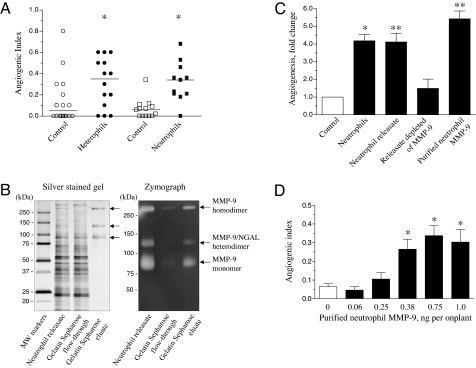
Heterophils have been shown previously to constitute the initial inflammatory cells that infiltrate angiogenic sites and provide a major cellular source of MMP-9 (31). However, the actual molecular components secreted by this analogue of mammalian neutrophils and contributing to CAM angiogenesis were not investigated mechanistically. In the present study, we analyzed the contribution of human neutrophils and their MMP-9 to angiogenesis. Purified human neutrophils and chicken embryo heterophils were incorporated at 5 × 10⁴ cells per collagen onplant. Both types of granulocytes induced significant physiologic angiogenesis (Fig. 1A). A substantial, 4- to 5-fold increase in angiogenesis over control was observed in collagen onplants supplemented with human neutrophils, indicating that isolated neutrophils can functionally replace exogenous heterophils as angiogenic inducers.
Fig. 1.
Angiogenic potential of neutrophils, neutrophil releasate, and purified neutrophil MMP-9. (A) Human neutrophils efficiently induce angiogenesis. Isolated chicken heterophils or human neutrophils were incorporated at 5 × 10⁴ per 30-μl collagen onplant. Control onplants were supplemented with only buffer. Presented is a scatter plot from one of two independent experiments for each cell type. Each point represents the angiogenic index of an individual onplant and horizontal lines are medians (*, P < 0.05). (B) Purification of neutrophil MMP-9. Gelatin Sepharose affinity chromatography was performed on the releasate prepared from PMA-treated neutrophils. Proteins from the original releasate, gelatin Sepharose flow-through fraction, and the DMSO-eluted fraction (eluate) were analyzed by silver staining (Left) and zymography (Right). Positions of molecular mass standards (kDa) are indicated to the left. Zymography analysis indicated gelatinolytic activity of three proteins (arrows) of molecular mass corresponding to the MMP-9 monomer (90–95 kDa), MMP-9/NGAL heterodimer (120–130 kDa), and MMP-9 homodimer (200–300 kDa). (C) MMP-9 is the major angiogenic moiety in neutrophil releasate. Collagen onplants were supplemented with neutrophils (average 1.5 × 10⁴ cells per onplant), neutrophil releasate, purified MMP-9, or releasate depleted of MMP-9 (equivalent of 1.5 × 10⁴ cells per onplant), whereas control onplants (empty bar) contained collagen alone. Bars are means ± SEM fold changes in angiogenesis from three independent experiments (*, P < 0.05; **, P < 0.001). (D) Neutrophil MMP-9 induces angiogenesis in a dose-dependent manner. Collagen onplants were supplemented with purified MMP-9 (0.06–1.0 ng per onplant) or buffer only (empty bar). Bars are means ± SEM angiogenic index from two independent experiments. *, P < 0.05.
Neutrophil MMP-9 Is a Major Angiogenic Contributor.
Immunocytochemical staining with specific MMP-9 antibody confirmed that purified neutrophils contained MMP-9 positive granules [supporting information (SI) Fig. 4 Left]. The neutrophils were induced to release their granule contents with 160 nM phorbol 12-myristate 13-acetate (PMA) at 37°C. Within 20 min the neutrophils degranulated and their MMP-9 immunoreactivity was greatly diminished (SI Fig. 4 Right). The granule contents, referred herein as releasate, were harvested and the neutrophil MMP-9 was then separated from the other releasate proteins by gelatin Sepharose affinity chromatography.
The neutrophil releasate, the flow-through fraction, and the isolated neutrophil MMP-9 were analyzed by SDS/PAGE using silver staining, zymography, and Western blotting (Fig. 1B and SI Fig. 5). Silver-stained gels showed that neutrophil releasate contained numerous proteins, >90% of which were retained in the unbound fraction after gelatin Sepharose chromatography (Fig. 1B Left). The eluate contained only three distinct protein bands with apparent molecular mass of 90–95, 120–130, and 200–300 kDa, characteristic of multiple neutrophil MMP-9 species (32). Parallel zymographic analysis indicated three matching gelatinolytic bands in both the neutrophil releasate and gelatin Sepharose eluate, whereas the unbound fraction was substantially depleted of gelatinase activity (Fig. 1B Right). Corresponding MMP-9-specific bands were identified in the neutrophil releasate and gelatin Sepharose eluate by Western blotting, thus indicating that all three gelatinolytic bands contained human MMP-9 (SI Fig. 5). Western blotting also demonstrated that the intermediate 120- to 130-kDa MMP-9 species contained human neutrophil gelatinase-associated lipocalin (NGAL), confirming isolation of a characteristic neutrophil MMP-9/NGAL heterodimer. Under reducing conditions, all high molecular mass forms of neutrophil MMP-9 collapsed to the 92-kDa proMMP-9 monomer (SI Fig. 5).
Neutrophil releasate and purified neutrophil MMP-9 were compared with intact neutrophils for their angiogenic potential in CAM onplants, and both proved to be potent inducers of physiologic angiogenesis (Fig. 1C). As low as 1.5 ng of purified neutrophil MMP-9 caused a 5-fold amplification in angiogenesis. Importantly, the neutrophil releasate selectively depleted of MMP-9 did not induce angiogenesis above the control levels, suggesting that neutrophil MMP-9 is the major proangiogenic moiety in neutrophil releasate. Furthermore, the purified neutrophil MMP-9 induced angiogenesis in a dose-dependent manner, yielding a 4- to 5-fold increase in angiogenesis at subnanogram levels (Fig. 1D), translating to concentrations in the onplant as low as 100–200 pM.
Activation and Activity of Neutrophil MMP-9 Is Required to Induce Physiologic Angiogenesis.
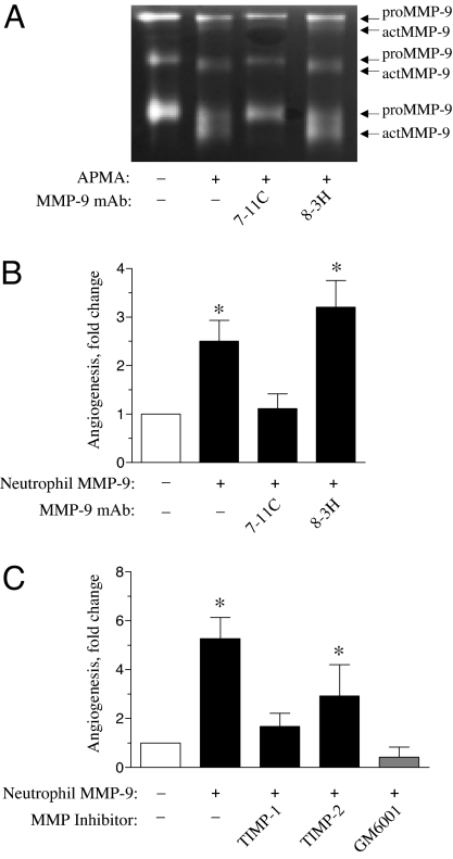
Because neutrophil MMP-9 is released as a zymogen, we analyzed whether subsequent activation of proMMP-9 is involved in the induction of angiogenesis by taking advantage of two human MMP-9-specific mAbs, i.e., 7-11C and 8-3H (33). The mAb 7-11C is capable of blocking activation of proMMP-9, whereas mAb 8-3H binds to proMMP-9, but does not inhibit its activation. Functional reactivity of these mAbs against neutrophil MMP-9 was confirmed with p-aminophenylmercuric acetate (APMA)-mediated activation in vitro (Fig. 2A). APMA induced the conversion of neutrophil proMMP-9 to lower molecular mass active forms as indicated by gelatin zymography. This conversion was blocked by the addition of mAb 7-11C, but not by mAb 8-3H.
Fig. 2.
Angiogenesis induced by neutrophil MMP-9 involves activation of MMP-9 zymogen and proteolytic activity of the activated MMP-9 enzyme. (A) MMP-9-specific mAb 7-11C is capable of blocking activation of neutrophil proMMP-9. A total of 0.4 μg of neutrophil proMMP-9 was activated for 1 h with 3 mM APMA in the presence or absence of 90 μg/ml MMP-9-specific mAb 7-11C or mAb 8-3H. Whereas mAb 8-3H does not prevent APMA-induced processing of proMMP-9, mAb 7-11C efficiently blocks the conversion of proMMP-9 into lower molecular mass activated species (actMMP-9). (B) Activation of neutrophil MMP-9 is required for the induction of angiogenesis. Collagen onplants containing (+) purified neutrophil MMP-9 (2 ng per onplant) were additionally supplemented with 30 ng of mAb 7-11C or mAb 8-3H. Control onplants (empty bar) were supplemented with buffer only (−). (C) Catalytic activity of MMP-9 is required for angiogenesis. TIMP-1 or TIMP-2 in a 5-fold molar excess to MMP-9 or a synthetic MMP inhibitor GM6001 at a final concentration of 5 μM was added to the collagen mixture containing purified neutrophil MMP-9 (2 ng per onplant). Bars are means ± SEM fold changes in angiogenesis over control (no MMP-9 added; empty bar) from three independent experiments. *, P < 0.01.
We next analyzed the effects of the MMP-9-specific mAbs on angiogenesis induced by neutrophil proMMP-9. The activation-blocking mAb 7-11C, added to collagen onplants containing neutrophil proMMP-9, caused a significant reduction in angiogenesis, whereas mAb 8-3H did not affect the angiogenesis levels (Fig. 2B). Importantly, mAb 7-11C also did not change the baseline angiogenesis in onplants containing no MMP-9 (data not shown). These results strongly indicate that activation of neutrophil proMMP-9 is required for the induction of angiogenesis.
Neutrophils are reported to express no TIMPs (32); therefore, neutrophil proMMP-9 would be released as TIMP-free proenzyme. Indeed, we failed to identify even trace levels of human TIMP-1 in the neutrophil releasate by Western blotting (data not shown). To confirm that the proteolytic activity of TIMP-free neutrophil MMP-9 is involved in the induction of physiologic angiogenesis, natural tissue inhibitors of MMPs, TIMP-1 and TIMP-2 were added to the collagen onplants at a 5-fold molar excess to the neutrophil proMMP-9. TIMP-1 caused a substantial decrease (85–90%) in the induced levels of angiogenesis (Fig. 2C), whereas the addition of TIMP-2 resulted in only a 40–45% diminishment, consistent with the reported differential in sensitivity of active MMP-9 to the respective TIMPs (34). The addition of the MMP inhibitor GM6001 almost completely inhibited angiogenesis, confirming further that MMP-9-stimulated angiogenesis depends on the catalytic activity of the activated enzyme.
Angiogenic Capacity of Neutrophil MMP-9 Depends on Its TIMP-Free Status.
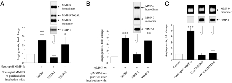
Because modulation of angiogenesis by natural MMP inhibitors was performed in large molar excess of inhibitors to MMP-9, we examined the angiogenic potential of MMP-9 stoichiometrically complexed with TIMP-1. Neutrophil-derived, TIMP-free proMMP-9 was incubated with either TIMP-1 or TIMP-2 in 5-fold molar excess, and then the protein mixture was passed over gelatin-Sepharose to remove excess inhibitor and purify MMP-9/TIMP complexes, which are known to form at a 1:1 stoichiometric ratio (34). After the elution of the bound fraction from the neutrophil MMP-9/TIMP-1 mixture, TIMP-1 was detected in the eluate by both silver staining (data not shown) and Western blotting with a TIMP-1-specific mAb (Fig. 3A Inset). Purified neutrophil proMMP-9/TIMP-1 complexes were analyzed in vivo for angiogenic properties and demonstrated to be completely incapable of inducing angiogenesis (Fig. 3A). TIMP-2, known to bind to activated MMPs but not to proMMP-9 (34, 35), was recovered in the gelatin Sepharose flow-through of the neutrophil MMP-9/TIMP-2 mixture and was not detected in the eluate, thus confirming that no catalytically active form of MMP-9 was present in the original preparation. Furthermore, the TIMP-2-free eluate retained angiogenic potential and induced angiogenesis levels similar to those induced by the repurified control neutrophil proMMP-9 that had been incubated with buffer alone (Fig. 3A).
Fig. 3.
TIMP-1-free proMMP-9 is required for the induction of angiogenesis. (A) Neutrophil proMMP-9 complexed with TIMP-1 loses its proangiogenic capacity. Purified neutrophil proMMP-9 was incubated with PBS or a 5-fold molar excess of TIMP-1 or TIMP-2. MMP-9 and MMP-9/TIMP complexes were purified by gelatin Sepharose affinity chromatography and incorporated at 2 ng (by MMP-9 content) per onplant. Presence of proMMP-9 species and TIMP-1 in the elution fraction from the proMMP-9/TIMP-1 mixture was confirmed by Western blotting with MMP-9- and TIMP-1-specific antibodies (Inset above the corresponding bar). (B) TIMP-free rpMMP-9 induces angiogenesis if not complexed with TIMP-1. Collagen onplants were supplemented with 2.5 ng of rpMMP-9 repurified by gelatin Sepharose affinity chromatography after incubation with PBS or 5-fold molar excess of TIMP-1 or TIMP-2. Composition of MMP-9 monomer/homodimer species and the presence of TIMP-1 in the elution fraction from the rpMMP-9/TIMP-1 mixture were verified by Western blotting with MMP-9- and TIMP-1-specific antibodies (Inset). (C) The proMMP-9/TIMP-1 complex naturally produced by human monocytic and tumor cells is inefficient in the induction of angiogenesis. Collagen onplants were supplemented with 2 ng of proMMP-9 purified from neutrophil releasate or conditioned medium from the U937 cells and HT-1080 fibrosarcoma. Gelatin zymography confirmed presence of proMMP-9 in all gelatin Sepharose elution fractions, whereas TIMP-1-specific mAbs confirmed the presence of TIMP-1 only in the proMMP-9 purified from U937 and HT-1080 conditioned media (Insets above corresponding bars). Bars are means ± SEM fold changes in angiogenesis over control (no MMP-9 added; empty bars) from three independent experiments. *, P < 0.03 in one-tailed Student's t test; ** and ***, P < 0.01 and P < 0.001 in two-tailed Student's t test, respectively.
Overall, these results demonstrate that proenzyme activation and the resulting catalytic activity of native TIMP-1-free neutrophil MMP-9 are required to induce angiogenesis and that neutrophil proMMP-9 stoichiometrically complexed with TIMP-1 dramatically loses its proangiogenic capability.
Angiogenic Capacity of Human proMMP-9 Purified from Different Sources.
Recombinant human proMMP-9 (rpMMP-9) includes mainly monomers and some homodimers, but no NGAL heterodimers characteristic of neutrophil MMP-9. In addition, like neutrophil MMP-9, rpMMP-9 is essentially TIMP-free. When substituted for neutrophil MMP-9, low nanogram levels of rpMMP-9 (1–3 ng) induced a significant, 4-fold increase in angiogenesis (Fig. 3B). However, if rpMMP-9 was complexed with TIMP-1, the purified stoichiometric rpMMP-9/TIMP-1 complex failed to induce angiogenesis. Furthermore, similar to neutrophil MMP-9, rpMMP-9 did not bind TIMP-2 during the preincubation with excess recombinant TIMP-2; thus, the resulting rpMMP-9 eluate yielded similar angiogenic levels as the repurified buffer control rpMMP-9 (Fig. 3B). These results indicate that TIMP-1-free proMMP-9 purified from distinctly separate sources is capable of causing physiologic angiogenesis.
The unique angiogenesis-inducing nature of TIMP-free proMMP-9 was further illustrated when the MMP-9 purified from neutrophils was directly compared with a MMP-9 isolated from both human monocytic and human tumor cells (Fig. 3C). Conditioned medium from PMA-treated U937 cells and HT-1080 fibrosarcoma was harvested, passed over gelatin Sepharose, and selectively eluted, yielding a purified preparation of human proMMP-9. However, unlike human neutrophil proMMP-9, the proMMP-9 produced by U937 and HT-1080 cells is secreted as, or subsequently forms, a stoichiometric complex with TIMP-1 (corresponding Insets in Fig. 3C). Addition of either monocytic- or tumor-derived MMP-9 into collagen onplants demonstrated that it was unable to induce angiogenesis compared with the 5- to 6-fold induction caused by equivalent catalytic amounts of neutrophil MMP-9 (Fig. 3C).
Collectively, these findings indicate that the unique levels of angiogenic activity exhibited by neutrophil MMP-9 could be attributed to its distinct TIMP-free nature. In contrast, monocytic proMMP-9 and tumor cell proMMP-9, both naturally complexed with TIMP-1, are clearly nonangiogenic, which illustrates a critical aspect of neutrophil MMP-9 in tumor angiogenesis and highlights a possible mechanism involved in regulating the overall angiogenic capacity of MMP-9.
Discussion
Despite general agreement that inflammatory neutrophils are an important source of MMP-9 at the angiogenic site during primary tumor development, the specific catalytic functions of neutrophil MMP-9 and their regulation have not been formally linked with the angiogenic switch in an in vivo model. Therefore, no underlying mechanism has been proposed to explain why neutrophil MMP-9 might function as a critical proangiogenic protease. Previously, we used a quantitative angiogenesis model in the chicken embryo (28) and demonstrated that avian inflammatory cells and their MMPs, including monocyte MMP-13 and heterophil MMP-9, were critical for the induction of both physiologic and tumor angiogenesis (30, 31). By taking advantage of this system, which allows for direct access to the angiogenic site and testing of intact viable cells and their delivered products, we directly analyzed for the involvement of functioning human neutrophils and the catalytic capabilities of their MMP-9 in angiogenesis.
First, as few as 1–5 × 10⁴ neutrophils per onplant can provide enough proangiogenic stimuli to increase the angiogenesis levels by 4- to 5-fold in as short a time period as 3 days. Because neutrophil response at the site of inflammation is associated with rapid granule release (24, 32), we verified that the neutrophil granule contents contained the majority, if not all, proangiogenic moieties. By separating via gelatin Sepharose affinity chromatography the only gelatinase produced by neutrophils, i.e., MMP-9, from the bulk of released proteins, we demonstrated directly that neutrophil MMP-9 was a critical proangiogenic molecule. Although neutrophils indeed contain many proangiogenic factors, including VEGF, the amount of VEGF released by stimulated neutrophils and retained in the MMP-9-depleted releasate, i.e., <0.5 pg per 5 × 10⁴ cells (36), could not induce angiogenesis in our model (28). In contrast, 1.5 ng of MMP-9, released by the same numbers of neutrophils, triggered a 5-fold increase in angiogenesis levels, indicating its unique proangiogenic potential. As a proof of principal, human recombinant MMP-9 was demonstrated to be proangiogenic at similar subnanogram concentrations (0.2–0.7 nM).
Using a specific antibody capable of inhibiting proteolytic processing of proMMP-9, we were able to demonstrate that proenzyme activation is required for induction of angiogenesis. However, as in many other in vivo model systems, the natural activators of proMMP-9 in our model system are unknown. Autoactivation by conformational alteration of proMMP-9 upon substrate binding has been demonstrated in vitro (37), but in vivo it remains an unexplored possibility. Direct activation of proMMP-9 by another protease such as MMP-3 appears more likely, considering that MMP-3 is highly capable of processing and activating the MMP-9 zymogen (38, 39).
Further experiments involving direct addition of natural and synthetic MMP inhibitors confirmed that the catalytic activity of the neutrophil MMP-9 enzyme is involved in angiogenesis. The differential inhibition of MMP-9-induced angiogenesis by 5-fold molar excess levels of TIMP-1 versus TIMP-2 was consistent with the known difference in the ki values of active MMP-9 for the two TIMPs (34) and additionally indicated that neutrophil proMMP-9 undergoes activation before catalytically stimulating angiogenesis. The possible substrates that active MMP-9 could enzymatically modify include a number of ECM components (e.g., native and denatured type IV and type I collagens) that upon cleavage might provide a remodeled scaffold for the sprouting endothelium. MMP-9 enzyme might also release sequestered growth factors or cytokines such as VEGF and basic FGF or directly generate cleaved products that are specifically proangiogenic (7). These MMP-9-generated angiogenic factors could continue to function even though MMP-9 may be subsequently cleared from the tissue. Conversely, active MMP-9 might also degrade and thus eliminate proteinaceous inhibitors of angiogenesis that normally maintain the angiostatic state. That any one of these MMP-9-mediated cleavage reactions occurs in vivo with the appropriate kinetics and is directly responsible for MMP-9-induced angiogenesis has not yet been demonstrated in any model system. The rapid and potent vascular response of the CAM tissue to subnanomolar levels of neutrophil MMP-9 indicates that this model system may help identify critical MMP-9 substrates.
The presence of previously characterized forms of neutrophil MMP-9, i.e., monomer, heterodimer with NGAL, and homodimer (40, 41), raised a question regarding which species are responsible for the angiogenic induction. All three forms have been found to be proteolytically active against gelatin and native collagens (40). Biochemically, both monomer and homodimer forms are equally susceptible to catalytic activation by MMP-3 and inhibition by TIMP-1, and enzymatically are quite similar in substrate proteolysis (35). The possibility that NGAL in the MMP-9/NGAL heterodimer was responsible for the unique proangiogenic properties of neutrophil MMP-9 was excluded in the experiments, demonstrating that recombinant MMP-9, which is NGAL-free, was angiogenically equal to neutrophil MMP-9. Furthermore, when purified NGAL was exogenously added alone or with recombinant MMP-9, it did not provide any significant additive or synergistic angiogenic activity (data not shown). It is possible however, that NGAL might partially contribute to the effectiveness of neutrophil MMP-9 by diminishing the turnover of the enzyme in targeted tissues as evidenced by the studies of Moses and colleagues (42), who demonstrated the stabilizing effect of NGAL on the MMP-9/NGAL heterodimer. Therefore, it appears that the native neutrophil MMP-9 monomer moiety, possibly assembling into multimers within collagen onplants, is sufficient to induce angiogenesis in our model.
Our further analysis was focused on a possible biochemical explanation for why MMP-9 released by neutrophils acts as a uniquely potent proangiogenic molecule. Two major types of inflammatory cells have been linked to the angiogenic switch at the site of primary tumor development, i.e., monocytes/macrophages and granulocytes, including neutrophils. These cell types can be stimulated to produce and secrete MMP-9, therefore facilitating an angiogenic switching mechanism. However, only neutrophils have been shown to produce little or no TIMP-1, thus releasing their prestored MMP-9 as a TIMP-free proenzyme (32). In contrast, monocytes synthesize MMP-9 de novo in response to chemotactic factors, cellular interactions, or adhesion to ECM (32, 43). Furthermore, the induction of MMP-9 synthesis in cells of monocytic origin is accompanied by production of TIMP-1 (32, 44) and generation of MMP-9/TIMP-1 complexes (45). By using affinity purification, we have affirmed that freshly isolated peripheral blood mononuclear cells stimulated with PMA indeed release proMMP-9 complexed with TIMP-1 (data not shown). Importantly, proMMP-9 complexed with TIMP-1 has been shown to be significantly impaired in its activation (45, 46), apparently because TIMP-1, bound to proMMP-9 at its C terminus, maintains its free N-terminal region available for inhibiting potential MMP-9 activators, such as MMP-3, or for complexing and blocking any active MMP-9 molecules generated via autoactivation.
To emphasize that the neutrophil MMP-9 is released as a TIMP-free zymogen, readily available for activation and further enzymatic functions, we generated and purified neutrophil MMP-9/TIMP-1 complex and tested its angiogenic potential. Neutrophil MMP-9 presented in the MMP-9/TIMP-1 complex, which recapitulates the presentation of MMP-9 by most other cell types, completely lost the capacity to induce angiogenesis. Similarly, a stoichiometric complex of recombinant MMP-9 and TIMP-1 was ineffective in the induction of angiogenesis. A most interesting finding in this regard is the observation that the MMP-9/TIMP-1 complex naturally produced by human monocytic U937 cells and tumor HT-1080 fibrosarcoma did not exhibit any proangiogenic properties when added at levels of equal catalytic potential to that of neutrophil MMP-9. These findings clearly indicate that the TIMP-free status of neutrophil MMP-9 appears to be a prerequisite of its unique angiogenic characteristics.
In conclusion, our study not only links directly the activation and catalytic functions of MMP-9 at the site of angiogenesis, but also explains biochemically why neutrophil MMP-9 may serve as a unique proangiogenic molecule at the sites of physiologic and tumor angiogenesis. Our results demonstrate that the TIMP-free status of neutrophil MMP-9, readily available for rapid release by neutrophils upon their influx into target tissue, is the major factor determining the high levels of its angiogenic functionality.
Materials and Methods
Recombinant Proteins and Antibodies.
rpMMP-9 and recombinant TIMP-1 and TIMP-2 were a generous gift from Rafael Fridman (Wayne State University School of Medicine, Detroit). The broad-range hydroxamate MMP inhibitor (GM6001) was purchased from Sigma. mAbs specific to human MMP-9 were generated in our laboratory (33). Murine mAbs against TIMP-1 (clone 7-6C1) and TIMP-2 (clone 67-4HIII) were purchased from EMD Biosciences, and rat mAb (clone 220310) to NGAL (lipocalin 2) was from R&D. Purified NGAL was kindly provided by Marsha Moses (Children's Hospital, Boston).
CAM Angiogenesis Assay.
CAM angiogenesis assay, a modification of the original assay designed by Folkman and colleagues (29), was performed by using shell-free COFAL-negative White Leggorn chick embryos essentially as described (28). To prepare onplants, type I rat tail collagen (BD Biosciences) was neutralized and used at a final concentration of 2.1 mg/ml. Tested ingredients, such as isolated granulocytes, neutrophil contents, or purified proteins, were incorporated into the collagen mixture at final concentrations indicated in Results and figure legends. Four to six onplants, containing 30 μl of collagen mixture polymerized within two grid meshes, were grafted on the CAM of each embryo. At least two independent experiments were performed for each condition with five to six embryos per variable. Angiogenesis was scored at 70–90 h with a stereomicroscope. Blood vessels visualized in the grids of the upper mesh were regarded as angiogenic. Angiogenesis level was determined either as an angiogenic index, i.e., percentage of angiogenic grids (number of grids containing blood vessels over the total number of grids scored) or as a fold difference of a variable (intact cells or cell components added into collagen) over control (collagen alone).
Isolation of Chicken Heterophils and Human Neutrophils.
Heterophils were isolated from the peripheral blood of 18-day-old chick embryos as described (31). Neutrophils were purified from heparinized human peripheral blood by 6% dextran sedimentation, followed by red blood cell lysis with 0.15 M NH4Cl and gradient centrifugation with 1.077 Histopaque. Purity of neutrophil isolation was 80–90% as confirmed by Hema 3 staining.
Cell Cultures and Treatments.
Human monocytic U937 cells (ATCC) were maintained in RPMI medium 1640/10% FCS. To induce MMP-9 production, the U937 cells were incubated for 72 h at 2 × 10⁶ cells/ml of serum-free medium supplemented with 160 mM PMA. Human HT-1080 fibrosarcoma (ATCC) was maintained in DMEM/10% FCS. To increase MMP-9 production, the HT-1080 cultures were treated for 24 h with 160 mM PMA in serum-free DMEM.
Purification of MMP-9 and MMP-9/TIMP Complexes.
Neutrophils were induced to release their granule contents by incubating for 1–2 h at 37°C with 160 nM PMA (Sigma). After centrifugation at 1,200 × g for 10 min at 4°C, the cell-free supernatant (neutrophil releasate) was collected and kept frozen at −80°C until use. To purify neutrophil MMP-9, neutrophil releasate was incubated with gelatin Sepharose (Amersham Biosciences) for 1–2 h at room temperature. Because U937 cells also produce MMP-2, the MMP-9 induced by PMA treatment was separated from MMP-2 by immunoprecipitation with anti-MMP-9 mAb 3-8H and protein G Sepharose. Bound IgG/MMP-9 complexes were eluted with glycine, pH 3.5, and immediately neutralized with Tris buffer, pH 7.4. Eluted MMP-9 was then separated from IgG by gelatin Sepharose chromatography. To isolate proMMP-9 produced by PMA-treated fibrosarcoma HT-1080 cells, conditioned medium was incubated with gelatin Sepharose. To isolate stoichiometric complexes of MMP-9 with TIMP-1 or TIMP-2, rpMMP-9 or purified neutrophil MMP-9 was incubated with a 5-fold molar excess of recombinant TIMP-1 or TIMP-2 for 1 h at room temperature, followed by incubation with gelatin Sepharose. Elution of bound proteins from gelatin Sepharose was performed with 10% DMSO. Proteins in the neutrophil releasate, the gelatin Sepharose flow-through, and eluate fractions were analyzed by silver staining, zymography, and Western blotting.
Silver Staining, Zymography, and Western Blotting.
Silver staining of the SDS/PAGE gels was performed as described (47). Quantitation of proteins in the gels was performed by using BSA standards within a 1- to 20-ng range. Gelatin zymography was performed as described (31). For Western blotting, proteins were separated by 4–20% SDS/PAGE and transferred to PVDF membranes. The membranes were blocked with 5% nonfat dry milk/PBS/0.1% Tween 20, incubated with 1 μg/ml of primary mAb, washed, and incubated with corresponding HRP-conjugated secondary goat anti-mouse (Pierce) or anti-rat (Invitrogen) antibodies. Specific bands were visualized after developing with SuperSignal West Pico Chemilumninescent Substrate (Pierce).
Immunocytochemistry.
Neutrophils were fixed on glass slides with cold methanol and treated with 0.3% H2O2. Nonspecific binding was blocked with 2% BSA and 2% normal goat serum. The slides were incubated with 10 μg/ml of human MMP-9-specific mAb 8-3H overnight at 4°C, followed by incubation with secondary goat anti-mouse biotinylated antibody for 30 min at room temperature and then with AvidinD-HRP conjugate (both from Vector). After washing, the slides were incubated with a diaminobenzidine chromogenic substrate and counterstained with Mayer hematoxylin.
Statistical Analysis.
Statistical analysis was performed by unpaired Student's t test for P < 0.05. Angiogenesis levels are presented either as scatter plots where each data point represents angiogenic index of individual onplant or as bar graphs representing the means ± SEM. Mean fold difference was calculated from the ratios of angiogenic index determined for individual onplants over the mean of angiogenesis index determined for control in each independent experiment.
Supplementary Material
ACKNOWLEDGMENTS.
We thank Dr. Rafael Fridman for recombinant human proteins, including MMP-2, MMP-9, TIMP-1, and TIMP-2; Dr. Marsha Moses for recombinant NGAL; and Chenxing Li and Matt Crawford for excellent technical assistance. This work was supported by National Institutes of Health Grants CA55852 and CA105412 (to J.P.Q.) and T32 HL07195 (to V.C.A.).
Footnotes
The authors declare no conflict of interest.
This article is a PNAS Direct Submission.
This article contains supporting information online at www.pnas.org/cgi/content/full/0706438104/DC1.
References
- 1.Carmeliet P, Jain RK. Nature. 2000;407:249–257. doi: 10.1038/35025220. [DOI] [PubMed] [Google Scholar]
- 2.Folkman J. Annu Rev Med. 2006;57:1–18. doi: 10.1146/annurev.med.57.121304.131306. [DOI] [PubMed] [Google Scholar]
- 3.Hanahan D, Folkman J. Cell. 1996;86:353–364. doi: 10.1016/s0092-8674(00)80108-7. [DOI] [PubMed] [Google Scholar]
- 4.Bergers G, Benjamin LE. Nat Rev Cancer. 2003;3:401–410. doi: 10.1038/nrc1093. [DOI] [PubMed] [Google Scholar]
- 5.Egeblad M, Werb Z. Nat Rev Cancer. 2002;2:161–174. doi: 10.1038/nrc745. [DOI] [PubMed] [Google Scholar]
- 6.Deryugina EI, Quigley JP. Cancer Metastasis Rev. 2006;25:9–34. doi: 10.1007/s10555-006-7886-9. [DOI] [PubMed] [Google Scholar]
- 7.Mott JD, Werb Z. Curr Opin Cell Biol. 2004;16:558–564. doi: 10.1016/j.ceb.2004.07.010. [DOI] [PMC free article] [PubMed] [Google Scholar]
- 8.Coussens LM, Werb Z. Nature. 2002;420:860–867. doi: 10.1038/nature01322. [DOI] [PMC free article] [PubMed] [Google Scholar]
- 9.Parks WC, Wilson CL, Lopez-Boado YS. Nat Rev Immunol. 2004;4:617–629. doi: 10.1038/nri1418. [DOI] [PubMed] [Google Scholar]
- 10.Elkington PT, O'Kane CM, Friedland JS. Clin Exp Immunol. 2005;142:12–20. doi: 10.1111/j.1365-2249.2005.02840.x. [DOI] [PMC free article] [PubMed] [Google Scholar]
- 11.Balkwill F, Charles KA, Mantovani A. Cancer Cell. 2005;7:211–217. doi: 10.1016/j.ccr.2005.02.013. [DOI] [PubMed] [Google Scholar]
- 12.Condeelis J, Pollard JW. Cell. 2006;124:263–266. doi: 10.1016/j.cell.2006.01.007. [DOI] [PubMed] [Google Scholar]
- 13.Giraudo E, Inoue M, Hanahan D. J Clin Invest. 2004;114:623–633. doi: 10.1172/JCI22087. [DOI] [PMC free article] [PubMed] [Google Scholar]
- 14.Huang S, Van Arsdall M, Tedjarati S, McCarty M, Wu W, Langley R, Fidler IJ. J Natl Cancer Inst. 2002;94:1134–1142. doi: 10.1093/jnci/94.15.1134. [DOI] [PubMed] [Google Scholar]
- 15.Witko-Sarsat V, Rieu P, Descamps-Latscha B, Lesavre P, Halbwachs-Mecarelli L. Lab Invest. 2000;80:617–653. doi: 10.1038/labinvest.3780067. [DOI] [PubMed] [Google Scholar]
- 16.Hao Q, Chen Y, Zhu Y, Fan Y, Palmer D, Su H, Young WL, Yang GY. J Cereb Blood Flow Metab. 2007;27:1853–1860. doi: 10.1038/sj.jcbfm.9600485. [DOI] [PubMed] [Google Scholar]
- 17.Nozawa H, Chiu C, Hanahan D. Proc Natl Acad Sci USA. 2006;103:12493–12498. doi: 10.1073/pnas.0601807103. [DOI] [PMC free article] [PubMed] [Google Scholar]
- 18.Coussens LM, Tinkle CL, Hanahan D, Werb Z. Cell. 2000;103:481–490. doi: 10.1016/s0092-8674(00)00139-2. [DOI] [PMC free article] [PubMed] [Google Scholar]
- 19.Acuff HB, Carter KJ, Fingleton B, Gorden DL, Matrisian LM. Cancer Res. 2006;66:259–266. doi: 10.1158/0008-5472.CAN-05-2502. [DOI] [PMC free article] [PubMed] [Google Scholar]
- 20.Bergers G, Brekken R, McMahon G, Vu TH, Itoh T, Tamaki K, Tanzawa K, Thorpe P, Itohara S, Werb Z, Hanahan D. Nat Cell Biol. 2000;2:737–744. doi: 10.1038/35036374. [DOI] [PMC free article] [PubMed] [Google Scholar]
- 21.Yang L, DeBusk LM, Fukuda K, Fingleton B, Green-Jarvis B, Shyr Y, Matrisian LM, Carbone DP, Lin PC. Cancer Cell. 2004;6:409–421. doi: 10.1016/j.ccr.2004.08.031. [DOI] [PubMed] [Google Scholar]
- 22.Jodele S, Blavier L, Yoon JM, DeClerck YA. Cancer Metastasis Rev. 2006;25:35–43. doi: 10.1007/s10555-006-7887-8. [DOI] [PubMed] [Google Scholar]
- 23.Owen CA, Campbell EJ. J Leukocyte Biol. 1999;65:137–150. doi: 10.1002/jlb.65.2.137. [DOI] [PubMed] [Google Scholar]
- 24.Van den Steen PE, Dubois B, Nelissen I, Rudd PM, Dwek RA, Opdenakker G. Crit Rev Biochem Mol Biol. 2002;37:375–536. doi: 10.1080/10409230290771546. [DOI] [PubMed] [Google Scholar]
- 25.Bjorklund M, Koivunen E. Biochim Biophys Acta. 2005;1755:37–69. doi: 10.1016/j.bbcan.2005.03.001. [DOI] [PubMed] [Google Scholar]
- 26.Hamano Y, Zeisberg M, Sugimoto H, Lively JC, Maeshima Y, Yang C, Hynes RO, Werb Z, Sudhakar A, Kalluri R. Cancer Cell. 2003;3:589–601. doi: 10.1016/s1535-6108(03)00133-8. [DOI] [PMC free article] [PubMed] [Google Scholar]
- 27.Heissig B, Hattori K, Dias S, Friedrich M, Ferris B, Hackett NR, Crystal RG, Besmer P, Lyden D, Moore MA, Werb Z, Rafii S. Cell. 2002;109:625–637. doi: 10.1016/s0092-8674(02)00754-7. [DOI] [PMC free article] [PubMed] [Google Scholar]
- 28.Seandel M, Noack-Kunnmann K, Zhu D, Aimes RT, Quigley JP. Blood. 2001;97:2323–2332. doi: 10.1182/blood.v97.8.2323. [DOI] [PubMed] [Google Scholar]
- 29.Nguyen M, Shing Y, Folkman J. Microvasc Res. 1994;47:31–40. doi: 10.1006/mvre.1994.1003. [DOI] [PubMed] [Google Scholar]
- 30.Zijlstra A, Aimes RT, Zhu D, Regazzoni K, Kupriyanova T, Seandel M, Deryugina EI, Quigley JP. J Biol Chem. 2004;279:27633–27645. doi: 10.1074/jbc.M313617200. [DOI] [PubMed] [Google Scholar]
- 31.Zijlstra A, Seandel M, Kupriyanova TA, Partridge JJ, Madsen MA, Hahn-Dantona EA, Quigley JP, Deryugina EI. Blood. 2006;107:317–327. doi: 10.1182/blood-2005-04-1458. [DOI] [PMC free article] [PubMed] [Google Scholar]
- 32.Opdenakker G, Van den Steen PE, Dubois B, Nelissen I, Van Coillie E, Masure S, Proost P, Van Damme J. J Leukocyte Biol. 2001;69:851–859. [PubMed] [Google Scholar]
- 33.Ramos-DeSimone N, Moll UM, Quigley JP, French DL. Hybridoma. 1993;12:349–363. doi: 10.1089/hyb.1993.12.349. [DOI] [PubMed] [Google Scholar]
- 34.Olson MW, Gervasi DC, Mobashery S, Fridman R. J Biol Chem. 1997;272:29975–29983. doi: 10.1074/jbc.272.47.29975. [DOI] [PubMed] [Google Scholar]
- 35.Olson MW, Bernardo MM, Pietila M, Gervasi DC, Toth M, Kotra LP, Massova I, Mobashery S, Fridman R. J Biol Chem. 2000;275:2661–2668. doi: 10.1074/jbc.275.4.2661. [DOI] [PubMed] [Google Scholar]
- 36.Schruefer R, Lutze N, Schymeinsky J, Walzog B. Am J Physiol. 2005;288:H1186–H1192. doi: 10.1152/ajpheart.00237.2004. [DOI] [PubMed] [Google Scholar]
- 37.Bannikov GA, Karelina TV, Collier IE, Marmer BL, Goldberg GI. J Biol Chem. 2002;277:16022–16027. doi: 10.1074/jbc.M110931200. [DOI] [PubMed] [Google Scholar]
- 38.Ogata Y, Enghild JJ, Nagase H. J Biol Chem. 1992;267:3581–3584. [PubMed] [Google Scholar]
- 39.Ramos-DeSimone N, Hahn-Dantona E, Sipley J, Nagase H, French DL, Quigley JP. J Biol Chem. 1999;274:13066–13076. doi: 10.1074/jbc.274.19.13066. [DOI] [PubMed] [Google Scholar]
- 40.Hibbs MS, Hasty KA, Seyer JM, Kang AH, Mainardi CL. J Biol Chem. 1985;260:2493–2500. [PubMed] [Google Scholar]
- 41.Kjeldsen L, Johnsen AH, Sengelov H, Borregaard N. J Biol Chem. 1993;268:10425–10432. [PubMed] [Google Scholar]
- 42.Yan L, Borregaard N, Kjeldsen L, Moses MA. J Biol Chem. 2001;276:37258–37265. doi: 10.1074/jbc.M106089200. [DOI] [PubMed] [Google Scholar]
- 43.Galt SW, Lindemann S, Medd D, Allen LL, Kraiss LW, Harris ES, Prescott SM, McIntyre TM, Weyrich AS, Zimmerman GA. Circ Res. 2001;89:509–516. doi: 10.1161/hh1801.096339. [DOI] [PubMed] [Google Scholar]
- 44.Lacraz S, Nicod LP, Chicheportiche R, Welgus HG, Dayer JM. J Clin Invest. 1995;96:2304–2310. doi: 10.1172/JCI118286. [DOI] [PMC free article] [PubMed] [Google Scholar]
- 45.Watanabe H, Nakanishi I, Yamashita K, Hayakawa T, Okada Y. J Cell Sci. 1993;104:991–999. doi: 10.1242/jcs.104.4.991. [DOI] [PubMed] [Google Scholar]
- 46.Ogata Y, Itoh Y, Nagase H. J Biol Chem. 1995;270:18506–18511. doi: 10.1074/jbc.270.31.18506. [DOI] [PubMed] [Google Scholar]
- 47.Mortz E, Krogh TN, Vorum H, Gorg A. Proteomics. 2001;1:1359–1363. doi: 10.1002/1615-9861(200111)1:11<1359::AID-PROT1359>3.0.CO;2-Q. [DOI] [PubMed] [Google Scholar]
Associated Data
This section collects any data citations, data availability statements, or supplementary materials included in this article.


